(Annals of the Brazilian Academy of Sciences) ISSN 0001-3765
www.scielo.br/aabc
High susceptibility of activated lymphocytes to oxidative
stress-induced cell death
GIOVANNA R. DEGASPERI, ROGER F. CASTILHO and ANIBAL E. VERCESI
Departamento de Patologia Clínica, Faculdade de Ciências Médicas, Universidade Estadual de Campinas (UNICAMP) 13083-887 Campinas, SP, Brasil
Manuscript received on August 31, 2007; accepted for publication on January 21, 2008;
contributed byANIBALE. VERCESI*
ABSTRACT
The present study provides evidence that activated spleen lymphocytes from Walker 256 tumor bearing rats are more susceptible than controls totert-butyl hydroperoxide (t-BOOH)-induced necrotic cell deathin vitro. The iron chelator and antioxidant deferoxamine, the intracellular Ca2+chelator BAPTA, the L-type Ca2+channel antagonist nifedipine or the mitochondrial permeability transition inhibitor cyclosporin A, but not the calcineurin inhibitor FK-506, render control and activated lymphocytes equally resistant to the toxic effects of t-BOOH. Incubation of activated lymphocytes in the presence of t-BOOH resulted in a cyclosporin A-sensitive decrease in mitochondrial membrane potential. These results indicate that the higher cytosolic Ca2+level in activated lymphocytes increases their susceptibility to oxidative stress-induced cell death in a mechanism involving the participation of mitochondrial permeability transition.
Key words: cell death, free radicals, immune response, lymphopenia, mitochondrial permeability transition, spleen
lymphocyte.
INTRODUCTION
Lymphocyte activation is an essential step in the im-mune response that generally occurs during tumor de-velopment; it requires T-cell recognition of specific tu-mor antigens through the interaction of the lymphocyte receptor with the major histocompatibility complex of antigen-presenting cells (Hermiston et al. 2002, Roy 2003, Grossman et al. 2004). Elevations in cytosolic Ca2+concentration ([Ca2+]
cyt) and reactive oxygen
spe-cies (ROS) production are characteristically observed in such activated lymphocytes, and are important factors associated with either a high rate of cell proliferation or mechanisms that lead to cell death (Buttke and Sand-strom 1995, Williams and Kwon 2004, Feske 2007). It is proposed that a high expression of anti-apoptotic fac-tors, including interleukin-2, mitochondrial uncoupling
*Member Academia Brasileira de Ciências Correspondence to: Anibal E. Vercesi E-mail: [email protected]
protein-2, and members of the Bcl-2 family, increases the resistance of activated lymphocytes to cell death, pushing the cell towards proliferation (Quintana et al. 2005, Arnold et al. 2006, Degasperi et al. 2006a, Kram-mer et al. 2007).
In the present study, the effects of the tert-butyl hydroperoxide (t-BOOH), a well known prooxidant, were evaluated on proliferation and death of activated spleen lymphocytes from Walker 256 tumor bearing (TB) rats (Quintana et al. 2005, Degasperi et al. 2006a, b) and lymphocytes from control (CT) rats. t-BOOH in-duces oxidative stress by exhausting cellular GSH and NADPH, substrates of the antioxidant enzymes gluta-thione peroxidase and glutagluta-thione reductase, respect-ively, thus favoring the accumulation of H2O2(Jocelyn and Dickson 1980, Bernardes et al. 1986, Castilho et al. 1995). In addition, t-BOOH promotes a Ca2+-stimulated
results from our group indicated that the enhanced and continuous production of ROS by mitochondria treated with Ca2+plus t-BOOH leads to a form of non-selective
permeabilization of the inner mitochondrial membrane know as the mitochondrial permeability transition (MPT) (Castilho et al. 1995, Kowaltowski et al. 2001).
The results presented here demonstrate that activ-ated TB rat lymphocytes are more susceptible to necrotic death induced by t-BOOH-mediated oxidative stress than are control cells.
MATERIALS AND METHODS
CHEMICALS
Thetert-butyl hydroperoxide (t-BOOH), carbonyl cya-nide m-chloro phenyl hydrazone (CCCP), cyclosporin A (CsA), oligomycin, deferoxamine, nifedipine and pro-pidium iodide (PI) were obtained from Sigma (St. Louis, MO, USA). BAPTA-AM (1,2-bis(2-aminophenoxy) ethane N,N,N′,N′-tetraacetic acid, tetraacetoxymethyl
ester), 3,3′-dihexyloxacarbocyanine iodide (DioC6(3)),
Fluo-3AM and Pluronic acid F-127 were purchased from Molecular Probes Inc. (Eugene, OR, USA). Ficoll-PaqueTM PLUS was obtained from Becton Dickinson
Biosciences (San Jose, CA, USA), RPMI 1640 (Roswell park medium) from Cultlab (Campinas, Brazil), and FK-506 from LC Laboratories (Woburn, MA, USA). Annexin V was purchased from the Laboratory of Im-munobiology of the University of São Paulo (São Paulo, Brazil). All other chemicals were standard commercial products of reagent-grade quality.
ANIMALTREATMENT
Nine-week-old male Wistar rats (Rattus novergicus al-binos) were obtained from the UNICAMP Central An-imal Breeding Center. Rats were kept under standard laboratory conditions (20-22◦C and 12 h/12 h light/dark
cycle) with free access to a standard diet (Labina/Purina, Campinas, SP, Brazil) and tap water. Animal experi-ments followed the University guidelines for the use of animals in experimental studies (protocol no. 487-1, ap-proved by the UNICAMP Ethics Committee in 2002) and followed the Guide for the Care and Use of Labora-tory Animals published by the US National Institute of Health (NIH publication no. 85-23, revised in 1996).
The Walker 256 tumor cell line (originally obtained
from the Christ Hospital Line, National Cancer Insti-tute Bank, Cambridge, USA) is kept frozen in liquid ni-trogen. For experimental procedures, Walker 256 cells were maintained by consecutive intraperitoneal inocu-lums, each with 20× 106 tumor cells in
phosphate-buffered saline solution (PBS). Walker 256 tumor cells with viability>98% isolated from the ascitic fluid were used for intraperitoneal inoculums, with viability esti-mated by the trypan blue exclusion method.
SPLEENLYMPHOCYTEISOLATION
Rat spleens were gently homogenized in a manual Dounce homogenizer. Spleen homogenates were over-laid onto a Ficoll-PaqueTMPLUS layer, with density
ad-justed to 1.076 g/ml, and centrifuged at 1000 g at room temperature for 25 min. The interface cell layer contain-ing lymphomonocytes was recovered by Pasteur pipette, washed twice in PBS, and centrifuged at 500 g for 10 min (Boyum 1976, Degasperi et al. 2006a, b). Cells were counted in a Neubauer chamber, and cell viability was determined by the trypan blue exclusion method. Cells were only used when viability was greater than 98%.
CELLCULTURE ANDTREATMENTS
The in vitro lymphocyte cultures were obtained by seeding isolated spleen lymphocytes at an initial den-sity of 106cells/ml in RPMI 1640 in 1.5 cm2plate wells in a humidified atmosphere (5% CO2at 37◦C). After 1 h,
lymphocytes from TB rats showed a more rapid growth rate than those of the CT rats. At this time, the TB rat lymphocytes were again counted and diluted to 106 cells/ml in RPMI 1640. CT and TB rat lymphocytes were than treated with t-BOOH in the presence of deferoxam-ine, nifedipdeferoxam-ine, cyclosporin A or FK-506, as indicated in the figure legends.
For BAPTA loading, the lymphocytes were pre-incubated with 20µM BAPTA-AM, after 1 h, the lym-phocytes were washed, counted and diluted to 106cells/ ml in RPMI 1640. BAPTA-loaded CT and TB rat lym-phocytes were than incubated under control conditions or treated with t-BOOH for up to 4 h.
FLOWCYTOMETRY
cytome-ter equipped with an argon laser and CellQuest software (Becton Dickinson, San Jose, CA, USA). Ten thousand events were acquired from each sample. The lymphocyte populations were identified by their light-scattering char-acteristics, enclosed in electronic gates, and analyzed for intensity of the fluorescent probe signal (Degasperi et al. 2006a).
ANALYSIS OFCELLVIABILITY BYANNEXIN-V ANDPI STAINING
Lymphocytes were labelled with annexin-V following the manufacturer’s instructions (Brumatti et al. 2003). Briefly, 106cells were harvested at each point in time, washed twice with PBS and resuspended in a binding buffer containing annexin V-FITC (1:500). After 20 min of incubation at room temperature, the lymphocytes were centrifuged at 1000 g for 5 min and resuspended in bind-ing buffer containbind-ing PI (1:50). Quantitative determi-nations of apoptotic and necrotic cells were carried out as previously described (Martin et al. 1995).
MEASUREMENT OFCYTOSOLICFREECA2+ CONCENTRATIONS
Cytosolic free Ca2+ concentrations ([Ca2+]
cyt) were
monitored with the green fluorescent probe Fluo-3AM (5µM), a single wavelength indicator, by flow cytome-try. Spleen lymphocytes (106cells/mL) in RPMI 1640 medium were loaded with 5µM Fluo-3AM containing 1µM pluronic acid F-127 (for proper dispersal) and 30µg/mL BSA in a humified CO2incubator (5% CO2) at 37◦C for 40 min. Nonhydrolyzed Fluo-3 AM was
re-moved by washing the cells with medium just before flu-orescence acquisition. Calibration was performed at the end of each experiment. [Ca2+]
cytwas calculated
con-sidering Kdof the Ca2+-Fluo-3 complex 390 nM (Mather
and Rottenberg 2002).
DETERMINATION OFMITOCHONDRIALMEMBRANE ELECTRICALPOTENTIAL(m) USINGFLOW CYTOMETRY
After experimental incubation, lymphocytes (106cells/ ml) were washed in PBS buffer and incubated with 0.2 nM DioC6(3) in 400µl of RPMI 1640 medium for 30 min at 37◦C in a humidified CO2 incubator (5%
CO2). One half of each cell sample (200µl) was sepa-rated into a new tube, with the addition of 50µM CCCP a
protonophore that dissipatesm (Rottenberg and Wu
1998, Campos et al. 2004). Both samples were incu-bated for a further 30 minutes. Oligomycin (1µg/ml) was added during the last 10 min prior to FACS analy-sis, as indicated in the figure. In non-saturated concen-trations, DioC6(3) binds preferentially to mitochondria, since electrical potential of the mitochondrial (m) is
much higher (–180 mV) than that of the plasma mem-brane (–60 mV). Results were normalized using the F/FCCCP ratio, where F is the mean fluorescence
inten-sity of DioC6(3) (maximum fluorescence) and FCCCPis
the mean fluorescence in the presence of CCCP (mini-mum fluorescence).
STATISTICALANALYSIS
Three independent experiments were performed, each in triplicate, with results displayed as average ± SD;
significance was assessed by ANOVA and a post-hoc Tukey test. The results of cell viability estimated by an-nexin-V and propidium iodide staining were analysed by a Mann-Whitney U test. Level of significance was set at p<0.05 using SAS software (Statistical Analysis System 6.12/SAS Institute Inc, 1989-1996, Cary, NC, USA).
RESULTS
HIGH SUSCEPTIBILITY OF ACTIVATEDLYMPHOCYTES TO T-BOOH-INDUCEDCELLDEATH
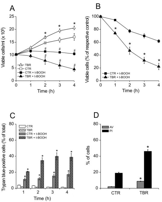
Fig. 1 – Increased susceptibility to t-BOOH-induced necrotic cell death of lymphocytes from tumor bearing rats (TBR) over that of control rats (CTR). (A) Lymphocytes were incubated for 4 h in the absence of 500µM t-BOOH (: CTR;△: TBR) or in its presence (: CTR + t-BOOH;: TBR + t-BOOH). *p<0.05vs.CTR without t-BOOH;#p<0.05vs.respective group without t-BOOH. (B) Effect of t-BOOH on the percentage of viable cells considering their respective controls at each time point (: CTR + t-BOOH;: TBR + t-BOOH). *p<0.05vs.CTR + t-BOOH. (C) Death of lymphocytes in the absence of 500µM t-BOOH (CTR: white bars; TBR: white hatched bars) or in its presence (CTR + t-BOOH: gray bars; TBR + t-BOOH: gray hatched bars). Viability was estimated by the trypan blue exclusion method repeated hourly for 4 h. *p<0.05
vs.respective group without t-BOOH. (D) Determination of apoptotic and necrotic death of lymphocytes incubated in the presence of t-BOOH. After 4 h, samples of 106cells/ml were annexin V-FITC labeled (AV; gray bars) and propidium iodide stained (PI; black bars) and then analyzed
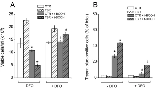
Fig. 2 – t-BOOH-induced death of CTR and TBR lymphocytes is prevented by deferoxamine (DFO). (A) Growth of DFO-treated CTR (white and gray bars) and TBR lymphocytes (white hatched and gray hatched bars) incubated for 4 h in the absence of 500µM t-BOOH (white and white hatched bars) or in its presence (gray and gray hatched bars). (B) Death of DFO-treated CTR and TBR lymphocytes incubated for 4 h in the absence of 500µM t-BOOH or in its presence. *p<0.05vs.respective group without t-BOOH in the absence of DFO.#p<0.05vs.respective group treated with t-BOOH in the absence of DFO.
was much larger than that of those positive to annexin-V (Fig. 1D), indicating that cell death under these condi-tions was predominantly necrotic.
T-BOOH-INDUCED LYMPHOCYTE DEATH IS PREVENTED BYDEFEROXAMINE
The involvement of oxidative stress in the process of t-BOOH-induced spleen lymphocyte death was assessed in vitro, by testing the effect of deferoxamine on cell pro-liferation and viability (Fig. 2). In the presence of this antioxidant/iron chelator (Halliwell 1989, Bartesaghi et al. 2004), t-BOOH did not significantly decrease the pro-liferation rate of CT and TB rat lymphocytes (Fig. 2A). Moreover, deferoxamine decreased at least 2.5-fold the number of trypan blue-positive cells after 4 h of incuba-tion in the presence of t-BOOH (Fig. 2B) indicating that lymphocyte death under these conditions is mediated by oxidative stress.
INCREASED SUSCEPTIBILITY OFTB RATLYMPHOCYTES TO T-BOOH-INDUCEDDEATH ISMEDIATED BY
HIGH[CA2+] CYT
The experiments presented in Figure 3 show that
[Ca2+]cytin TB rat spleen lymphocytes is at least double
that of CT rat lymphocytes when both cells were incu-bated during 3 h in RPMI 1640 medium. When lym-phocytes were incubated in a medium containing 10µM nifedipine, a L-type Ca2+channel antagonist, no such
significant difference in [Ca2+]
cytwas observed.
Fig. 3 – The increase in cytosolic free Ca2+concentration ([Ca2+]cyt)
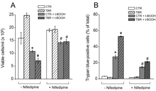
t-BOOH-induced death of CT and TB rat lympho-cytes incubated in the presence of nifedipine (Fig. 4) or loaded with the Ca2+chelator BAPTA (Fig. 5) was also
investigated. The cytoprotective effects of nifedipine and BAPTA were similar to those obtained with deferoxam-ine. Thus, these findings indicate that [Ca2+]
cytplays
an important role in the higher susceptibility of TB rat lymphocytes to t-BOOH.
T-BOOH-INDUCEDLYMPHOCYTENECROSIS IS MEDIATED BYMPT
Both cyclosporin A and FK-506 inhibit calcineurin, but only cyclosporin A inhibits MPT (Crompton et al. 1988, Griffiths and Halestrap 1991, Galat 2003). Here, the presence of cyclosporin A (Figs. 6A-C) almost com-pletely prevented the decrease in cell proliferation and viability, as well as the increase in the number of try-pan blue-positive cells induced by t-BOOH. A possible decrease in cell proliferation by the immunosuppressant drug cyclosporin A was not observed probably because the activated lymphocytes already contained high lev-els of IL-2 during the time course of our experiments. In contrast, protective effects were not observed in the presence of FK-506 (Figs. 6D-F). Moreover, the results shown in Figure 7 indicate that incubation in the pres-ence of t-BOOH for 4 h induces a decrease in m
especially in TB rat lymphocytes. CT rat lymphocytes treated with t-BOOH showed only a∼20% decrease
in the F/Fcccp ratio during this incubation time (results
not shown). The t-BOOH-induced decrease in m
was prevented by simultaneous incubation in the pres-ence of cyclosporin A plus oligomycin, but not in that of oligomycin alone, supporting the notion that t-BOOH promotes MPT in these cells.
DISCUSSION
The present study analyses the effects of t-BOOH on the proliferation and death of lymphocytes obtained from TB and CT rats. The TB rat lymphocytes have been ac-tivated as seen by the presence of high levels of interleu-kin 2 (IL-2) (Degasperi et al. 2006a). Cell proliferation was stimulated doubling the number of activated lym-phocytes in four hours (Fig. 1A). The activated TB rat lymphocytes overexpress the anti-apoptotic protein Bcl-2 and mitochondrial uncoupling protein-Bcl-2 (Degasperi et
al. 2006a) which protects the cells by preventing the con-version of high levels of ROS and [Ca2+]
cytinto a
path-way leading to mitochondrial dysfunction and cell death. Despite the protective effect of these proteins, TB rat lymphocytes display a higher susceptibility to cell death induced by t-BOOH than do the control cells (Fig. 8). t-BOOH induces oxidative stress by exhausting cellular GSH and NADPH, and promotes a Ca2+-stimulated
gen-eration of methyl,t-butoxyl, andt-butylperoxyl radicals as detected by electron paramagnetic resonance (EPR) signals (Kennedy et al. 1992, Castilho et al. 1995). Both nifedipine or BAPTA, that decrease [Ca2+]
cytof control
and activated lymphocytes render them equally resistant to the toxic effects of t-BOOH. This indicates that the higher [Ca2+]
cytin activated lymphocytes renders these
cells more susceptible to exogenous prooxidants. Although both cyclosporin A and FK-506 inhibit the activity of peptidylprolyl cis/trans isomerases or imu-nophilins, only cyclosporin A is an inhibitor of MPT (Crompton et al. 1988, Griffiths and Halestrap 1991, Friberg et al. 1998, Galat 2003). Therefore, the reduc-tion in t-BOOH-induced necrotic lymphocyte death by cyclosporin A, but not by FK-506, indicates that MPT is involved in the mechanism of cell death and excludes activation of the calcineurin pathway. Inner membrane permeabilization caused by MPT results in loss of ma-trix components, impairment of mitochondrial function and substantial mitochondrial swelling, with consequent outer membrane rupture and release of pro-apoptotic mi-tochondrial proteins (Zoratti and Szabo 1995, Green and Reed 1998, Lemasters et al. 1998, Crompton 1999, Kowaltowski et al. 2001, Green and Kroemer 2004). As a result, MPT actively participates in events that initiate either necrotic or apoptotic cell death.
The protection conferred by deferoxamine against t-BOOH-induced lymphocyte death indicates the partic-ipation of ROS under these conditions. Deferoxamine, approved by FDA for the removal of iron in conditions involving iron overload, such asβ-thalassemia (Giardini et al. 1993), has been used primarily as a metal chelat-ing agent to block the iron-dependent hydroxyl radical (Goldstein and Czapski 1990). It may involve an addi-tional or alternative antioxidant mechanism which would directly scavenge free radicals, such as superoxides (O.−
Fig. 4 – t-BOOH-induced death of CTR and TBR lymphocytes is prevented by nifedipine. (A) Growth of nifedipine-treated CTR (white and gray bars) and TBR lymphocytes (white hatched and gray hatched bars) incubated for 4 h in the absence of 500µM t-BOOH (white and white hatched bars) or in its presence (gray and gray hatched bars). (B) Death of nifedipine-treated CTR and TBR lymphocytes incubated for 4 h in the absence of 500µM t-BOOH or in its presence. *p<0.05 vs respective group without t-BOOH in the absence of nifedipine.#p<0.05vs.respective group treated with t-BOOH in the absence of nifedipine.
Fig. 5 – t-BOOH-induced death of CTR and TBR lymphocytes is prevented by intracellular Ca2+chelation. (A) Growth of BAPTA-loaded CTR (white and gray bars) and TBR lymphocytes (white hatched and gray hatched bars) incubated for 4 h in the absence of 500µM t-BOOH (white and white hatched bars) or in its presence (gray and gray hatched bars). (B) Death of BAPTA-loaded CTR and TBR lymphocytes incubated for 4 h in the presence of 500µM t-BOOH or in its absence. *p<0.05vs.respective group without t-BOOH, in the absence of BAPTA.#p<0.05vs.
respective group treated with t-BOOH, in the absence of BAPTA.
al. 1982), peroxyls (Hartley et al. 1990), peroxynitrite-derived carbonate species (CO.−
3 ) or nitrogen dioxide (.NO2)radicals (Bartesaghi et al. 2004). Indeed, it is
Fig. 6 – t-BOOH-induced lymphocyte cell death is prevented by cyclosporin A. (A; D) CTR and TBR lymphocytes were incubated for 4 h in the presence of 1µM cyclosporin A (A) or 0.5µM FK-506 (D) without other additions (: CTR;△: TBR) or with 500µM t-BOOH (: CTR + t-BOOH;: TBR + t-BOOH). *p<0.05vs.CTR without t-BOOH;#p<0.05vs.respective group with t-BOOH. (B; E) Effect of t-BOOH on the percentage of viable cells considering their respective controls at each time point when the incubations were conducted in the presence of 1µM cyclosporin A (B) or 0.5µM FK-506 (E) (: CTR + t-BOOH;: TBR + t-BOOH). *p<0.05vs.CTR + t-BOOH. (C; F) Death of CTR and TBR lymphocytes incubated with 1µM cyclosporin A (C) or 0.5µM FK-506 (F), in the absence of 500µM t-BOOH (CTR: open bars; TBR: open
hatched bars) or in its presence (CTR: gray bars; TBR: gray hatched bars). Viability was estimated by the trypan blue exclusion method repeated hourly for 4 h. *p<0.05vs.respective group without t-BOOH.
neurons and astrocytes (Abe and Saito 1998) or cardiac myocytes (Daly et al. 1991).
These results indicate that activated lymphocytes are more susceptible to oxidative stress-induced death than are controls. This observation may help the under-standing of the mechanisms underlying activated lym-phocytes deathin vivoin patients treated by the anthra-cyclines doxorubicin or daunorubicin. The lymphope-nia caused by these antineoplastic agents is associated with increased susceptibility to opportunistic infections (Mackall et al. 1994, Sthanke et al. 2001). In addition
to interacting with DNA, these compounds also give rise to ROS, since they undergo intracellular redox-cycling (Doroshow 1983, Wallace 2003, Conklin 2004). There-fore, it seems that activated lymphocytes would be more susceptible to death caused by antineoplastic agents such as anthracyclines, alkylating agents, or camptothecins (Conklin 2004).
cytotox-Fig. 7 – Mitochondrial permeability transition mediates t-BOOH-inducedmdecrease in TBR lymphocytes.min CTR and TBR
lympho-cytes (106cells/ml) was determined after 4 h incubation in the presence of 500µM t-BOOH. Where indicated, 1µM oligomycin (Oligo) and/or
1µM cyclosporin A (CsA) were added to the incubation medium.mwas determined as the ratio of DioC6(3) fluorescence (F) in the absence
of 50µM CCCP to that in its presence (F/FCCCP). *p<0.05vs.respective CTR group;#p<0.05vs.lymphocytes treated only with oligomycin.
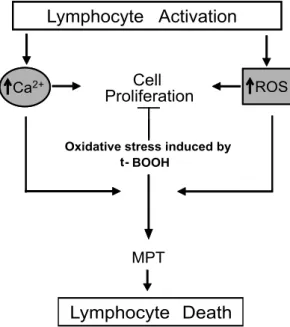
Fig. 8 – Summary of the effects of t-BOOH-induced oxidative stress on pathways involved in proliferation and death of activated lymphocytes. The process of lymphocyte activation involves both ROS overproduction and increases in [Ca2+]cyt. Treatment with agents that induces cellular
icity seems to be mediated by increased [Ca2+] cytand
MPT (Fig. 8). These findings may contribute to the un-derstanding of increased fragility of the immunological system to chemotherapy-generated oxidative stress.
ACKNOWLEDGMENTS
The authors thank Edilene S. Siqueira and Elisangela J. Gomes for their technical assistance and Linda El-Dosh for the linguistic revision of the manuscript. The work was supported by Fundação de Amparo à Pesquisa do Estado de São Paulo (FAPESP), Conselho Nacional de Desenvolvimento Científico e Tecnológico (CNPq) and Coordenação de Aperfeiçoamento de Pessoal de Nível Superior (CAPES).
RESUMO
O presente estudo demonstra que linfócitos ativados de baço de ratos portadores do tumor de Walker 256 são mais susceptíveis à morte celular necrótica induzida por tert-butil hidroperó-xido (t-BOOH)in vitroquando comparados aos controles. O quelante de ferro e antioxidante deferoxamina, o quelante in-tracelular de Ca2+BAPTA, o antagonista de canal de Ca2+ nifedipina ou o inibidor da transição de permeabilidade mi-tocondrial ciclosporina-A, mas não o inibidor de calcineurina FK-506, inibiram de maneira similar a morte celular induzida por t-BOOH em linfócitos ativados e controles. Os linfóci-tos ativados apresentaram redução do potencial de membrana mitocondrial induzida por t-BOOH num mecanismo sensível a ciclosporina-A. Nossos resultados indicam que o aumento da concentração de Ca2+citosólico em linfócitos ativados au-menta a susceptibilidade dos mesmos à morte celular induzida por estresse oxidativo, num mecanismo envolvendo a partici-pação do poro de transição de permeabilidade mitocondrial.
Palavras-chave:morte celular, radicais livres, resposta imune,
linfopenia, transição de permeabilidade mitocondrial, esplenó-citos.
REFERENCES
ABEKANDSAITOH. 1998. Characterization of t-butyl hy-droperoxide toxicity in cultured rat cortical neurones and astrocytes. Pharmacol Toxicol 83: 40–46.
ARNOLD R, BRENNER D, BECKER M, FREY CR AND KRAMMERPH. 2006. How T lymphocytes switch be-tween life and death. Eur J Immunol 36: 1654–1658.
BARTESAGHIS, TRUJILLOM, DENICOLA A, FOLKESL, WARDMANPANDRADIR. 2004. Reactions of desfer-rioxamine with peroxynitrite-derived carbonate and nitro-gen dioxide radicals. Free Radic Biol Med 36: 471–483. BARTOLIGM, PICCIONIE, AGOSTARAG, CALVIELLOG
ANDPALOZZA P. 1994. Different mechanisms of tert-butyl hydroperoxide-induced lethal injury in normal and tumor thymocytes. Arch Biochem Biophys 312: 81–87. BERNARDESCF, PEREIRA DASILVALANDVERCESIAE.
1986. t-Butylhydroperoxide-induced Ca2+efflux from liver mitochondria in the presence of physiological con-centrations of Mg2+ and ATP. Biochim Biophys Acta 850: 41–48.
BOYUMA. 1976. Isolation of lymphocytes, granulocytes and macrophages. Scand J Immunol Suppl 5: 9–15.
BRUMATTI G, WEINLICH R, CHEHAB CF, YON M AND AMARANTE-MENDESGP. 2003. Comparison of the anti-apoptotic effects of Bcr-Abl, Bcl-2 and Bcl-x(L) follow-ing diverse apoptogenic stimuli. FEBS Lett 541: 57–63. BUTTKETMANDSANDSTROMPA. 1995. Redox regulation
of programmed cell death in lymphocytes. Free Radic Res 22: 389–397.
CAMPOSCB, DEGASPERI GR, PACIFICO DS, ALBERICI LC, CARREIRA RS, GUIMARÃES F, CASTILHO RF ANDVERCESIAE. 2004. Ibuprofen-induced Walker 256 tumor cell death: cytochrome c release from functional mitochondria and enhancement by calcineurin inhibition. Biochem Pharmacol 68: 2197–2206.
CASTILHO RF, KOWALTOWSKI AJ, MEINICKE AR, BECHARA EJANDVERCESIAE. 1995. Permeabiliza-tion of the inner mitochondrial membrane by Ca2+ions is stimulated by t-butyl hydroperoxide and mediated by reactive oxygen species generated by mitochondria. Free Radic Biol Med 18: 479–486.
CONKLIN KA. 2004. Chemotherapy-associated oxidative stress: impact on chemotherapeutic effectiveness. Integr Cancer Ther 3: 294–300.
CROMPTONM, ELLINGERHANDCOSTIA. 1988. Inhibi-tion by cyclosporin A of a Ca2+-dependent pore in heart mitochondria activated by inorganic phosphate and oxida-tive stress. Biochem J 255: 357–360.
CROMPTONM. 1999. The mitochondrial permeability tran-sition pore and its role in cell death. Biochem J 341: 233–249.
DEGASPERI GR, VELHO JA, ZECCHIN KG, SOUZA CT, VELLOSO LA, BORECKÝ J, CASTILHO RF AND VERCESI AE. 2006a. Role of mitochondria in the im-mune response to cancer: a central role for Ca2+. J Bioenerg Biomembr 38: 1–10.
DEGASPERIGR, ZECCHIN KG, BORECKY J, CRUZ-HO-FLING MA, CASTILHO RF, VELLOSO LA, GUIMA-RÃESFANDVERCESIAE. 2006b. Verapamil-sensitive Ca2+ channel regulation of Th1-type proliferation of splenic lymphocytes induced by Walker 256 tumor de-velopment in rats. Eur J Pharmacol 549: 179–184. DOROSHOW JH. 1983. Anthracycline antibiotic-stimulated
superoxide, hydrogen peroxide, and hydroxyl radical production by NADH dehydrogenase. Cancer Res 43: 4543–4551.
FESKES. 2007. Calcium signalling in lymphocyte activation and disease. Nat Rev Immunol 7: 690–702.
FRIBERGH, FERRAND-DRAKEM, BENGTSSONF, HALES-TRAPAPANDWIELOCHT. 1998. Cyclosporin A, but not FK 506, protects mitochondria and neurons against hypoglycemic damage and implicates the mitochondrial permeability transition in cell death. J Neurosci 18: 5151– 5159.
GALAT A. 2003. Peptidylprolyl cis/trans isomerases (im-munophilins): biological diversity – targets – functions. Curr Top Med Chem 3: 1315–1347.
GIARDINI C, LA NASA G, CONTU L, GALIMBERTI M, POLCHIP, ANGELUCCIE, BARONCIANID, BARBANTI I, MURETTOPANDLUCARELLIG. 1993. Desferriox-amine therapy induces clearance of iron deposits after bone marrow transplantation for thalassemia: case report. Bone Marrow Transplant Suppl 1: 108–110.
GOLDSTEINSANDCZAPSKIG. 1990. Transition metal ions and oxygen radicals. Int Rev Exp Pathol 31: 133–164. GREENDRANDKROEMERG. 2004. The pathophysiology
of mitochondrial cell death. Science 305: 626–629. GREENDRANDREEDJC. 1998. Mitochondria and
apopto-sis. Science 281: 1309–1312.
GRIFFITHSEJANDHALESTRAPAP. 1991. Further evidence that cyclosporin A protects mitochondria from calcium overload by inhibiting a matrix peptidyl-prolyl cis-trans isomerase. Implications for the immunosuppressive and toxic effects of cyclosporin. Biochem J 274: 611–614. GROSSMAN Z, MIN B, MEIER-SCHELLERSHEIM MAND
PAULWE. 2004. Concomitant regulation of T-cell acti-vation and homeostasis. Nat Rev Immunol 4: 387–395. HALLIWELL B. 1989. Protection against tissue damage in
vivo by desferrioxamine: what is its mechanism of action? Free Radic Biol Med 7: 645–651.
HARTLEYA, DAVIESMANDRICE-EVANSC. 1990. Desfer-rioxamine as a lipid chain-breaking antioxidant in sickle erythrocyte membranes. FEBS Lett 264: 145–148 HERMISTONML, XUZ, MAJETIRANDWEISSA. 2002.
Reciprocal regulation of lymphocyte activation by tyrosine kinases and phosphatases. J Clin Invest 109: 9–14. HOES, ROWLEYDAANDHALLIWELLB. 1982. Reactions
of ferrioxamine and desferrioxamine with the hydroxyl radical. Chem Biol Interact 41: 75–81.
JOCELYNPC ANDDICKSONJ. 1980. Glutathione and the mitochondrial reduction of hydroperoxides. Biochim Bio-phys Acta 590: 1–12.
KENNEDYCH, CHURCHDF, WINSTONGWANDPRYOR WA. 1992. tert-Butyl hydroperoxide-induced radical production in rat liver mitochondria. Free Radic Biol Med 12: 381–387.
KOWALTOWSKI AJ, CASTILHO RF AND VERCESI AE. 2001. Mitochondrial permeability transition and oxida-tive stress. FEBS Lett 495: 12–15.
KRAMMERPH, ARNOLDR AND LAVRIKIN. 2007. Life and death in peripheral T cells. Nat Rev Immunol 7: 532–542.
LEMASTERSJJET AL. 1998. The mitochondrial permeability transition in cell death: a common mechanism in necrosis, apoptosis and autophagy. Biochim Biophys Acta 1366: 177–196.
MACKALLCL, FLEISHERTA, BROWNMR, MAGRATHIT, SHADAT, HOROWITZME, WEXLERLH, ADDE MA, MCCLURELLANDGRESSRE. 1994. Lymphocyte de-pletion during treatment with intensive chemotherapy for cancer. Blood 84: 2221–2228.
MARTINSJ, REUTELINGSPERGER CPM, MCGAHON AJ, RADER J, VAN SCHIE RCAA, LAFACE DM AND GREENDR. 1995. Early redistribution of plasma mem-brane phosphatidylserine is a general feature of apoptosis regardless of the initiating stimulus: inhibition by overex-pression of Bcl-2 and Abl. J Exp Med 182: 1–12. MATHERMWANDROTTENBERGH. 2002. The inhibition of
calcium signaling in T lymphocytes from old mice results from enhanced activation of the mitochondrial permeabil-ity transition pore. Mech Ageing Dev 123: 707–724. NIEMINENAL, BYRNEAM, HERMANBANDLEMASTERS
QUINTANAA, GRIESEMERD, SCHWARZECANDHOTH M. 2005. Calcium-dependent activation of T-lympho-cytes. Pflugers Arch 450: 1–12.
ROTTENBERGHANDWUS. 1998. Quantitative assay by flow cytometry of the mitochondrial membrane potential in intact cells. Biochim Biophys Acta 1404: 393–404. ROYCR. 2003. Immunology: professional secrets. Nature
425: 351–352.
STAHNKE K, FULDA S, FRIESEN C, STRAUSS G AND DEBATINKM. 2001. Activation of apoptosis pathways in peripheral blood lymphocytes by in vivo chemotherapy. Blood 98: 3066–3073.
WALLACE KB. 2003. Doxorubicin-induced cardiac mito-chondrionopathy. Pharmacol Toxicol 93: 105–115. WILLIAMSMSANDKWONJ. 2004. T cell receptor
stimu-lation, reactive oxygen species, and cell signaling. Free Radic Biol Med 37: 1144–1151.
![Estudo dose-resposta e perfil de expressão gênica do herbicida diuron [3-(3,4-diclofenil)-1,1-dimetiluréia] em bexiga urinária de ratos wistar machos](data:image/gif;base64,R0lGODlhAQABAIAAAP///wAAACH5BAEAAAAALAAAAAABAAEAAAICRAEAOw==)