Functional insights into the plant-specific SR45 splicing
factor
Vera Alexandra Sacramento Nunes
Dissertação para obtenção do Grau de Mestre em
Engenharia Agronómica
Orientador: Paula Duque Magalhães Santos
Coorientadores: Raquel Fonseca de Carvalho
Sara Barros Queiroz Amâncio
Júri:
Presidente: Doutora Cristina Maria Moniz Simões de Oliveira, Professora Associada com agregação do Instituto Superior de Agronomia da Universidade de Lisboa.
Vogais: Doutor Jorge Alexandre Matos Pinto de Almeida, Professor Associado do Instituto Superior de Agronomia da Universidade de Lisboa; Doutora Paula Duque Magalhães Santos, Investigadora Principal do Instituto Gulbenkian de Ciência da Fundação Calouste Gulbenkian.
i
ACKNOWLEDGMENTS
To my supervisor Paula Duque, first of all for giving me the opportunity to do this thesis in the PMB lab, which was a wonderful experience, and for all the support, motivation and knowledge transmitted during this period.
To Raquel Carvalho, for the ceaseless guidance, tutoring and motivation as my co-supervisor, always answering (several times!) to all my questions and doubts with infinite patience and always with a smile.
To Sara Amâncio, for accepting to be my co-supervisor and for all shown availability and help during this thesis.
To Elena Baena-González and Paula Duque, not only for “sponsoring” my last tuition, but for the encouragement and availability that I always felt since I joined IGC.
To all members of the PMB group for all the helpful comments, encouragement and for all the reasons I will mention below. A special thanks to Estelle Remy, even though abroad, her support was always near.
To all my wonderful past and present IGC colleagues, who always supported and motivated me during this thesis, for being such good persons, allowing an easier and pleasant job as a plant technician. A really great group to work and share time with.
To all my friends, for all the motivation during this thesis, for listening and for always providing such great joy to my life!
À minha família, por estarem sempre presentes, pelo incansável apoio, amizade e sorrisos, mesmo nos meus momentos de mau-humor!
ii
ABSTRACT
Alternative splicing is a post-transcriptional regulatory mechanism that generates transcriptome and proteome diversity. The SR protein family of splicing regulators is essential for the execution and regulation of both constitutive and alternative pre-mRNA splicing. The SR-related protein SR45 is an Arabidopsis bona fide splicing factor that plays a key role in plant development as well as in sugar and abscisic acid (ABA) signaling during early seedling development. The Arabidopsis 5PTase13, belonging to a group of inositol phosphatases involved in plant growth, development and stress responses, is a splicing target of SR45 previously implicated in hormonal and light signaling pathways. The 5PTase13 gene is alternatively spliced, generating two isoforms shown here to be ubiquitously expressed in Arabidopsis. The 5PTase13 loss-of-function mutant we isolated exhibits shorter roots when grown under low light conditions. Moreover, overexpression of either the 5PTase13.1 or the
5PTase13.2 isoforms in the sr45-1 background enhances the glucose hypersensitivity during early
seedling development conferred by the sr45-1 mutation. Finally, confocal microscopy data suggests that both 5PTase13 isoforms are located in the cytoplasm, with 5PTase13.1 appearing mostly associated with the plasma membrane. This work provides new insight into the in vivo roles of the SR45 splicing factor and its functional connections to the 5PTase13 phosphatase.
iii
RESUMO
O splicing alternativo é um mecanismo de regulação pós-transcricional que gera diversidade transcritómica e proteómica. As proteínas SR desempenham um papel essencial na execução e regulação do splicing constitutivo e alternativo do pré-mARN. A proteína SR45 é um factor de splicing genuíno em Arabidopsis, desempenhando um papel chave no desenvolvimento da planta e na sinalização dos açúcares e do ácido abscísico (ABA) durante o desenvolvimento inicial da plântula. A fosfatase 5PTase13 de Arabidopsis, pertencente a um grupo de inositol fosfatases envolvidas no crescimento e desenvolvimento vegetal bem como respostas ao stress, é um alvo de splicing da SR45 tendo sido previamente implicada nas vias de sinalização hormonais e da luz.
O gene 5PTase13 sofre splicing alternativo, gerando duas isoformas aqui descritas como sendo ubiquamente expressas em Arabidopsis. O mutante knockout de 5PTase13 que isolámos apresenta raízes mais curtas em condições de baixa luminosidade. Além disso, a sobre-expressão das isoformas
5PTase13.1 ou 5PTase13.2 no background do mutante sr45-1 aumenta a hipersensibilidade à glucose
durante o desenvolvimento inicial da plântula, fenótipo este conferido pela mutação sr45-1. Por fim, dados de microscopia confocal sugerem que ambas as isoformas da 5PTase13 se localizam no citoplasma com a 5PTase13.1 parecendo mais associada à membrana plasmática. Este trabalho fornece novas perspectivas sobre as funções in vivo do fator de splicing SR45 e suas conexões funcionais à fosfatase 5PTase13.
iv
RESUMO-ALARGADO
A maioria dos genes dos eucariotas autotróficos contêm regiões codificantes, denominadas exões, interrompidas por regiões não-codificantes, os intrões. Splicing é o processo pelo qual os intrões do pre-mARN são removidos e os exões ligados entre si, de forma a obter um mARN maduro. Através do splicing alternativo múltiplas formas de mARN maduro podem ser obtidas a partir de um único pre-mARN. Assim, um único gene pode produzir mais que uma proteína originando diversidade transcritómica e proteómica. Em plantas, estima-se que 61% dos genes com intrões sofram splicing alternativo, tendo a sua relevância biológica vindo a revelar-se nos últimos anos, nomeadamente numa variedade de processos fisiológicos e de desenvolvimento.
As proteínas SR (ricas em serina e arginina) constituem uma das famílias mais conservadas de factores de splicing, sendo essenciais à execução e regulação do splicing constitutivo e alternativo do pre-mARN. Ao serem capazes de se ligar ao ARN e interagir com componentes do spliceossoma direccionando-os para os sítios de splicing, têm um papel crucial no splicing alternativo, determinando a escolha dos sítios de splicing em função da sua concentração. O factor de splicing específico de plantas SR45, recentemente excluído da família de proteínas SR após uma revisão da definição da nomenclatura desta família, desempenha um papel-chave no normal desenvolvimento da planta, bem como na sinalização de açúcares e da fitohormona ácido abscísico (ABA).
Em plantas, embora o conhecimento de um grupo específico de inositol fosfatases denominado 5PTases ainda seja limitado, estas estão implicadas em diversas funções relacionadas com o crescimento e desenvolvimento vegetal, bem como a resposta ao stress ambiental. Estudos de caracterização funcional da fostatase 5PTase13 em Arabidopsis indicam a sua importância no desenvolvimento vegetal bem como em vias de sinalização hormonais e de luz, tendo também sido implicada na sinalização de nutrientes/energia. O gene que codifica esta fosfatase sofre splicing alternativo, originando duas isoformas distintas como resultado da retenção de um intrão na isoforma 5PTase13.1. Neste trabalho, mostramos que ambas as isoformas são ubiquamente expressas em plântulas, tecidos aéreos e raízes de Arabidopsis. Verificamos tambem que a isofoma
5PTase13.2 é o transcrito mais abundante em plântulas e nos diferentes tecidos analisados.
Estudos anteriores mostraram que a 5PTase13 era siginificamente induzida por ABA e ferimentos, e ligeiramente induzida pelo frio e sal, sugerindo o seu envolvimento na resposta ao stress. De facto, demonstrou-se também que mutantes knockout da 5PTase13 apresentam alterações na resposta ao stress induzido por ABA e a açúcares. Um dos objectivos do presente trabalho foi verificar se a produção das duas isoformas da 5PTase13 tem impacto funcional na resposta das plantas aos
v açúcares e ABA. Começámos por isolar um novo mutante knockout da 5PTase13, 5ptase13-3, e verificámos que em condições de pouca luz o mutante exibe raízes mais curtas.
Este trabalho veio confirmar também que o gene 5PTase13 é um alvo do factor de splicing SR45 e quisemos investigar se as isoformas da 5PTase13 teriam algum efeito no modo de acção da SR45. Verificámos que plantas transgénicas sobre-expressando cada uma das isoformas da 5PTase13 no mutante sr45-1, exibem um fenótipo de hipersensibilidade à glucose mais acentuado do que o previamente descrito para o mutante sr45-1 relativamente à germinação e desenvolvimento inicial de plântulas.
O último objectivo do presente trabalho foi estudar a localização subcelular de cada uma das isoformas da 5PTase13. Observámos através de microscopia confocal que ambas as isoformas se localizam no citoplasma e que a isofoma 5PTase13.1 poderá estar mais associada à membrana plasmática do que a 5PTase13.2.
Este trabalho fornece novas perspectivas relativamente às funções desempenhadas pelo factor de
vi
ABBREVIATIONS LIST
ABA abscisic acid
EEP exonuclease/endonuclease/phosphatase
ESR exonic splicing regulator
GFP green fluorescent protein
HXK hexokinase
Ins(1,4,5)P3 inositol (1,4,5) triphosphate
Ins(1,3,4,5)P4 inositol (1,3,4,5) tetrakisphosphate
ISR intronic splicing regulator
mRNA messenger RNA
NMD nonsense-mediated decay
PLC phospholipase C
pre-mRNA precursor-mRNA
PTC premature termination codon
PtdIns(3,4,5)P3 phosphatidylinositide (3,4,5) triphosphate
PtdIns(4,5)P2 phosphatidylinositide (4,5) biphosphate
PyTract polypyrimidine tract
RNP ribonucleoprotein
ROS reactive oxygen species
RRM RNA recognition motif
RS arginine/serine-rich
RT-PCR reverse-transcription polymerase chain reaction
S/RS serine and arginine serine-rich
SNF1 sucrose non-fermenting 1
SnRK SNF1-related kinase
snRNP small nuclear ribonucleoprotein
SPXR serine-proline-x-arginine motif
SF splicing factor
SR serine/arginine-rich
SS splice site
TAIR The Arabidopsis Information Resource
U2AF U2 auxiliary factor
vii
TABLE OF CONTENTS
ACKNOWLEDGMENTS ... i ABSTRACT ...ii RESUMO ... iii RESUMO-ALARGADO ... iv ABBREVIATIONS LIST ... vi I. INTRODUCTION ... 11. Pre-mRNA splicing in plants ... 1
2. The Arabidopsis SR45 splicing factor ... 4
3. Inositol polyphosphate 5-phosphatases ... 7
3.1. Arabidopsis 5PTase13 ... 10
4. Objectives ... 11
II. MATERIALS AND METHODS ... 12
1. Plant materials and growth conditions ... 12
2. Isolation of T-DNA insertion mutants ... 12
3. Phenotypical analyses ... 13
4. Generation of transgenic lines ... 14
5. RNA extraction and RT-PCR analyses ... 14
6. Confocal laser scanning microscopy... 15
III. RESULTS ... 16
1. Expression and splicing pattern of the 5PTase13 gene in Arabidopsis ... 16
2. Isolation of a null 5PTase13 loss-of-function mutant ... 17
3. Phenotypical analysis of the 5PTase13-3 loss-of-function mutant ... 19
3.1. Effect of glucose and ABA on seed germination under normal and low light conditions ... 20
3.2. Root growth under low light conditions ... 22
4. Functional relevance of each 5PTase13 isoform in the sr45-1 background ... 22
viii IV. DISCUSSION ... 28 V. CONCLUSIONS AND FUTURE PERSPECTIVES ... 32 VI. REFERENCES ... 33
ix
TABLE INDEX
Table 1. T-DNA insertion lines and corresponding gene-specific primers used for mutant line
genotyping ... 13 Table 2. Gene-specific primers used in the analysis of the expression and splicing patterns of the Arabidopsis 5PTase13 gene. ... 15
x
FIGURES INDEX
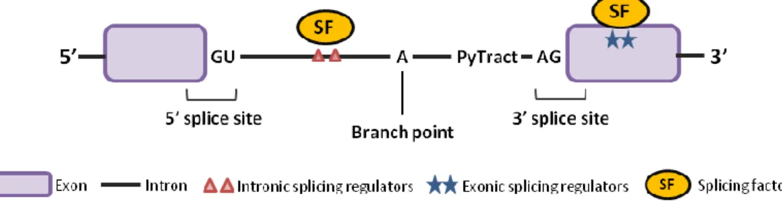
Figure 1. Regulatory elements that contribute to the regulation of alternative splicing ... 1
Figure 2. Frequency of the basic types of alternative splicing events in humans and Arabidopsis ... 3
Figure 3. Domain organization of the Arabidopsis SR45 protein ... 5
Figure 4. Inositol signaling and its termination via 5PTases... 8
Figure 5. Schematic representation of the domain organization of Arabidopsis 5PTases ... 9
Figure 6. Structure of the Arabidopsis 5PTase13 gene and its alternative transcripts, as well as domain organization of their encoded protein isoforms ... 16
Figure 7. Expression and splicing pattern of the Arabidopsis 5PTase13 gene ... 17
Figure 8. Molecular characterization of two T-DNA insertion mutants for the 5PTase13 gene ... 18
Figure 9. Isolation of the 5ptase13-3 null mutant ... 19
Figure 10. Germination of 5ptase13-3 and 5ptase13-2 mutant seeds under normal light ... 20
Figure 11. Germination of 5ptase13-3 and 5ptase13-2 mutant seeds under low light ... 21
Figure 12. Root growth of the 5ptase13-3 and 5ptase13-2 mutants under low light ... 22
Figure 13. 5PTase13 transcript levels in sr45-1 mutant plants expressing the individual 5PTase13 splice variants ... 23
Figure 14. Glucose-related phenotypes of transgenic plants expressing the individual 5PTase13 splice variants in the sr45-1 mutant background ... 24
Figure 15. Subcellular localization of the SR45.1 isoform in Arabidopsis roots ... 25
Figure 16. Subcellular localization of the 5PTase13.1 and 5PTase13.2 isoforms in Arabidopsis sr45-1 mutant roots ... 26
Figure 17. Subcellular localization of SR45.1 as well as 5PTase13.1 and 5PTase13.2 isoforms in protoplasts ... 27
Functional insights into the plant-specific SR45 splicing factor
1
I. INTRODUCTION
1. Pre-mRNA splicing in plants
Most genes in autotrophic eukaryotes contain coding regions, named exons, interrupted by stretches of non-coding sequences, called introns. Splicing is a fundamental post-transcriptional gene regulation process that removes the introns from the precursor-mRNA (pre-mRNA) and joins the exons in order to obtain a mature mRNA that may later be translated into protein. This process occurs cotranscriptionally and is carried out by a large ribonucleoprotein (RNP) complex, the spliceossome, composed of five small nuclear ribonucleoproteins (snRNPs) and a large number of non-snRNP proteins. There are four intron-defining splicing signals that are important for accurate splicing of the pre-mRNA – the 5’ and 3’ splice sites (SSs) with the consensus sequences GU and AG, respectively, that mark the border between exons and introns; the branch point, a sequence near the 3’SS that is defined by an adenoside residue; and the polypyrimide tract (PyTract), a stretch of pyrimidines downstream of the branch point site (Figure 1). These consensus sequences are crucial in the splicing process, as a change in only one of the conserved nucleotides may inhibit intron splicing.
Figure 1. Regulatory elements that contribute to the regulation of alternative splicing
Pre-mRNA splicing is directed by cis elements, including the splice sites, the branch point and the polypyrimidine tract. The selection of alternative splice sites is affected by trans-acting factors binding to auxiliary exonic and intronic cis splicing regulators.
The splicing reaction involves two sequential trans-esterification reactions. First, there is the nucleophilic attack by the hydroxyl group of the adenosine in the branch point site to the 5’ SS phosphate. This will create a bond between the first nucleotide of the intron and the branch point’s adenosine, forming a looped structure known as lariat, and leaving a free 3’ hydroxyl group at the 5’ exon. The second reaction involves another nucleophilic attack of the newly freed 3’ hydroxyl group to the phosphodiester at the 3’ SS, which results in the junction of the two exons and the release of the lariat containing the intron.
Functional insights into the plant-specific SR45 splicing factor
2 Alternative splicing occurs when different combinations of splice sites are recognized by the spliceossome, thus allowing for different rearrangements of the genes’ coding fragments and resulting in the generation of multiple forms of mature mRNA from the same pre-mRNA molecule. In this way, a single gene may lead to the production of more than one polypeptide, upscaling the genome’s coding capacity. It is indeed the most prevalent mechanism to generate transcriptome and proteome diversity in metazoans (Graveley, 2001; Maniatis & Tasic, 2002). The most fascinating example is the Dscam gene from Drosophila melanogaster that can give rise to over 38000 alternatively-spliced variants (Schmucker et al., 2000). Alternative splicing may therefore have important consequences to the cell. It allows a single gene to give rise to multiple proteins, with different sequence and domain arrangement and hence potentially different subcellular localization, stability or function (reviewed in Syed et al., 2012). Moreover, it can also regulate transcript levels by introducing premature termination codons (PTCs) that then target the mRNAs for degradation by nonsense-mediated decay (NMD) (reviewed in Nicholson et al. 2010), a surveillance mechanism that prevents accumulation of truncated and potentially harmful proteins. It has been shown that alternative splicing coupled to NMD potentially regulates 13% of intron-containing Arabidopsis genes (Kalyna et al., 2012).
Alternative splicing can be constitutive or subject to regulation. In the first case, multiple isoforms are produced in constant amounts in all or in the majority of cell types, while in the latter case, different splicing patterns occur according to different cell environmental conditions. In fact, regulated alternative splicing can be tissue-specific, dictated by different developmental stages or modulated by external stimuli, with splice site usage being controlled by fundamental biochemical mechanisms, such as spliceossome composition and concentration of splicing factors, as well as chromatin structure and transcription rates (reviewed in Nilsen and Graveley, 2010; Staiger & Brown, 2013).
Most alternative splicing events can be classified into four basic types – exon skipping/inclusion, selection of alternative 5’ or 3’ splice sites and intron retention (Figure 2). In Arabidopsis the most prominent alternative splicing event is intron retention ( 40%), followed by alternative 3’ splice site and 5’ splice site, with exon skipping events being relatively rare (Figure 2) (reviewed in Carvalho et al. 2013; Reddy et al., 2013). By contrast, in humans intron retention is the least frequent (<5%) event and exon skipping the most common (>40%) (Figure 2) (reviewed in Carvalho et al. 2013; Reddy et al., 2013).
Functional insights into the plant-specific SR45 splicing factor
3
Figure 2. Frequency of the basic types of alternative splicing events in humans and Arabidopsis
Comparison of the proportion of the basic types of alternative splicing events between humans and Arabidopsis. Adapted from Reddy et al. (2013).
In the last decade, there have been great improvements regarding alternative splicing analyses, particularly in plants, where studies had been limited to individual genes. Up to 95% of human multiexon genes are estimated to undergo alternative splicing (Pan et al., 2008). In plants, recent high coverage RNA-seq transcriptome analyses estimated that at least 61% of intron-containing genes of Arabidopsis (Marquez et al., 2012), 48% of rice, Oryza sativa (Lu et al., 2010) and 30% of grape, Vitis vinifera (Vitulo et al., 2014) undergo alternative splicing. With the sequencing of other plant genomes, and using expressed transcript sequence alignments, it is also estimated that alternative splicing occurs in 55% of barley, Hordeum vulgare (International Barley Genome Sequencing et al., 2012), 40% of maize, Zea mays (Thatcher et al., 2014), 5% of potato, Solanum
tuberosum (Potato Genome Sequencing et al., 2011) and 6% of the grass Brachypodium distachyon
(Walters et al., 2013) genes. These estimates are bound to rise as RNA-seq data becomes available from various cell types, organs, and developmental stages, and from plants grown under different biotic and abiotic stresses. In humans, the misregulation of alternative splicing can lead to developmental abnormalities and a large number of diseases, such as breast and ovarian cancer, spinal muscular atrophy (SMA), cystic fibrosis or Alzheimer’s disease (reviewed in Blencowe, 2006; Tazi et al., 2009; Venables et al., 2009; Ward & Cooper, 2010). Therapeutic strategies based on modulating alternative splicing are currently being developed to lighten the symptoms of some of these diseases (Nlend Nlend et al., 2010). In plant systems the biological relevance of alternative splicing is just beginning to unfold, with the last couple of years having seen an increasing number of examples of functional alternative splicing events in plants. Such reports have implicated alternative
Functional insights into the plant-specific SR45 splicing factor
4 splicing in a variety of developmental and physiological processes, such as photosynthetic and starch metabolism, hormone signaling, seed germination, root growth, flowering, circadian clock, natural variation, as well as in biotic and abiotic stress responses (reviewed in Carvalho et al., 2013; Staiger & Brown, 2013; Reddy et al., 2013).
The consensus sequences at the splice and branch point sites essential for splice site recognition are short and degenerate. Therefore, other cis-acting regulatory sequences in exons (exonic splicing regulators, ESRs) and/or in introns (intronic splicing regulators, ISRs) are required for an accurate splice site selection. These determine the interaction of additional proteins designated splicing factors, which bind to them and in this way direct the splicing machinery to the correct splice sites (see Figure 1) (Matlin et al., 2005; Nilsen & Graveley, 2010; Reddy, 2007; Wachter et al., 2012). One of the main conserved families of such splicing factors is the SR (serine/arginine-rich) family of RNA-binding proteins, which are essential for the execution and regulation of pre-mRNA splicing, playing vital roles in both constitutive and alternative splicing. SR proteins are present in metazoans and plants and share a characteristic structural organization, consisting of one or two N-terminal RNA-recognition motifs (RRMs) that bind to regulatory sequences in the pre-mRNA, and a C-terminal arginine/serine-rich (RS) domain that not only promotes protein-protein interactions with other components of the splicing machinery but also promotes pre-spliceosome assembly by contacting directly with the pre-mRNA via the branch-point and the 5’ splice site (Barta et al., 2010; Hertel & Graveley, 2005; Shen & Green, 2004; Shen et al., 2004; reviewed in Duque, 2011; Ram & Ast, 2007; Reddy & Shad Ali, 2011). By being able to bind the RNA and interact with spliceosomal components directing them to the correct splice sites, SR proteins are important players in alternative splicing, determining splice site selection under different conditions in a concentration-dependent manner (Graveley, 2000; Manley & Tacke, 1996). In animals, these RNA-binding proteins have also been implicated in mRNA transport (Huang & Steitz, 2001), mRNA stability and mRNA nonsense-mediated decay (NMD) (Zhang & Krainer, 2004) and translation (Sanford et al., 2004). In plants, a multitude of SR proteins has been identified in Arabidopsis (Lorkovic & Barta, 2002), rice (Iida & Go, 2006; Isshiki et al., 2006), Brachypodium (International Brachypodium, 2010), maize and sorghum (Rauch et al., 2014). Based on the recent updated definition for plant SR proteins, the rice SR family comprises of 22 members while Arabidopsis has 18 (Barta et al., 2010).
2. The Arabidopsis SR45 splicing factor
The Arabidopsis SR45 is a plant specific SR-related protein. It functions as an essential splicing factor, being able to complement in vitro an animal splicing extract deficient in SR proteins (Ali et al., 2007).
Functional insights into the plant-specific SR45 splicing factor
5 Since it displays a highly atypical SR protein structure, i.e. one RRM flanked by two distinct RS domains (Figure 3), and due to the recent revision of the plant SR family classification criteria (Barta et al., 2010), SR45 has been excluded from this family of splicing regulators. The two RS domains of SR45 present different complexity levels with the N-terminal domain (RS1) being rich in serine and serine repeats (S/RS) and the more complex C-terminal domain (RS2) containing arginine-serine repeats (RS) and arginine-serine-proline-x-arginine motifs (SPXR) (Figure 3) (Zhang & Mount, 2009).
Figure 3. Domain organization of the Arabidopsis SR45 protein
Schematic diagram showing the domain organization of the SR45 protein, including the N-terminal RS1 domain, the middle RRM and the C-terminal RS2 domain. RRM (RNA Recognition Motif), RS1 and RS2 (Arginine/Serine rich domains 1 and 2).
The SR45 gene (At1g16610) comprises twelve exons and is reported to generate three splice variants. In 2007, a comprehensive analysis of the Arabidopsis SR protein gene family revealed two SR45 isoforms that differ by a 21-nucleotide sequence present in isoform 1 (SR45.1) and missing in isoform 2 (SR45.2) due to an alternative 3’ splice site event occurring in intron 6 (Palusa et al., 2007). The proteins encoded by these isoforms are very similar, with SR45.1 including an additional 7-amino acid segment (T218S219P220Q221R222K223T224). The third splice variant, SR45.3, has been annotated in The Arabidopsis Information Resource (TAIR) database (http://www.arabidopsis.org). This transcript arises from selection of an alternative 5’ splice site in intron 9 leading to the inclusion of a 33-nt sequence that is absent from the other two splice variants sequence, encoding a protein with 11 additional amino acids. All the alternatively spliced segments are in frame, so none of the SR45 splice variants give rise to a truncated form of the protein.
The SR45 splicing factor has been extensively characterized in plants. It was first isolated in a Yeast-2-Hybrid screen where it was shown to interact with the U1 snRNP 70K protein, known to function in 5' SS selection, as well as with the SCL33 SR splicing factor (Golovkin & Reddy, 1999). In 2012, Day et al. provided evidence that besides U1-70K, SR45 also interacts with another spliceosomal protein, U2AF35, responsible for 3’SS selection, and directly binds SR30 splicing factor. Their results suggest that SR45 modulates 5’ and 3’ site selection and may bridge the 5’ and 3’ components of the spliceossome.
Expression of SR45 fused with GFP in cultured onion and tobacco cells and transgenic Arabidopsis plants has shown that SR45 is localized to the nucleus, diffusely distributed in the nucleoplasm or concentrated in speckles of different sizes and shapes (Ali et al., 2003). These speckles correspond to
Functional insights into the plant-specific SR45 splicing factor
6 interchromatin granule clusters that serve as storage/re-cycling sites for splicing factors (Misteli, 2000; Misteli et al., 1997) and deliver them to nearby active sites of transcription (Misteli & Spector, 1998). A loss-of-function mutant for SR45, sr45-1, was first reported by Ali et al. (2007), and its characterization showed that this splicing factor plays an important role in normal plant development. The authors found that depletion of SR45 levels leads to pleiotropic developmental phenotypes, such as narrow leaves, altered number of petals and stamens, delayed root growth and late flowering. In addition, the alternative splicing pattern of five SR genes was changed in the sr45-1 mutant, indicating that the observed phenotypes could be due to altered levels of these SR protein isoforms, which in turn would regulate the splicing of other transcripts (Ali et al., 2007). In 2012, Ausin et al. reported that sr45-1 mutants exhibit impaired DNA methylation establishment capacity and therefore are unable to methylate the FWA gene, a homeodomain-containing transcription factor that controls flowering. This could in part explain the flowering phenotype of the sr45-1 mutant. Interestingly, Zhang and Mount (2009) demonstrated that two of the alternatively-spliced SR45 isoforms (SR45.1 and SR45.2) have distinct biological functions. The authors generated stable transgenic lines expressing SR45.1-GFP and SR45.2-GFP constructs in the sr45-1 mutant background and observed that the SR45.1-GFP rescues the flower petal phenotype but not the root growth, while the plants overexpressing the second splice variant only complement the root growth phenotype. These results indicate that SR45.1 has a major role in flower petal development and SR45.2 in root growth. Very recently, Zhang et al. (2014) showed that a single phosphorylation of T218 in the 21nt-sequence present in SR45.1 but absent from SR45.2, is key to the distinct function of the former isoform in petal formation. The authors also revealed that phosphorylation of this amino acid is required for the function of SR45.1 in promoting splicing of its direct target, the SR30 mRNA.
In 2010, Carvalho et al. presented evidence that the SR45 splicing factor acts as negative regulator of sugar and abscisic acid (ABA) signaling during early seedling development. The authors observed that the sr45-1 mutant is hypersensitive to glucose, displaying impaired cotyledon greening and expansion when grown under the presence of this sugar. The mutant also exhibits hypersensitivity to the stress hormone ABA along with enhanced glucose-induced ABA accumulation, which is responsible for the observed glucose oversensitivity. The same authors have subsequently found that disruption of the Hexokinase (HXK) 1 gene, encoding a conserved glucose sensor, does not suppress the glucose phenotype of the sr45-1 mutant, indicating that the mode of action of SR45 in the sugar signalling pathway is independent of this sensor (Carvalho et al., submitted). Interestingly, SR45 control of sugar responses largely depends on the serine/threonine SnRK1.1 kinase, since disruption of the SnRK1.1 gene leads to a significant rescue of the sr45-1 glucose phenotype (Carvalho et al., submitted). Moreover, despite unchanged transcript levels, sr45-1 leaves contain significantly higher
Functional insights into the plant-specific SR45 splicing factor
7 amounts of SnRK1.1 in the presence of glucose as a result of reduced proteasomal degradation of this protein. The SnRK1.1 kinase has been previously described as a sensor and signaling component of stress-associated energy deprivation (Baena-González et al., 2007) and is involved in sugar and ABA signaling (Jossier et al., 2009). An inositol polyphosphate 5-phosphatase, 5PTase13, interacts with and stabilyzes SnRK1.1 in vitro (Ananieva et al., 2008) and is proposed to act in a nutrient-dependent manner. Under limited sugar supply, 5PTase13 binds SnRK1.1 protecting it from proteasomal degradation. Unpublished data by Carvalho et al. show that the splicing pattern of this phosphatase gene is markedly altered in the sr45-1 mutant, suggesting that SR45 regulates SnRK1.1 levels in response to sugars by modulating alternative splicing of the 5PTase13 gene.
3. Inositol polyphosphate 5-phosphatases
In order to adapt and survive to a constantly changing environment, living organisms evolved a series of receptors and signaling pathways, responsible for transmitting information within the cell. External stimuli arrive at the cell surface, usually in the form of a chemical signal, and are perceived by membrane-bound receptors that convert them into intracellular secondary messengers, which then amplify and relay the signals to target molecules (Berridge, 1993). Many organisms use the inositol triphosphate (Ins(1,4,5)P3) signaling pathway (Figure 4). This pathway is initiated by
signal-induced activation of phospholipase C (PLC), which then cleaves the phosphatidylinositide (4,5) biphosphate (PtdIns(4,5)P2) substrate producing the secondary messenger Ins(1,4,5)P3 (Figure 4). This
secondary messenger stimulates a transient release of calcium (Ca2+) from intracellular stores that will then trigger a series of downstream biological responses (Figure 4). The signal induced by Ins(1,4,5)P3 ends with its consecutive dephosphorylation into free inositol. The first of these
dephosphorylation reactions is catalyzed by a group of specific inositol phosphatases referred to as inositol polyphosphate 5-phosphatases (5PTases) (Figure 4). These enzymes remove specifically the 5-position phosphate from the inositol ring of Ins(1,4,5)P3, as well as of other inositol-containing
secondary messengers, such as the water-soluble inositol polyphosphates Ins(1,3,4,5)P4 and/or the
phosphoinositides PtdIns(4,5)P2 and PtdIns(3,4,5)P3, all important signaling molecules in eukaryotic
organisms (Majerus et al., 1999). Phosphoinosites are ubiquitous phospholipid constituents of cell membranes, and their phosphorylation generates a collection of signaling molecules with discrete functions. By regulating the levels of specific phosphoinositides, 5PTases allow the regulation of several cellular processes, such as protein trafficking, phagocytosis and synaptic vesicle recycling, with their gain or loss of function having been implicated in human disease (reviewed in Ooms et al., 2009).
Functional insights into the plant-specific SR45 splicing factor
8
Figure 4. Inositol signaling and its termination via 5PTases
Termination of signaling in mammals can occur by hydrolysis of Ins(1,4,5)P3 and three other inositol-containing
secondary messengers by 5PTases. R, putative receptor; PLC, phospholipase C; PtdIns(4,5)P2,
phosphatidylinositide (4,5) biphosphate; PtdIns(3,4,5)P3, phosphatidylinositide (3,4,5) triphosphate; Ins(1,4,5)P3, inositol (1,4,5) triphosphate; Ins(1,3,4,5)P4, inositol tetrakisphosphate. Adapted from Berdy et al.
(2001).
5PTases belong to the large Exonuclease/Endonuclease/Phosphatase (EEP) superfamily (Marchler-Bauer et al., 2013) and are characterized by the presence of a conserved 5PTase catalytic domain of around 350 amino acid residues, in which there are two conserved catalytic motifs that are essential for the 5PTase activity (Whisstock et al., 2002). There are four known substrates for 5PTases: the water-soluble Ins(1,4,5)P3 and Ins(1,3,4,5)P4, and the lipids PtdIns(4,5)P2 and PtdIns(3,4,5)P3. Majerus
et al. (1999) have classified mammalian 5PTases according to their substrate specificity into four groups. Type I 5PTases hydrolyze the phosphate from Ins(1,4,5)P3 and Ins(1,3,4,5)P4 and are the most
active of all 5PTases toward water-soluble inositol polyphosphates, while Type II can hydrolyze the phosphate of both water-soluble inositol polyphosphates and lipid phosphoinositides. Whereas Type
III 5PTases can hydrolyze phosphates from PtdIns(3,4,5)P3 and Ins(1,3,4,5)P4 (substrates with a
3-position phosphate group), Type IV 5PTases can only specifically dephosphorylate PtdIns(3,4,5)P3.
Based on sequence homology to known 5PTases of other organisms, Berdy et al. (2001) predicted that there are at least 15 genes in Arabidopsis thaliana encoding conserved 5PTases. These authors identified two classes of plant 5PTases according to their phylogenetic relationship: group A comprises 11 smaller 5PTases (5PTase1 to 5PTase11) that only contain the conserved 5PTase catalytic domain; while Group B contains four larger 5PTases (FRA3 and 5PTase12 to 5PTase14) that in addition to the 5PTase catalytic domain also incorporate a WD-repeat (or WD40) domain, with 5 to 6 WD-repeats (Zhong & Ye, 2004), a novel combination only present in plant 5PTases (Figure 5). The
Functional insights into the plant-specific SR45 splicing factor
9 WD-repeat is a conserved structural motif that adopts a beta propeller structure thought to create a stable platform to which several proteins can bind to reversibly, hence allowing sequential and/or simultaneous interactions with different protein sets (Smith et al., 1999; reviewed in Stirnimann et al., 2010).
Figure 5. Schematic representation of the domain organization of Arabidopsis 5PTases
Both classes of 5PTases have a conserved 5PTase catalytic domain. Class A (5PTase1-11) only has this calatytic domain, while class B (5PTase12-14 and FRA3) also includes a WD-repeat domain (novel combination only present in plant 5PTases).
In vitro, when expressed as recombinant proteins, most of the Arabidopsis 5PTases hydrolyze both InsP and PtdInsP substrates. The exceptions are 5PTase11 (Ercetin & Gillaspy, 2004) and 5PTase6/ CVP2 (Carland & Nelson, 2009), which can only hydrolyze PtdInsP, and 5PTase12 and 5PTase13, capable of only hydrolyzing Ins(1,4,5)P3 (Zhong & Ye, 2004).
In plants, knowledge of the function of 5PTases is still very limited, but genetic approaches have allowed the functional characterization of several 5PTases in Arabidopsis, revealing diverse roles of various aspects of plant growth and development as well as in stress responses. For instance, a mutation in the FRAGILE FIBER3 (FRA3) 5PTase gene in Arabidopsis causes an accentuated reduction in secondary wall thickness and altered actin organization in fiber cells resulting in a decrease in stem strength (Zhong et al., 2004), while mutants for the COTYLEDON VASCULAR PATTERN 2 (CVP2)/5PTase6 gene exhibit an abnormal venation pattern in their leaves and cotyledons, as well as ABA hypersensitivity (Carland & Nelson, 2004). The MORPHOGENESIS OF ROOT HAIR 3 (MRH3)/5PTase5 protein is involved in root hair development, being required for restricting both the size of the root‐hair initiation site and the width of the root hairs during the transition to tip growth (Jones et al., 2006). In addition, the 5PTase1, 5PTase2 and 5PTase11 proteins have been shown to be required for growth regulation in seedlings (Ercetin et al., 2008; Gunesekera et al., 2007). Loss of function mutants for 5PTase11 display a delay in germination and a decrease in hypocotyl growth when grown in the dark (Ercetin et al., 2008) while, in the same conditions, mutants for 5PTase1 and
5PTase2 germinate faster and have longer hypocotyls than wild-type plants (Gunesekera et al.,
Functional insights into the plant-specific SR45 splicing factor
10 hypersensitive to exogenous ABA. Recent studies have implicated 5PTase7 and 5PTase9 phosphatases in plant responses to salt stress, with knockout mutants for each of these genes showing increased salt sensitivity associated with a reduction in the production of reactive oxygen species (ROS) and altered stress-responsive gene expression (Golani et al., 2013; Kaye et al., 2011). In 2012, Wang et al., showed that a mutation in the 5PTase12 gene is responsible for precocious pollen germination. Interestingly, loss of function of 5PTase13 enhances this phenotype, with the
5ptase12 5ptase13 double mutants presenting an even higher precocious pollen germination rate, as
well as higher levels of Ins(1,4,5)P3/Ca2+. These results suggested that control of Ins(1,4,5)P3/Ca2+by
5PTases is crucial for maintaining pollen dormancy. Finally, it is worth noting that many of the reported 5PTases (5PTase1, 5PTase2, CVP2, 5PTase11 and 5PTase13) are implicated in ABA signaling (Ananieva et al., 2008; Burnette et al., 2003; Carland & Nelson, 2004; Ercetin & Gillaspy, 2004; Gunesekera et al., 2007; Lin et al., 2005; Sanchez & Chua, 2001; Zhong & Ye, 2004).
3.1. Arabidopsis 5PTase13
As mentioned above, inositol polyphosphate 5-phosphatase 13 (5PTase13 - At1g05630), belongs to the group B of 5PTases, harboring both a catalytic domain and a WD-repeat domain (Zhong & Ye, 2004) (Figure 6). Although it presents the highest sequence similarity to type II 5PTases in animals, biochemically it belongs to Type I, as it exhibits phosphatase activity specifically toward the water-soluble Ins (1,4,5)P3 (Zhong & Ye, 2004).
Functional characterization studies on 5PTase13 showed that this protein plays important roles in plant development as well as in hormonal and light signaling pathways. For instance, Lin et al. (2005) have reported the involvement of 5PTase13 in the regulation of auxin signaling and homeostasis, with 5PTase13 loss-of-function mutants presenting cotyledon vein development and patterning defects that are completely rescued by exogenous auxin. In addition, 5PTase13 has also been implicated in auxin transport (Wang et al., 2009). PIN proteins are essential for auxin distribution and root gravitropism and Wang et al. (2009) observed that 5PTase13 knockout mutants display enhanced sensitivity to gravistimulation, which is related to an expanded PIN2 expression in mutant roots as well as altered vesicle transport. In 2012, the same authors showed that 5PTase13 function is also important in pollen dormancy maintenance, as previously mentioned. Chen et al. (2008), have reported that 5PTase13 is also involved in blue light responses in Arabidopsis thaliana. Disruption of the 5PTase13 gene results in shortened hypocotyls and expanded cotyledons under blue light, accompanied by increased calcium levels, with epistatic analyses showing that 5PTase13 functions downstream of PHOTOTROPIN 1 (PHOT1), a blue light receptor controlling plant phototropism. Although unable to reproduce the vascular pattern phenotype described by Lin et al. (2005),
Functional insights into the plant-specific SR45 splicing factor
11 Ananieva et al. (2008) have implicated 5PTase13 in nutrient/energy signaling, showing that the WD40 repeat region of 5PTase13 interacts with the SnRK1.1 energy sensor. When compared with wild type seedlings, the 5ptase13-1 and 5ptase13-2 loss-of-function mutants display increased SnRK1.1 activity under no nutrient conditions. The opposite was observed when seedlings were grown under low nutrients or in the presence of 6% glucose, suggesting that 5PTase13 acts as a negative regulator of SnRK1.1 activity in the absence of nutrients, and as a positive regulator under low nutrients or 6% glucose. These mutants also display reduced root growth under no nutrient or low nutrient conditions, while germination assays indicate them to be insensitive to high sugar concentrations as well as to ABA (Ananieva et al., 2008). In 2004, Zhong and Ye showed that
5PTase13 was significantly induced by ABA and also wounding.Finally, localization studies show that GFP-tagged 5PTase13 is localized to the nucleus (Ananieva et al., 2008).
4. Objectives
This present work had as main goals:
1) To investigate whether the production of the two 5PTase13 splice variants have in vivo functional impact in plant sugar and ABA responses;
2) To assess potential functional interactions between the two 5PTase13 isoforms and SR45, a negative regulator of sugar and ABA signaling;
Functional insights into the plant-specific SR45 splicing factor
12
II. MATERIALS AND METHODS
1. Plant materials and growth conditions
Arabidopsis thaliana (L.) Heyhn, ecotype Columbia-0 (Col-0) was used as the wild type in all
experiments. A homozygous line for the sr45-1 T-DNA insertion mutant (SALK_004132) was previously isolated by Carvalho et al. (2010) and seeds homozygous for T-DNA insertions within the
5PTase13 gene, 5ptase13-1 (SAIL_350_FO1) and 5ptase13-2 (SALK_081991) were kindly provided by
Dr. Glenda Gillaspy from Virginia Tech, USA (Ananieva et al., 2008).
Seeds were surface-sterilized for 10 min in 50% (v/v) bleach and 0.028% (v/v) Tween 20 under continuous shaking and then washed three times in sterile water. Following stratification for three days in the dark at 4oC (to break dormancy), seeds were sown in Petri dishes containing Murashige & Skoog (1 X MS) salts (Duchefa Biochemie) supplemented with 0.5 mM myo-inositol and 2.5 mM MES, adjusted to pH 5.7 with 1 M KOH and solidified by 0.8% agar. The plates were then placed in a controlled environment reach-in chamber under long-day conditions, 16 h light (100 μmol.m2.s-1, cool white fluorescent light), 22oC / 8 h dark, 18oC, and 60% relative humidity. After 2-3 weeks, plants were transferred to soil in individual pots and placed in a growth chamber under the conditions described above.
2. Isolation of T-DNA insertion mutants
The T-DNA insertion lines SALK_083676, SALK_140382, GABI_028H03 and GABI_187B05 in the
5PTase13 (At1g05630) gene (Table 1) were identified from the Arabidopsis insertion mutant library
“TDNA-express: Arabidopsis gene mapping tool” (http://signal.salk.edu/cgi-bin/tdnaexpress) and purchased from the Nottingham Arabidopsis Stock Centre (NASC). Seeds from these insertion lines were surface sterilized as described above and selected for kanamycin (SALK lines) or sulfadiazine (GABI lines) resistance by plating seeds on 1 X MS medium supplemented with 50 μg/mL of kanamycin (K4378; Sigma) or 7.5 g/mL sulfadiazine (S8626; Sigma), respectively. As a precaution, in case of silencing of kanamycin/sulfadiazine resistance, some seeds were plated on 1 X MS medium alone. As none of the lines exhibited silencing of the marker gene, only the seedlings that grew on the specific resistance antibiotic were selected and transferred to soil.
The progeny of the mutant lines was genotyped by PCR using primers specific to the T-DNA vector and the 5PTase13 gene (Table 1). All PCR products were separated and visualized on 1% agarose gels. To confirm the exact insertion location, PCR products from homozygous mutant were sequenced (AB DNA Sequencer 3130xl).
Functional insights into the plant-specific SR45 splicing factor
13
Table 1. T-DNA insertion lines and corresponding gene-specific primers used for mutant line genotyping
T-DNA insertion lines for the Arabidopsis 5PTase13 gene obtained from the SALK and GABI-Kat collections, as well as antibiotic resistance marker and sequences of the primers used.
Gene
name Locus ID Line
Antibiotic
resistance Primers
5PTase13 At1g05630 SALK_083676 Kan F2: 5’-TCGAAGAAGAAGACGAAGAGGC-3’
R4: 5’- ATCTCCCAGTCTGCGTCTCCC-3’
SALK_140382 Kan F: 5’-CTCTCTCTCTCCCACCACC-3’
R2: 5’- CAGGACCACGATGAGCTAGCC-3’
GABI_028H03 Sulf F: 5’-GTGGTCATGTTCTGAAGGAGG-3’
R: 5’-CACATACCATCCTCGTACTCC-3’
GABI_187B05 Sulf F: 5’-GTGGTCATGTTCTGAAGGAGG-3’
R: 5’-CACATACCATCCTCGTACTCC-3’
T-DNA segment LBd: 5’-CCTGATAGACGGTTTTTCGCCC-3’
Kan, Kanamycin; Sulf, Sulfadiaziazine
3. Phenotypical analyses
Plants of different genotypes were sown and grown simultaneously under identical conditions. Seeds from fully matured siliques of dehydrated plants of the same age were collected and stored in the dark at room temperature. All assays were performed using seeds from comparable lots stored for 1-24 months.
For seed germination assays, seeds were sterilized (as described above) and water imbibed in the dark for 3 days at 4oC. After stratification, 80-100 seeds of each genotype were sown in triplicate in Petri dishes containing 0.5 X MS medium (MS salts, 2.5 mM MES pH 5.7, 0.5 mM myo-inositol and 0.8% agar), supplemented or not with the appropriate concentrations of D-glucose (Sigma) or ABA (mixed isomers, A1049; Sigma). ABA was dissolved in 100% ethanol and added to the cooled, sterile medium at the desired concentration. Plates were then placed in a controlled environment reach-in chamber under long-day photoperiod (16 h light, 22oC / 8 h dark, 18oC) and 60% relative humidity under standard light (100 μmol.m2.s-1) or low light (40 μmol.m2.s-1) conditions.
Germination (defined as the protrusion of the radicle from the seed coat), cotyledon greening and cotyledon expansion were scored every day after transfer to the growth chamber, with cotyledon greening and expansion rates being calculated over the total of germinated seeds. Average percentages were calculated with SEs of the triplicates.
For root growth assays, approximately 40 seeds per genotype were sterilized and sown in duplicate on square plates containing 0.5 X MS medium (MS salts, 2.5 mM MES pH 5.7, 0.5 mM myo-inositol and 0.8% agar). Plates were kept in the dark, vertically oriented, for 3 days at 4oC for stratification,
Functional insights into the plant-specific SR45 splicing factor
14 being transferred after that period to a controlled environment reach-in chamber with the same settings described above under low light (40 μmol.m2.s-1) conditions. Root tip positions were scored every 2 days. Primary root length was measured on scanned images using ImageJ (http://rsbweb.nih.gov/ij). All assays were repeated at least three times with similar results.
4. Generation of transgenic lines
Constructs previously cloned in the lab containing the 5PTase13.1- or 5PTase13.2-GFP-tagged wild-type coding sequence under the control of the 35S promoter were introduced into the
Agrobacterium tumefaciens strain EHA105 for floral dip transformation (Clough & Bent, 1998) of sr45-1 homozygous plants (sr45-1/35S:5PTase13). Wild-type (Col-0) and mutant (sr45-1) plants were
also transformed with the empty GFP vector (Col-0 Ø and sr45-1 Ø). T1 transformants were selected in BASTA-containing medium, grown to maturity and selfed. At least six independent transgenic lines of each construct were selected and all phenotypical analyses were conducted at the T2 generation.
5. RNA extraction and RT-PCR analyses
Total RNA was extracted using the innuPREP Plant RNA kit (Analytik Jena), according to the manufacturer’s protocol, and all RNA samples were digested with DNaseI (Promega) and phenol-chloroform purified. Reverse transcription was then performed on 1 g of total RNA with M-MLV reverse transcriptase (Promega) as instructed by the manufacturer.
For RNA analysis of homozygous mutants, as well as of sr45-1/35S:5PTase13 transgenic plants, total RNA was extracted from 2 week-old plantlets.
For the tissue-specific expression and splicing patterns, and expression analysis at the different developmental stages, plant material was harvested from seedlings/plantlets at different timepoints after stratification (7, 14 and 21 days) or from one individual 8-week old plant. Total RNA from siliques, stems, senescent leaves and roots was extracted using the innuPREP Plant RNA kit mentioned above, whereas the RNA from the remaining organs (young leaves, cauline leaves, adult leaves, flowers, seedlings and plantlets) was extracted with TriReagent (SIGMA) according to the manufacturer’s instructions. All RNA was DNAse-treated and phenol-chloroform purified before being reverse transcribed, as described above.
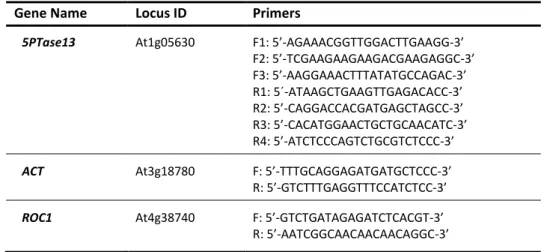
The 5PTase13 gene-specific primer pairs used are listed in Table 2. For PCR amplification, 35 cycles were performed, except for the housekeeping genes β-ACTIN (ACT) and CYCLOPHILIN (ROC1) (Table 2), which were detected after 23 and 28 cycles, respectively. All PCR products were separated on 1%
Functional insights into the plant-specific SR45 splicing factor
15 agarose gels and quantification of the amplified bands was performed with ImageJ (http://rsbweb.nih.gov/ij).
Table 2. Gene-specific primers used in the analysis of the expression and splicing patterns of the Arabidopsis
5PTase13 gene
Primers specific for the 5PTase13 gene (annealing in exonic sequences) as well as for two housekeeping genes,
β-ACTIN (ACT) and CYCLOPHILIN (ROC1).
Gene Name Locus ID Primers
5PTase13 At1g05630 F1: 5’-AGAAACGGTTGGACTTGAAGG-3’
F2: 5’-TCGAAGAAGAAGACGAAGAGGC-3’ F3: 5’-AAGGAAACTTTATATGCCAGAC-3’ R1: 5´-ATAAGCTGAAGTTGAGACACC-3’ R2: 5’-CAGGACCACGATGAGCTAGCC-3’ R3: 5’-CACATGGAACTGCTGCAACATC-3’ R4: 5’-ATCTCCCAGTCTGCGTCTCCC-3’
ACT At3g18780 F: 5’-TTTGCAGGAGATGATGCTCCC-3’
R: 5’-GTCTTTGAGGTTTCCATCTCC-3’
ROC1 At4g38740 F: 5’-GTCTGATAGAGATCTCACGT-3’
R: 5’-AATCGGCAACAACAACAGGC-3’
6. Confocal laser scanning microscopy
Confocal images were obtained with an LSM 510 laser-scanning microscope equipped with a Meta detector (Zeiss). For cell wall staining, roots were incubated in 10 μg/mL iodide propidium for 10 min. Excitation wavelengths used to detect fluorescence were 488 nm for GFP and 543 nm for iodide propidium. Emitted fluorescence was monitored at detection wavelengths between 500 and 550 nm for GFP and longer than 560 nm for iodide propidium.
Functional insights into the plant-specific SR45 splicing factor
16
III. RESULTS
1. Expression and splicing pattern of the 5PTase13 gene in Arabidopsis
The structure of the Arabidopsis 5PTase13 gene (At1g05630) is shown in Figure 6. The current genome annotation (TAIR10; www.arabidopsis.org) indicates that this gene generates two distinct splice variants due to an intron retention event. The first splice variant (5PTase13.1) results from inclusion of the 102-nt long intron, which is spliced out from the second splice variant (5PTase13.2) (Figure 6). As this alternatively-spliced fragment is in frame, the two predicted 5PTase13 protein isoforms differ only in 34 amino acids. Although no recognizable protein domain is included in this region, it falls within the catalytic domain of the phosphatase. Both splice forms exhibit the five WD40 repeats, the 5PTase catalytic domain and the two conserved motifs in the same position (Figure 6). However, the longer protein isoform (5PTase13.1) contains one extra N-myristoylation site (Figure 6).
Figure 6. Structure of the Arabidopsis 5PTase13 gene and its alternative transcripts, as well as domain organization of their encoded protein isoforms
Exon/intron organization of the 5PTase13 gene, where the boxes represent exons with UTRs in grey and lines represent introns. The arrows indicate the location of 5ptase13-specific primers used in the RT-PCR shown in Figure 7. The structure of the two splice variants (5PTase13.1 and 5PTase13.2) and of the corresponding predicted protein isoforms is also shown — the WD40 repeats and the 5PTase domain are represented by the blue and green boxes, respectively, the two black bars within the 5PTase domain mark the position of two conserved motifs (Zhong & Ye, 2004) and the dashed pink bar indicates the N-myristoylation site that is missing in the 5PTase13.2 protein isoform.
To evaluate the 5PTase13 development- and tissue-specific expression pattern in wild-type (Col-0) plants, RT-PCR analysis was conducted. Primers flanking the alternatively-spliced segment were
Functional insights into the plant-specific SR45 splicing factor
17 designed for the detection of both splice variants simultaneously (F1 and R1, Table 2, Figure 6). The amplified bands were then quantified and the ratio between 5PTase13.1/5PTase13.2 (SV1/SV2) calculated. Our results show that both 5PTase13 transcripts are ubiquitously expressed in Arabidopsis, being present in seedlings and different aged plantlets, as well as in different aerial organs (leaves of different ages, stems, flowers and siliques) and roots, with the shorter splice variant, 5PTase13.2, being by far the most abundant (Figure 7). Interestingly, both transcripts are less expressed in young (seven-day-old) seedlings. The ratio between the transcript levels of 5PTase13.1 and 5PTase13.2 is very similar in all organs and in all different aged seedlings and plantlets except for 14 day-old plantlets where expression of 5PTase13.1 is markedly higher when compared with 7-day-old seedlings and 21-day-7-day-old plantlets, as well as with all the other analyzed organs (Figure 7).
Figure 7. Expression and splicing pattern of the Arabidopsis 5PTase13 gene
RT-PCR analysis of the transcript levels of the two 5PTase13 splice variants, 5PTase13.1 and 5Ptase13.2, at different developmental stages and in different organs of Arabidopsis wild-type (Col-0) plants. The location of the F1 and R1 primers used is shown in Figure 6. Expression of the CYCLOPHILIN (ROC1) gene was used as a loading control. Bands were quantified and the histogram shows 5PTase13.1/5PTase13.2 ratios for each sample.
2. Isolation of a null 5PTase13 loss-of-function mutant
There are very few plant genes for which the production of multiple splice isoforms has been reported to have a clear in vivo functional impact. Since the 5PTase13 gene undergoes alternative splicing, one of our goals was to investigate the potential function of the two 5PTase13 isoforms by means of their individual expression in the 5PTase13 null background. Although there were already two previously isolated mutants for this gene – 5ptase13-1 and 5ptase13-2 (Ananieva et al., 2008; Lin et al., 2005) (Figure 8a), the lines kindly provided by Dr. Glenda Gillaspy from Virginia Tech, USA, were knockdown mutants. As seen in Figure 8b, RT-PCR analyses show that 5PTase13 expression in each mutant is not completely abolished. This could explain why we had difficulties in reproducing most of the phenotypes reported by Ananieva et al. (2008) (data not shown) and underscored the
Functional insights into the plant-specific SR45 splicing factor
18 need for the isolation of a true genetic null 5PTase13 mutant. To this end, after checking the available options at “TDNA-express: Arabidopsis gene mapping tool” (http://signal.salk.edu/cgi-bin/tdnaexpress), several lines carrying a T-DNA insertion within the 5PTase13 gene were obtained from the SALK and GABI-Kat collections (Table 1). A schematic representation of the 5PTase13 gene and the location of all screened T-DNA insertion lines is shown in Figure 9a.
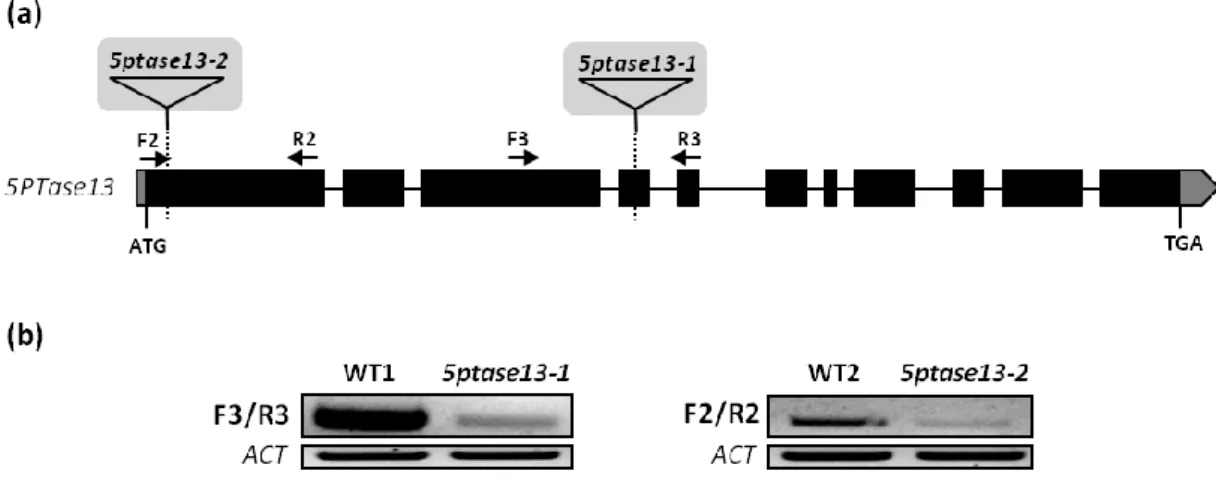
Figure 8. Molecular characterization of two T-DNA insertion mutants for the 5PTase13 gene
(a) Schematic representation of the 5PTase13 gene showing the T-DNA insertion sites (triangles) in the 5ptase13-1 and 5ptase13-2 mutants described by Ananieva et al. (2008). Boxes indicate exons with UTRs in
grey, lines between boxes represent introns, and arrows indicate the location of the F2, R2, F3 and R3
5ptase13-specific primers. (b) RT-PCR analysis of 5PTase13 gene expression in Col-0 wild-type (WT1 and WT2)
and mutant (5ptase13-1 and 5ptase13-2) plants. The location of the F2, R2, F3 and R3 primers used is shown in (a). Expression of the ACTIN (ACT) gene was used as a loading control.
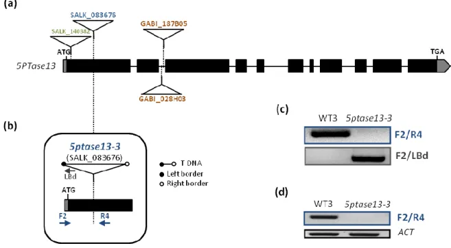
The obtained mutant lines were first screened for resistance to the specific selection marker for T-DNA insertions – kanamycin for SALK lines or sulfadiazine for GABI-Kat lines (Table 1). All resistant plants were then genotyped using gene-specific primers flanking the insertion site reported in the SALK database and the T-DNA-specific primer LBd that anneals at the end of the left border (LB) of the T-DNA (Table 1). This screen allowed the isolation of a homozygous mutant line for SALK_083676 containing the T-DNA insertion in exon 1, which was named 5ptase13-3 (Figure 9). Regarding the other mutant lines, GABI_187B0585, GABI_028H03 and SALK_140382 (Figure 9a), a total of 60, 85 and 19 plants, respectively, were genotyped but no plants homozygous for the insertion were retrieved.
PCR-based genotyping using gene-specific primers flanking the insertion in SALK_083676 (Figure 9b, Tables 1 and 2) amplified the expected size product for Col-0 (WT3), but not for the homozygous line, confirming gene disruption in the 5PTase13 gene (Figure 9c). On the contrary, PCR using the LBd primer only amplified a product in the mutant line and not in the corresponding Col-0 wild type
Functional insights into the plant-specific SR45 splicing factor
19 (WT3), confirming the presence of the insertion (Figure 9c). The obtained fragment was then sequenced to verify the exact insertion site.
Figure 9. Isolation of the 5ptase13-3 null mutant
(a) Exon/intron organization of the 5PTase13 gene (boxes represent exons with UTRs in grey and lines
represent introns) and location of the T-DNA insertion sites (triangles) in the mutant lines obtained from the SALK and GABI-Kat collections. (b) Schematic representation of the 5’UTR (grey box) and first exon (black box) of the 5PTase13 gene, showing the site of insertion and orientation of the T-DNA in the 5ptase13-3 mutant (SALK_083676). Arrows indicate the location of the F1 and R4 5ptase13-specific primers and the LBd1 primer annealing specifically at the left border of theT-DNA. (c) PCR-based genotyping of a 5ptase13-3 homozygous insertion line. The location of the F1, R4 and LBd primers is shown in (b). (d) RT-PCR analysis of 5PTase13 expression in Col-0 wild-type (WT3) and mutant (5ptase13-3) plants. The location of the F1 and R4 primers used is shown in (b). Expression of the ACTIN (ACT) gene was used as a loading control.
To examine the effect of the T-DNA insertion on the expression of the disrupted 5PTase13 gene, RT-PCR analysis of 5PTase13 expression in the 5ptase13-3 mutant line was performed. Using gene-specific primers flanking the insertion site (Figure 9b, Table 2), no expression was detected in the mutant (Figure 9d). It therefore appears that 5ptase13-3 is a true genetic null mutant, at least incapable of producing a full-length 5PTase13 transcript.
3. Phenotypical analysis of the 5PTase13-3 loss-of-function mutant
Bearing in mind future studies to investigate potential different functions of the 5PTase13 isoforms in the plant, it was imperative to check whether disruption of the 5PTase13 gene would lead to any evident phenotype. Thus, phenotypical analyses were conducted on the 5ptase13-3 loss-of-function
Functional insights into the plant-specific SR45 splicing factor
20 mutant alongside the previously reported 5ptase13-2 mutant (Ananieva et al., 2008). Since both these mutants harbor the T-DNA insertion in the first exon, we wanted to evaluate whether both would display the same phenotypes despite the latter being a leaky mutant. Phenotypical analyses included germination rates under normal and low light, in the presence or absence of glucose or ABA, as well as root growth under low light conditions.
3.1. Effect of glucose and ABA on seed germination under normal and low light conditions
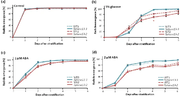
The germination of 5ptase13-3 and 5ptase13-2 mutant seeds on 0.5 X MS control medium under normal or low light conditions occurred at the same time as their respective wild types (WT3 and WT2) with full germination being attained one day after stratification (Figure 10a and 11a). The presence of 5% glucose in the media led to a delay in germination for all genotypes, with full germination rates occurring only around day 4 after stratification for 5PTase13-3 and WT3, and around day 6 for 5PTase13-2 and WT2, both under normal and low light conditions (Figure 10b and 11b). Although no differences in germination were perceived between 5ptase13-3 and WT3, the presence of glucose produced a slight delay in the germination of 5ptase13-2 mutant seeds, with the mutant never reaching the same germination rate as its corresponding wild type (WT2). This very slight delay occurred under both normal and low light conditions.
Figure 10. Germination of 5ptase13-3 and 5ptase13-2 mutant seeds under normal light
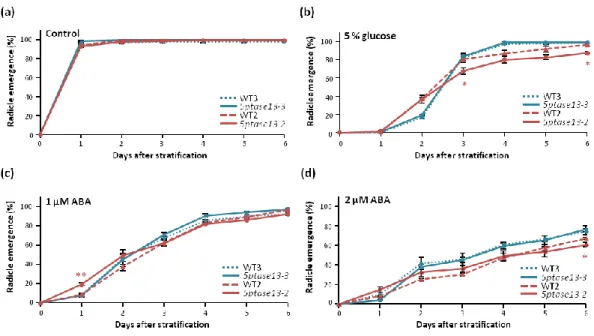
Germination (radicle emergence) rates of 5ptase13-3 (blue lines) and 5ptase13-2 (red lines) mutant seeds, as well as of the corresponding Col-0 wild-type (dashed lines) seeds, grown under standard light conditions (100 µmol s-1 m-2 ) in (a) control conditions or in the presence of (b) 5% glucose, (c) 1 μM ABA and (d) 2 μM ABA (means ± SE, n=3). Asterisks indicate significant differences between the 5ptase13-3 or 5ptase13-2 mutants and their corresponding wild-types (*p<0.05; **p<0.01) according to Student’s t-test.
Functional insights into the plant-specific SR45 splicing factor
21 The exogenous application of 1 μM and 2 µM of ABA delayed germination of wild-type and mutant seeds to a similar extent under both light intensity conditions (Figures 10c-d, 11c-d). The only exception was for the 5ptase13-2 mutant, which in the presence of 1 µM ABA under normal light in the first two days was less affected than the wild type (WT2) (Figure 10c). All genotypes reached their full germination rate six days after stratification in the presence of the lower concentration of ABA in normal light conditions (Figure 10c) however, under low light, a full germination rate was attained slightly earlier — 5ptase13-3 and WT3 at day 4 and 5ptase13-2 and WT2 at day 5 after stratification (Figure 11c). The delay in germination caused by ABA is more drastic in the presence of a higher concentration of this hormone (2 µM). As can be seen in Figures 10d and 11d, with the exception of 5ptase13-3 and WT3 under low light that were able to be fully germinated by day 6, under both light conditions neither of the genotypes was ever able to reach a 100% germination rate in this same 6-day time frame.
Figure 11. Germination of 5ptase13-3 and 5ptase13-2 mutant seeds under low light
Germination (radicle emergence) rates of 5ptase13-3 (blue lines) and 5ptase13-2 (red lines) mutant seeds, as well as of the corresponding Col-0 wild-type (dashed lines) seeds, grown under low light (40 µmol s-1 m-2 ) in (a) control conditions or in the presence of (b) 5% glucose, (c) 1 μM ABA and (d) 2 μM ABA (means ± SE, n=3). Asterisks indicate significant differences between the 5ptase13-3 or 5ptase13-2 mutants and their corresponding wild-types (*p<0.05) according to Student’s t-test.
These results indicate that the 5ptase13-3 and 5ptase13-2 mutants are neither affected by glucose nor ABA, either under normal or low light conditions.