Characterization of an
Aspergillus nidulans
mutant with abnormal
distribution of nuclei in hyphae, metulae, phialides and conidia
Marisa Vieira de Queiroz
a;*, Joaìo Luècio de Azevedo
ba
Departamento de Microbiologia/BIOAGRO, Universidade Federal de Vic°osa, 36571-000, Vic°osa, MG, Brazil
bEscola Superior de Agricultura ``Luiz de Queiroz'', Departamento de Geneètica, Caixa Postal 83, 13400-970, Saìo Paulo, SP, Brazil
Received 5 January 1998 ; revised 10 July 1998 ; accepted 10 July 1998
Abstract
The V10 deteriorated variant ofAspergillus nidulanshas hyphae, metulae, phialides and conidia with abnormal nuclear distributions. The alterations observed were : increase in the number of nuclei in hyphae, metulae and phialides, presence of anucleate, uninucleate and multinucleate conidia, abnormal vegetative growth and defective conidiation. When 0.5 M NaCl was added to the medium, an increase in the number of conidia was observed but their morphology and number of nuclei were not modified. The gene responsible for these alterations was namedanuA1. TheanuA1 gene is located on linkage group VII and is possibly involved in nuclear migration to hyphae, metulae, phialides and conidia. z1998 Federation of European
Microbiological Societies. Published by Elsevier Science B.V. All rights reserved.
Keywords : Aspergillus nidulans; Nuclear distribution ; Conidial development
1. Introduction
The fungus Aspergillus nidulans presents parallel vegetative and sexual reproductive cycles. The germi-nation of a vegetative haploid spore, the conidium, produces septated hyphae with multinucleate cells called conidiophores [14]. The vesicles are multinu-cleate and are formed at the conidiophore tips. A layer of metulae is formed on the surface of the vesicle and the phialides are produced from metulae by a single division of the nucleus. Metulae and phialides are referred to as sterigmata (primary and secondary, respectively). Conidia are formed by re-peated mitotic division of the phialide nucleus. A spore wall is formed around the distal nucleus, which
is arrested in the G1 phase of the cell cycle [14,18]. The metula, phialide and conidium are uninucleate.
Nuclear migration is very important for proper growth and development in both higher and lower eukaryotes. Anucleate primary sterigmata (aps) mu-tants ofA. nidulansare partially blocked in conidia-tion due to failure of the organized migraconidia-tion of nuclei into the conidiophore metulae. The mutants fall into two complementation groups, apsA and apsB, mapping on chromosomes IV and VI, respec-tively [4^6]. Mutation in the bncA gene leads to bi-nucleate conidiospores and abnormal distribution of nuclei in the metulae [13]. Another class of mutants with abnormal nuclear distribution was observed by Morris [9]. The gene symbol nud has been assigned to these mutants. The nudA, nudC and nudF genes were cloned and sequenced [11,20,21].
0378-1097 / 98 / $19.00 ß 1998 Federation of European Microbiological Societies. Published by Elsevier Science B.V. All rights reserved. PII : S 0 3 7 8 - 1 0 9 7 ( 9 8 ) 0 0 3 1 0 - 3
A. nidulans duplication strains produced sectors showing various degrees of phenotypic improvement and sectors with deteriorated morphology [2,10]. The improved sectors arose from nuclei with spontaneous deletions by an intrachromosomal process in either duplicate segment [2]. The deteriorated sectors ana-lyzed have a linear growth rate usually about that of the parent duplication strains, reduced conidiation, and di¡erently increased degrees of pigmentation. Di¡erent kinds of deteriorated variants were ob-served as follows : types with deletions from linkage
group I ; those with mutations in linkage group I or the II-I complex ; and those with mutations else-where in the genome. Deteriorated strains are well suited to looking for conidiophore and conidia alter-ations.
In this paper nuclear distribution and conidio-phore morphology were studied in deteriorated strain of A. nidulans denoted V10M. This mutant has a disordered nuclear distribution in the hyphae, metulae, phialides and conidia.
Table 1
Number of conidia produced by Aspergillus nidulans strains AM and V10M grown on complete medium (CM) and complete medium with 0.5 M NaCl (CMNaCl)
Strains Genotypes Number of conidia (U106)a
CM CMNaCl
AM proA1,pabaA1,yA1 140.00 320.00
V10M proA1,pabaA1,yA1 ;anuA1 2.10 16.00
aThe number of conidia was estimated by transferring pieces of the colonies (three pieces of 0.6 cm diameter from each colony tested) to 2 ml
0.1% (v/v) Tween 80.
2. Material and methods
2.1. Strains and culture conditions
The strains of A. nidulans used in this work were from Glasgow stocks. The strains V3, V10 and V17 were isolated by Azevedo and Roper [2] as sponta-neous sectors of the duplicated strains A, Dp(I-II) [10,15]. The strains AM and V10M were isolated as improved sectors of the strains A and V10, respec-tively. The strain V10W (wA1 ; pyroA4 ; riboB2; anuA) was a segregant of the V10MUMSE cross. The MSE strain of A. nidulans [7] has markers in each chromosome (su1adE20 ; adE20 ; wA1 ; galA1 ; pyroA4; facA303 ; sB3; nicB8; riboB2). Mutant al-leles of importance in this work were the following : yA2 and wA1, white and yellow conidia, respec-tively ; facA303 and galA1, unable to grow on ace-tate and galactose as the only source of carbon, re-spectively ; adE20, nicB8, pabaA1, pyroA4, proA1, riboB2, sB3, requiring adenine, nicotinic acid, p-ami-nobenzoic acid, pyridoxine, proline, ribo£avin and thiosulfate, respectively ; andsu1adE20, a suppressor ofadE20. The minimal medium (MM) and complete medium (CM) used were described by Pontecorvo et al. [14]. The CMNaCl medium corresponded to CM with 0.5 M NaCl added.
2.2. Genetic analyses
Genetic analyses were performed as described by Pontecorvo et al. [14]. Diploids were prepared by Roper's [16] technique.
2.3. Growth rate and conidiation
Colony growth rates were measured with a ruler on the back of the dish after 6 days of incubation in CM and CMNaCl at 37³C. The number of conidia was estimated by transferring pieces of the colonies (three pieces of 0.6 cm diameter from each colony tested) to 2 ml 0.1% (v/v) Tween 80. Counts were made in a haemocytometer with four replicates per strain tested.
2.4. Nuclear staining and microscopy
Cultures for microscopic examination consisted of
a ¢lm of complete medium with 106
conidia/ml on a coverslip incubated at 37³C for 19.5 h. Coverslips with adherent hyphae were transferred to methanol and incubated at room temperature for 5 min. The coverslips were incubated for 10 min at 63³C in 1 N HCl solution, rinsed in distilled water and trans-ferred to 100 mM phosphate bu¡er (pH 7.0) prior to staining. For staining, the coverslips were incu-bated for 5 min at room temperature in a Giemsa solution [17] with 0.5 mg/ml Trypan Blue (INLAB). The coverslips were then rinsed in 100 mM phos-phate bu¡er (pH 7.0) and mounted on clean glass slides and observed.
The conidial nuclei were stained with Giemsa based on the method of Tanaka et al. [17]. The cov-erslips were observed using an Olympus BHS micro-scope coupled with an Olympus PM-10AD photo-micrography system, when necessary.
3. Results and discussion
This paper results from our interest in studying the morphological variations in deteriorated sectors spontaneously isolated from duplicated strains Dp(I-II) ofA. nidulans [2,10]. The deteriorated var-iants are produced at a frequency of approximately
1 in ten colonies analyzed [1]. The deteriorated strains show poor conidiation and di¡erent degrees of pigmentation. Three variants isolated by Azevedo and Roper [2] were chosen for analysis (V3, V10 and V17). The deteriorated variant V10 was the only one that showed morphological alterations in the coni-dia, metulae and phialides. To eliminate duplication interference an improved sector of V10, designated V10M, was isolated. This strain had brown mycelia and poor conidiation when grown on CM. When 0.5 M NaCl was added to CM, the conidiation rate was increased, but it was not similar to the rate of sporulation of the AM strain grown in the same conditions (Table 1).
The yield of conidia from strains containing multi-ple genetic lesions that conidiate poorly can often be increased by incorporating high levels of supple-ments into the growth medium and by increasing the osmotic pressure of the medium through the ad-dition of 1.2 M D-sorbitol or 0.7 M KCl [19]. In the
presence of 0.6 M KCl, four deteriorated variants isolated as sectors from duplicated strains of A. ni-dulansdisplayed normal morphology [8]. The deter-iorated variant V10M showed a 7.6-fold increase in the rate of sporulation in the presence of 0.5 M NaCl compared to a 2.3-fold increase for AM. However, this increase of conidiation in the presence of the salt was not su¤cient to consider V10M as a phenocopy of the AM strain, since V10M continued to have morphological alterations.
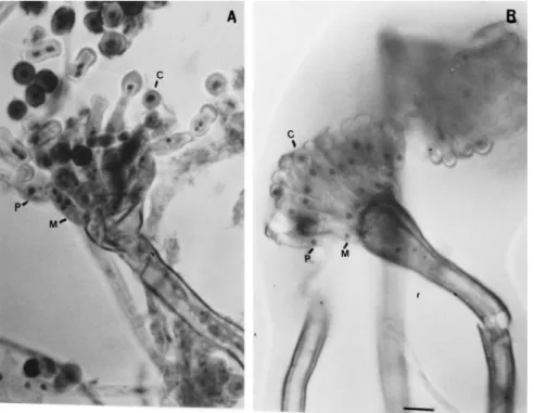
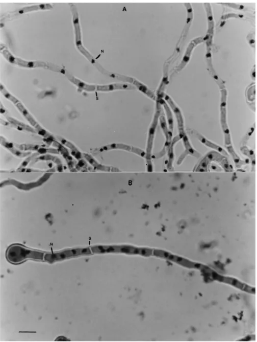
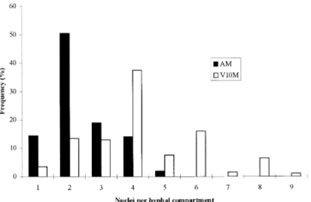
The observation of conidia, metulae and phialides of the V10M variant by microscopy showed a
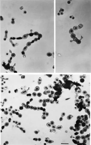
num-ber of di¡erences from the wild-type (Figs. 1 and 2). The distribution of nuclei in hyphal compartments is investigated in Figs. 3 and 4. V10M shows 83% of the compartments containing more than two nuclei compared to 35% in the wild-type. The irregular dis-tribution of nuclei in vegetative hyphae was observed by Clutterbuck [5] in theapsA6 mutant of A. nidu-lans. All the aps mutants examined exhibited high concentrations of nuclei in hyphal tip cells with dis-tribution of nuclei in clusters separated by conspic-uous gaps.
The metulae and phialides observed in V10M were long and swollen (Fig. 1). Some of the conidia of V10M variants were large, oval or shaped in the form of small bottles (Figs. 1 and 2). About 1^3 nuclei were present per metula, phialide or conidium and anucleate conidia were also observed. Table 2 shows the number of anucleate, uninucleate and multinucleate conidia found in V10M compared to other strains, a heterokaryon and a diploid between V10M and MSE strains. The anucleate conidia were often in an intermediate position in the chain of spores and sometimes black conidia were observed, possibly resulting from degeneration of anucleate conidia (Fig. 2). Normally, after the developing coni-dium had received one nucleus and reached about 3 Wm in diameter a septum formed separating the conidium from the phialide. However, it was possible to see phialides completely connected with conidia without the presence of a septum and two conidia united in the V10M variant, as if the spore separa-tion step had failed (Figs. 1 and 2).
A heterokaryon between strains V10M and MSE, which has genetic markers in each chromosome ofA. nidulans, also showed anucleate and multinucleate conidia, but at a rate lower than that of the V10M variant (Table 2). The cleistothecia produced from the heterokaryon between strains V10M and V10W were examined and of the 60 analyzed none showed ascospore production. The mutantapsA18 analyzed by Clutterbuck [5] was also sexually sterile but all otherapsA mutants appeared normal. Diploid strain contained uninucleate, binucleate and anucleate con-idia (results not shown). After analysis of nuclear distribution in hyphae, conidia, metulae and phia-lides of the V10M mutant, the gene responsible for this phenotype was named anuA1 (anucleate). The gene anuA was mapped by the parasexual methods
to chromosome VII (results not shown) and is evi-dently the same gene as that formerly designatedv10 by Azevedo and Roper [2] as conferring a mutant V10 deteriorated phenotype. A diploid strain homo-zygous foranuA1 was very unstable, sometimes pro-duced sectors and appeared to be a heterokaryon.
Mutants defective in nuclear migration have been described and studied in A. nidulans [3^6,9,11^13]. The genes anuA1 and bncA1 [5,13] share similar characteristics such as the presence of multinucleate conidia and alterations in the metulae and phialides. However, the mutation in bncA did not produce anucleate conidia andbncA was mapped to a di¡er-ent chromosome [13].
This paper represents the ¢rst step in the study of a gene of A. nidulans possibly involved in nuclear migration of hyphae, metulae, phialides and conidia.
Acknowledgments
This work was supported by Brazilian agencies CNPq, Conselho Nacional de Desenvolvimento Cient|è¢co e Tecnoloègico; RHAE; and FAPESP, Fundac°aìo de Amparo aé Pesquisa do Estado de Saìo Paulo.
References
[1] Azevedo, J.L. (1975) Altered instability due to genetic changes in a duplication strain ofAspergillus nidulans. Genet. Res. 26, 55^61.
[2] Azevedo, J.L. and Roper, J.A. (1970) Mitotic non-conformity in Aspergillus nidulans: successive and transposable genetic changes. Genet. Res. 16, 79^93.
[3] Chiu, Y. and Morris, N.R. (1995) Extragenic suppressors of
nudC3, a mutation that blocks nuclear migration in Aspergil-lus nidulans. Genetics 141, 453^464.
[4] Clutterbuck, A.J. (1977) The genetics of conidiation in Asper-gillus nidulans. In : Genetics and Physiology of Aspergillus
(Smith, J.E. and Pateman, J.A., Eds.), pp. 305^317. Academic Press, London.
[5] Clutterbuck, A.J. (1994) Mutants ofAspergillus nidulans de¢-cient in nuclear migration during hyphal growth and conidia-tion. Microbiology 140, 1169^1174.
[6] Fischer, R. and Timberlake, W.E. (1995)Aspergillus nidulans apsA (anucleated primary sterigmata) encodes a coiled-coil protein necessary for nuclear positioning and completion of asexual development. J. Cell Biol. 128, 485^498.
[7] McCully, K.S. and Forbes, E. (1965) The use of p-£uoro-phenylalanine with ``master'' strains of Aspergillus nidulans
form assigning genes to linkage groups. Genet. Res. 6, 352^ 359.
[8] Molina, S.M.G. and Azevedo, J.L. (1994) Phenotypic rever-sion of Aspergillus nidulans morphological deteriorated var-iants in the presence of osmotic stabilisers. FGN 41, 62. [9] Morris, N.R. (1976) Mitotic mutants ofAspergillus nidulans.
Genet. Res. 26, 237^254.
[10] Nga, B.H. and Roper, J.A. (1968) Quantitative intrachromo-somal changes arising at mitosis inAspergillus nidulans. Ge-netics 58, 193^209.
[11] Osmani, A.H., Osmani, S.A. and Morris, N.R. (1990) The molecular cloning and identi¢cation of a gene product specif-ically required for nuclear movement inAspergillus nidulans. J. Cell Biol. 111, 543^551.
[12] Pascon, R.C. (1994) Isolamento e caracterizac°aìo de setores deteriorados da linhagem Abnc deAspergillus nidulans. PhD Thesis, Univ. of Saìo Paulo, Piracicaba.
[13] Pizzirani-Kleiner, A.A. and Azevedo, J.L. (1986) Character-ization and genetical analysis of anAspergillus nidulansstrain that produces multinucleate conidia. Trans. Br. Mycol. Soc. 86, 123^130.
[14] Pontecorvo, G., Roper, J.A., Hemmons, L.M., McDonald, K.D. and Bufton, A.W.J. (1953) The genetics ofAspergillus nidulans. Adv. Genet. 5, 141^238.
[15] Pritchard, R.H. (1956) A genetic investigation of some ad-enine-requiring mutants ofAspergillus nidulans. Ph.D. thesis, The University of Glasgow, Glasgow.
[16] Roper, J.A. (1952) Production of heterozygous diploids in ¢lamentous fungi. Experientia 8, 14^15.
[17] Tanaka, Y., Murata, N. and Kato, H. (1979) Behavior of nuclei and chromosomes during ascus development in the mating between either rice-strain or weeping lovegrass-strain and ragi-strain ofPyricularia. Ann. Phytopathol. Soc. Jpn. 45, 182^191.
[18] Timberlake, W.E. and Clutterbuck, A.J. (1995) Genetic regu-lation of conidiation. In : Aspergillus nidulans, 50 Years on (Martinelli, S.D. and Kinghorn, J.R., Eds.), pp. 383^478. Elsevier, Amsterdam.
[19] Timberlake, W.E. and Hamer, J.E. (1986) Regulation of gene activity during conidiophore development inAspergillus nidu-lans. In : Genetic Engineering Principles and Methods (Setlow, Table 2
Number of nuclei present in the conidia of the AM, V10M, MSE, heterocaryon (V10+MSE) and diploid (V10M/MSE) strains ofAspergillus nidulans
Strains Number of nuclei (%)
0 1 2 3
J.K. and Hollander, A., Eds.), Vol. 8, pp. 1^29. Plenum Press, New York.
[20] Xiang, X., Beckwith, S. and Morris, N.R. (1994) Cytoplasmic dynein is involved in nuclear migration inAspergillus nidulans. Proc. Natl. Acad. Sci. USA 91, 2100^2104.