CONIDIOGENESIS OF DETERIORATED VARIANT
OF THE STRAIN Abnc OF
Aspergillus nidulans
Ágata Cristiane Huppert Giancoli1; Aline Aparecida Pizzirani-Kleiner2*
1
USP/ESALQ - Programa de Pós-Graduação em Genética e Melhoramento de Plantas - Depto. de Genética, C.P. 83 - 13400-970 - Piracicaba, SP - Brasil.
2
USP/ESALQ - Depto. de Genética.
*Corresponding author <[email protected]>
ABSTRACT: The Abnc strain of A. nidulans carries the bncA1 gene (binucleated conidia), which induces the formation of binucleate and trinucleate conidia, displaying a chromosome I duplicated area and shifted to the chromosome II (IàII), and bringing forth genetic instability with degenerated sectors. This work has considered in a cytological level the conidiogenesis of the deteriorated variants isolated from the Abnc strain of A. nidulans, observing the event at the level of structural alterations, which composes the conidiophore, and the variations in the number of sterigmata and conidia nuclei. Cytogenetic analyses of conidiogenesis were accomplished in predetermined periods, under Giemsa stain, to observe the nuclei and analysis through the Scanning Electronic Microscopy, and also the structures that composes the conidiophore. The analyzed, deteriorated variants presented alterations in the cell-foot, metulae and phialides structure, conidiophore number and conidias reduction, and the formation of secondary conidiophores. These alterations can be related to genes for the development, bristle, and activities of NIMA e NINXcdc2 (involved in morphogenesis regulatory cycle) that induce the expression of brislte, establishing the link to the regulation and expression of the genes control throughout the conidiophore development.
Key words: bristle, conidiophore, developmental mutants
CONIDIOGÊNESE DE VARIANTES DETERIORADOS DA
LINHAGEM Abnc DE
Aspergillus nidulans
RESUMO: A linhagem Abnc de A. nidulans é portadora do gene bncA1 (“binucleated conidia”), que confere a característica de conídios bi e trinucleados, além de apresentar uma região duplicada do cromossomo I e translocada para o cromossomo II (IàII), gerando instabilidade genética com formação de setores deteriorados. Este trabalho analisou citologicamente a conidiogênese dos variantes deteriorados isolados da linhagem Abnc de A. nidulans, observando a ocorrência de alterações das estruturas que compõe o conidióforo e as variações no número de núcleos dos esterígmas e conídios. As análises citogenéticas da conidiogênese foram realizadas com períodos pré-determinados, sendo efetuada a coloração de Giemsa para observação dos núcleos, e análise por Microscopia Eletrônica de Varredura, para observação das estruturas que compõe o conidióforo. Os variantes deteriorados analisados apresentaram alterações na estrutura das células-pé, métulas e fiálides, redução do número de conidióforos e conídios, e formação de conidióforos secundários. Estas alterações no conidióforo podem estar relacionadas com os genes para o desenvolvimento, bristle e com atividade das proteínas NIMA e NINXcdc2 (envolvidas com o ciclo regulatório da morfogênese) as quais induzem a expressão de brislte, estabelecendo o caminho para a regulação e controle da expressão dos genes durante o desenvolvimento do conidióforo.
Palavras chave: bristle, conidióforo, mutantes para o desenvolvimento
INTRODUCTION
The fungus Aspergillus nidulans presents a life cycle three phases, Vegetative Cycle, Asexual Cycle or Conidiogenesis and Sexual Cycle or Ascosporogenesis. The vegetative growth begins with the spore germination that can be a conidia or an ascospore (Lee & Adams, 1994). The ramified air hyphae differs in a conidiophore, a generative structure of the asexual cycle (Adams et al., 1998). This differentiation and development process con-stitute the Conidiogenesis. The conidiophore morphology
phialides apex original primary conidia and arranging conidia chains (Oliver, 1972; Bergen & Morris, 1983).
The structural and temporal development pro-cesses are controlled by groups of regulatory genes and the activation sequence involves the genes bristle (brlA),
abacus (abaA) and wet-white (wetA), establishing the regulatory pathway and genes expression control during the conidiophore development (Adams et al., 1998). Other two genes, stund (stuA) and medusa (medA), interact with the regulators genes (Miller, 1990; Miller et al., 1992).
The strain A of A. nidulans, with double chro-mosome segment (chrochro-mosome I) and transferred (chro-mosome II) (Nga & Roper, 1968; 1969) presents mitotic instability, which leads to deteriorated sections from to-tal or partial loss of the chromosomic duplication, with subsequent insert of that segment in any of the eight groups of junction of A. nidulans (Azevedo & Roper, 1970). Several factors affect the genetic instability: chemical factors, as the caffeine (Azevedo & Santana, 1975), physical factors, such as temperature (Lieber, 1976), and genetic factor, such as the effect of the bncA1 gene, that originates conidia bi and trinucleted (Pizzirani-Kleiner & Azevedo, 1986a, 1994b; Pascon, 1994) and reduce the mitotic instability in the Abnc strain, with an increase in the frequency of deteriorated sections (Pascon, 1994). The bncA1 gene causes abnor-malities during the formation of the conidiophore such as the reduced number and phialides and metulae septa (Pascon et al., 2001).
This article presents the study of the conidiogenisis of the deteriorated variants originated from the Abnc strain of A. nidulans. Structural and temporary alterations were observed in the conidiophore of the de-teriorated variants, VB7, VB8, VB9, VB10 and VB11. These structural alterations present structural likeness with mutants bristle.
MATERIAL AND METHODS
Strains
The MSE of A. nidulans strain belongs to the Glasgow (England) stock, and presents the marks wA3 (II), facA303 (V), galA1 (III), yA1(I), pyroA4 (IV), sB3 (VI), nicB6 (VII) and riboB2 (VIII) (McCully & Forbes, 1965). The deteriorated variants VB7, VB8, VB9, VB10 and VB11 were isolated in the laboratory of Microorgan-isms of Genetics (USP/ESALQ) out of spontaneous Abnc strain sections which present the marks proA1 (I),
pabaA6 (I), Dp (I-II), and the bncA1 gene (IV), that gen-erates conidia bi and trinucleated (Pizzirani, 1977).
Medium Culture and Conidiogenesis.
Conidia of the VB 7, VB8, VB9, VB10 and VB11 strain were incubated in complete medium (Pontecorvo et al., 1953), and over dialysis membranes and subsequent
incubation at a temperature of 37oC for 20, 32 and 72 h (Giancoli, 2000).
Stain of nucleus and optical microscopy
After the established periods, the coverslips with adherent hyphae were transferred to etanol and acetic acid, lactic acid solution (6:1:1), and incubated at room temperature for 15 min. Then, coverslips were incubated for 12 min. at 63o
C in 1 mol L-1
HCl solution, rinsed in distilled water and transferred to 100 mmol L-1
phosfhate buffer (pH 7.0) for 1 hour, prior to staining after incuba-tion for 20 min. at room temperature in a Giemsa solu-tion (Tanaka et al., 1979).
Scanning Electron Microscopy (SEM)
After the pre established periods the cover-slips adherent hyphae and conidiophores were transferred to glutaraldehyde 25% solution, paraformaldehyde 10% in caccodilate buffer 0.2% pH 7.2 and CaCl2 0,1 mol L-1
. After 1 –2 hours the cove-rslips were washed for 10 min with buffer and post fixed to 1 hour in similarly buffered 1% OsO4. Cover slips were then washed in distilled wa-ter and dehydrated in a graded acetone series. Cover slips were dried to a critical point (Balzers CPD 030), coated with gold (MED 010 - Balzers), and examined under a Scanning Electron Microscope (Leo 435 VP).
RESULTS AND DISCUSSION
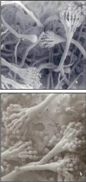
Starting from 20 hours of cultivation MSE and Abnc strain, it was possible to observe indifferentiated hy-phae, and in some cases differentiated hyphae in cell-foot with the stem of the conidiophore already formed. At 32 hours, ripe phialides with conidia development were de-tected. These data are similar to those reported by Axelrod et al. (1973) and Champe et al. (1981), demonstrated that the temporary and space development of the conidiophores occurred after 20 hours of cultivation. Starting from 72 hours, it was possible to observe the conidiophores with long conidia chains (Figures 1a and 1b). The Abnc strain bearing of the bncA1 gene (Pizzirani-Kleiner, 1981), be-sides presenting conidia bi and trinucleated, shows other interesting aspects: the disposition of the conidia chains, where they are just found conidia chains uninucleated and or just binucleated, are rarely found mixed chains, what agrees with the observations obtained by Pascon (1994) and Pascon et al. (2001). The irregular organization of the metulae and phialides along the vesicle of the conidio-phore, was noticed in a small number of structures when compared with the strains that don’t show the mutation
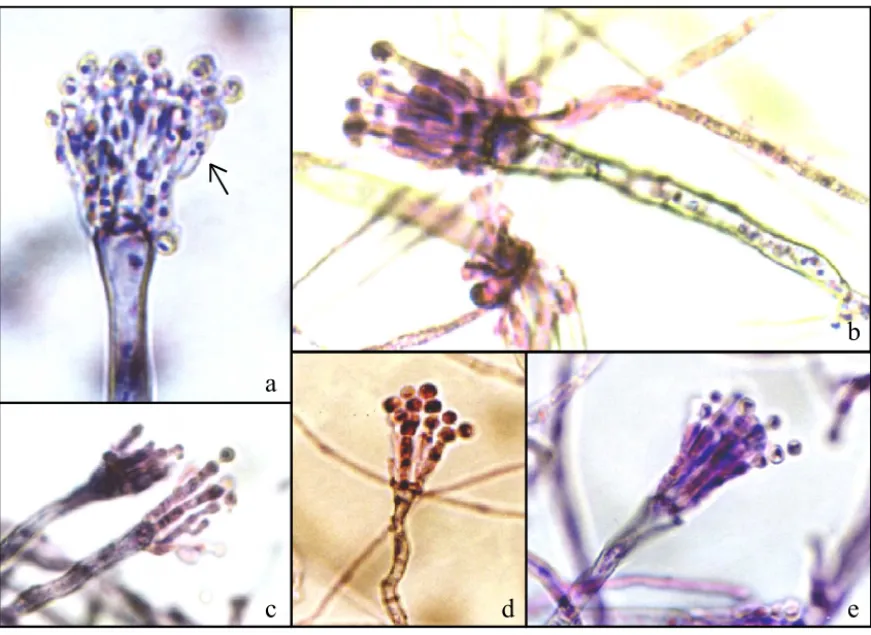
bncA1 (not given presented); the phialides are septate with until three nuclei in its interior (Figure 3a).
affect the formation of the conidiophore and conidia, in-side of the Classe II - are subdivided more four types of genes (bristle, abacus, stund and medusa). The bncA1 gene probably is into the mutants’ categories with modi-fication of the conidiophore (Miller et al., 1992; Pascon et al., 2001), which are combined by the genes stuA and medA, that cause specific alterations in the morphology and space organization of the conidiophore, but do not avid the conidia development
The deteriorated variants were produced with the approximate frequency of one in ten analyzed colonies (Giancoli, 2000). The deteriorated variants analyzed, VB7, VB8, VB9, VB10 and VB11 presented little conidiation and different degrees pigmentation.
The deteriorated variant VB7 presented time of compatible development with the pattern strain (MSE), with differentiation of the hyphae starting from 20 hours of development. The conidia’s growth occurred within 32 hours, being observed small conidia chains uni and binucleated. VB7 presented a smaller amount of conid-iophore if compared with the MSE strain. The temporary development of the conidiophore was late, the metulae and phialides demonstrated characteristic alterations,
ob-served in the strain with the bncA1 gene (Figure 3a). The metulae and phialides present extended and unlikely structures (Figure 2a) with lack of conidia. Normal con-idiophores were observed. We could observe the forma-tion of secondary conidiophore that is a characteristic of the bristle and medusa genes (Figure 2b).
The deteriorated variant VB8 has shown a space and temporary abnormal organization outstanding when compared to the other deteriorated variants. The hyphae differentiation, followed by the vesicles growth was ob-served starting from 32 hours. There was a characteristic abnormal spatial organization, with extended and unlikely metulae. (Figures 2c, 2d and 3b). Some vesicles present
Figure 1 - MSE strain (a. 1000 x) and Abnc (b. 2000 x) A. nidulans, 72 hours of growth (SEM).
Figure 2 - SEM (a). VB7 sterigmata without differentiation, metulae and phialides prolonged after 32 hours of cultivation (4600 x). (b). VB7 formation of secondary conidiophore with 72 hours of cultivation (4500 x). (c, d) VB8 sterigmata without differentiation, metulae and phialide prolonged ofter 32 hours and 72 hours of cultivation respectively (4000 x and 5030 x). (e and f) VB9 and VB10 sterigmata without differentiation, metulae and phialides prolonged with 32 hours of cultivation (3580 x and 3870 x). (g and h). VB11 abnormality in the formation of the vesicle with of 72 hours of cultivation (3110 x; h. VB11) sterigmata without differentiation, metulae and phialides prolonged with 72 hours of cultivation (3140 x).
a b
c d
f e
irregularities in their formats and the amount of conidio-phores are small. That deteriorated variant presented an extremely high number of hülle cells, when compared with the MSE strain (data not available).
The cytological analysis of VB9 presented like-ness with VB7. The period of temporary development was similar to the established ones for the pattern strain. There was a characteristic abnormal spatial organization, with the extension and diferentiation for metulae (Figures 2a and 2e). The amount of conidia is reduced. For carrying the mutation in the bncA1 gene it presents septated and smaller amount of metulae and phialides. Normal conid-iophores were observed.
The deteriorated variants VB 10 and VB11 pre-sented great likeness. There was a delay in the process of temporary-spatial development of the conidiophore. In these deteriorated variants occurred abnormal spatial or-ganization, with metulae extension (Figures 2f, 2g and 3d and 3e). VB11 presented some vesicles with irregular format (Figure 2f). The beginning of the differentiation of the hyphae occurred about 32 hours and presented a reduced number of conidiophores.
The morphologic alterations observed in the structure of the conidiophore are characteristic for de-velopment genes. These genes should cause conidia al-terations (wetA); alterations in the conidiophore (stuA and medA), which don’t affect the conidia formation; mutants that block the conidia formation (brlA and abaA) and mutants that affect the pigmentation of the conidia (ivoA and ivoB) (Timberlake & Clutterbuck, 1994). The main characteristics of the conidiophore de-velopment are due to the central control of the bristle gene. However, the activation of the protein Brl is not well studied, but the occurrence of the interactions is known among the genes stu (stunted) and med (medusa). Ye et al. (1999) and Schier et al. (2001), while study-ing the relationship between the regulation of the cel-lular cycle and the morphogenetic development of A. nidulans, they noticed that exists a deep relationship between the two cycles, being the morphogeneses de-pendent of the increase of the quinase activity of NIMA and NINXcdc2, so this way can occur the activation and regulation in cascade of genes, that take the normal for-mation of the conidiophore.
Figure 3 - Giemsa stain. (a). VB7 showing alteration in the nucleus number in phialides caused by the mutation bncA1, growth of 72 hours (1200 x); (b, c, d, e) VB 8 VB9 VB10 and VB11 respectively, sterigmata without differentiation, prolongated metulae and phialides, growth of 72 hours (1200 x).
a
c
d
e
The microscopic analyses resulted in the detection of abnormalities formation of the conidiophore, as the pres-ence of secondary conidiophore starting from metulaes ab-normal, besides extended metulae and phialide. This phe-notype is characteristic so much of mutants light bristle or middleman (leaky) as of medula mutants. These last ones, however, present a pigment decrease of the conidiophore and metulae chains (Miller, 1993), two absent character-istics in the analyzed deteriorated variants.
In analyzing the intermediate conidiophore we always find morphological vesicles and metula alter-ations. Adams et al. (1998) observed that after the first synthesis of the protein Brl (BrlA), there is a differentia-tion of the vesicle and the germinadifferentia-tion of the metulae, so the only nuclei then migrate for the metulae and dif-ferentiation is concluded in an autonomous way. These results suggest that the amount of protein Brl that is aimed for each metulae altogether with the cytoplasm during its germination should induce the genes for the final stage of the fiálides differentiation. However, when there is little protein expression Brl in the vesicles, the metulae could receive an insufficient amount for the beginning of the induction, tending to an abnormal behavior, differ-entiating in a stem. This abnormal stem could originate a new secondary conidiophore. This interpretation would also justify the fact that some vesicles not present differ-entiated metulae, growing as secondary stem, beside dif-ferentiated metulae, with fialides and conidia. The syn-chronism lack can be the result of different amounts of protein Brl of each metulae. These observations indicate that the synchronism usually found in the differentiation of the structures of the conidiophore depend on the mini-mum of protein Brl produced in the vesicle, guarantee-ing surpluses in the metulae, enough to control small de-viations in the distribution of this product during the ger-mination (Adams et al., 1998).
The cytological analyses suggest that the deterio-rated variants present the characteristic phenotype of the bristle gene. However, these alterations can be influenced by regulatory genes of the cell cycle avoiding the cor-rect activation of the bristle gene (Ye et al., 1999 and Schier et al., 2001). These were the first steps in the study of this group of deteriorated variants that are going along with the investigation at a molecular level.
ACKNOWLEDGMENTS
Dr. E. W. Kitajima and Dr. B. Leite for allow the execution of the observations of Scanning Electron Mi-croscopy (SEM) in NAP/MEPA - ESALQ/USP.
REFERENCES
ADAMS, T.H.; WIESER, J.K.; YU, J.H. Asexual sporulation in Aspergillus nidulans. Microbiology and Molecular Biology Reviews, v.62, p.35-54, 1998.
AXELROD, D.E.; GEALT, M.; PASTUSHOK, M.; Gene control of developmental competence in Aspergillus nidulans. Developmental Biology, v.34, p.9-15, 1973.
AZEVEDO, J.L.; ROPER, J.A. Mitotic non-conformity in Aspergillus nidulans: successive and transposable genetic changes. Genetical Research, v.16, p.79-93, 1970.
AZEVEDO, J.L.; SANTANA, E.P. The use of cloroneb to obtain haploid segregants from heterozygous diploid of Aspergillus nidulans.
Aspergillus Newsletter, v.13, p.6, 1975.
BERGEN, L.G.; MORRIS, N.R. Kinetics of nuclear division cycle of Aspergillus nidulans. Journal of Bacteriology, v.156, p.155-160, 1983. CHAMPE, S.P.; KURTZ, M.; YAGER, I.; BUTNICK, N.; AXELROD, D. Spore formation in Aspergillus nidulans: competence and other developmental processes. In: HOHL, H.R.; TURIAN,G (Ed.) The fungal spore morphogenetic control. New York: Academic Press, 1981. p.255-276.
CLUTTERBUCK, J. A mutational analysis of conidial development in Aspergillus nidulans. Genetics, v.63, p.317-327, 1969.
CLUTTERBUCK, J. A variegated position effect in Aspergillus nidulans.
Genetical Research, v.16, p.303-316, 1970.
CLUTTERBUCK, J. The genetics of conidiation in Aspergillus nidulans. In: PATERMAN, J.A.; SMITH, J.E. (Ed.) Genetics and physiology of
Aspergillus. New York: Academic Press, 1977. p.305-317.
GIANCOLI, A.C.H. Caracterização citológica e genética de Linhagens de Aspergillus nidulans portadoras de duplicação cromossômica e do gene bncA1. Piracicaba: USP/ESALQ, 2000. 103p. (Dissertação - Mestrado). LEE, B.N.; ADAMS, T.H. The Aspergillus nidulans fluG gene is required for production of an extracelular developmental signal. Genes and Development, v.8, p.641-651, 1994.
LIEBER, M.M. The effects of temperature on genetic instability in Aspergillus nidulans. Mutation Research, v.34, p.93-122, 1976. McCULLY, K.S.; FORBES, E. The use of p-fluorphenilalanine with “master”
strain of Aspergillus nidulans form assigning gene of to linkage groups.
Genetical Research, v.6, p.352-359, 1965.
MILLER, B.L. The developmental genetics of asexual reproduction in Aspergillus nidulans. Developmental Biology, v.1, p.207-219, 1990. MILLER, B. Brushing up on bristles: complex genes and morphogenesis in
molda. Trends in Genetics, v.9, p.293-295, 1993.
MILLER, K.Y.; WU, J.; MILLER, B.L. Stu A is required for cell pattern formation in Aspergillus. Genes and Development, v.6, p.1770-1782, 1992.
MIMS, C.W.; RICHARDSON, E.A.; TIMBERLAKE, W.E. Ultrastructural analysis of conidiophore development in the fungus Aspergillus nidulans using freeze-substitution. Protoplasma, v.144, p.132-141, 1988. NGA, B.H.; ROPER, J.A. Quantitative intrachromosomal changes arising
at mitosis in Aspergillus nidulans. Genetics, v.5, p.193-209, 1968. NGA, B.H.; ROPER, J.A. A system generation spontaneous in
intrachromosomal changes at mitosis in Aspergillus nidulans. Genetical Research, v.14, p.63-70, 1969.
OLIVER, P.T. Conidiophore and spore developmental in Aspergillus nidulans. Journal of General Microbiology, v.73, p.45-54, 1972. PASCON, R.C. Isolamento e caracterização de setores deteriorados da
linhagem Abnc de Aspergillus nidulans. Piracicaba: USP/ESALQ, 1994. 140p. (Dissertação - Mestrado).
PASCON, R.C.; PIZZIRANI-KLEINER, A.A.; MILLER, B.L. The Aspergillus nidulans bncA1 mutation causes defects en the cell division cycle, nuclear and developmental morphogenesis. Molecular and General Genetics, v.264, p.546-554, 2001.
PIZZIRANI, A.A. Características, propriedades e estabilização de dissômicos em Aspergillus nidulans. Piracicaba: USP/ESALQ, 1977. 111p. (Dissertação - Mestrado).
PIZZIRANI-KLEINER, A.A. Efeito do gene bncA1 na formação de conídios de Aspergillus nidulans. Piracicaba: USP/ESALQ, 1981. 141p. (Tese -Doutorado).
PIZZIRANI-KLEINER, A.A.; AZEVEDO, J.L. Characterization and genetical analysis of an Aspergillus nidulans strain that produces mutinucleate conidia. Transaction of the British Mycological Society, v.86, p.123-130, 1986a.
Received February 20, 2003 Accepted April 28, 2004 PONTECORVO, G.; ROPER, J.A.; HEMMONS, L.M.; MacDONALD,
K.D.; BUFTON, A.W.J. The genetics of Aspergillus nidulans. Advances in Genetics, v.5, p.141-238, 1953.
TANAKA, Y.; MURATA, N.; KATO, H. Behavior of nuclei and chromosomes during ascus development in the mating between either rice-strain or weeping lovegrass-strain and ragi-strain of Pyricularia.
Annual Phythopathology Society Japan, v.45, p.182–191, 1979. SCHIER, K.; LIESE, R.; FISCHER, R. A Pcl-Like Cyclin of Aspergillus
nidulans is Transcriptionally Activated by Developmental Regulators and Is Involved in Sporulation. Molecular and Cellular Biology, v.21, p.4075–4088, 2001.
TIMBERLAKE, W.E.; CLUTTERBUCK, A. Genetic regulation of Conidiation. In: MARTINELLI, S.D.; KINGHORN, J.R. Aspergillus
50 years on. Amsterdam: Elservier, 1994. v.29, cap.16, p.383-427. YE, X.; LEE, S.; WOLKOW, T.; McGUIRE, S.; HAMER, J.; WOOD, G.;