CHROMOSOMIC DUPLICATION
Ágata Cristiane Huppert Giancoli1*; João Lúcio de Azevedo2; Aline Aparecida Pizzirani-Kleiner2
1
Laboratório de Produtos Bioativos de Origem Microbiana, Departamento de Ciências Fundamentais e Desenvolvimento Agrícola,
Faculdade de Ciências Agrárias, Universidade Federal do Amazonas, Manaus, AM, Brasil; 2Laboratório de Genética de
Microrganismos, Departamento de Genética e Melhoramento de Plantas, Universidade de São Paulo, Piracicaba, SP, Brasil.
Submitted: March 09, 2009; Returned to authors for corrections: May 04, 2009; Approved: July 24, 2009.
ABSTRACT
A development mutant, named V103, was obtained spontaneously from the A strain of A. nidulans. The A
strain contains a duplicated segment of chromosome I that has undergone translocation to chromosome II
(I II). It is mitotically unstable and generates phenotypically deteriorated types, some with enhanced
stability. The deteriorated variants of A. nidulans show abnormal development,exhibiting slower colony
growth, variations in colony pigmentation and changes in conidiophore structure. The alterations observed
in the conidiophore include fewer metulae and phialides, further elongation and ramification of these
structures, delayed nuclear migration and the presence of secondary conidiophores.
Key words:Aspergillus nidulans, Conidiogenesis, Nuclear migration, Asexual Cycle, Mitotic Instability
INTRODUCTION
The life cycle of the fungus Aspergillus nidulans is marked
by important developmental events, which include the
germination of asexual, uninucleate mitotic spores (conidia),
nuclear migration through the mycelium and the establishment
of a highly polarized growth pattern that gives rise to
multinucleate hyphae (6). Nuclear migration is an essential
feature for the growth of filamentous fungi and the process of
conidiation involves temporal and special regulation of gene
expression, cell specialization, and intercellular communication
(1). In this context, several classes of mutants with abnormal
nuclear distribution that impairs hyphal extension have been
observed. For instance, nuclear distribution (nud) mutants contain
nuclei that divide at a normal rate but fail to move from the spore
end of the growing germ tube (12, 13). In nud mutants, gene
products essential for nuclear distribution in hyphae have lost
function, resulting in abnormal conidiophore morphology (24).
Furthermore, in bim mutants (blocked in mitosis – cell cycle
mutants), a wide range of cell cycle functions involved in mitotic
spindle formation are impaired (4, 8).
The germination of conidia produces septated hyphae with
conidiophores (17). The vesicles are multinucleate and develop at
the conidiophore tips. A layer of metulae forms on the surface of
the vesicle and the metulae produce phialides by a single division
of nucleus. Repeated mitotic division of the phialide nucleus
generates conidia. (10, 21). A limited set of regulatory loci
controls the coordinates temporal and special expression of
hundreds of genes required for morphogenesis. The sequential
and wet-white (wetA), establishes the central regulatory pathway
for control of gene expression observed during conidiophore
development (5). Two other genes, stuA and medA, interact with
key regulatory genes and may be considered morphological
modifiers. Mutants for these genes are oligosporous because of
abnormalities in their conidiophores (1).
In this study, we have characterized a new, spontaneously
obtained mutant of Aspergillus nidulans with substantial
developmental changes. We investigated conidiophore
development and the cell cycle of V103, a strain derived from the
A strain of A. nidulans, which is mitotically unstable because
of a partial chromosomal duplication followed by translocation
(I II). We have shown that this development mutant exhibits
significant changes in colony growth, colony pigmentation and
conidiophore structure. In V103, the conidiophore contained
fewer metulae and phialides, with further elongation and
ramification of these structures. We have also observed
delayed nuclear migration and the presence of secondary
conidiophores.
MATERIALS AND METHODS
Strains
The haploid strains of A. nidulans were derived from
Glasgow stocks. For this study, the deteriorated strain V103 was
isolated from spontaneous sectors of the duplicated A strain, Dp
(I-II). The MSE strain of A. nidulans (9) has the following
markers in each chromosome: wA3 (II), facA303 (V), galA1 (III),
yA1 (I), pyroA4 (IV), sB3 (VI), nicB6 (VII) and riboB2 (VIII).
The A strain of A. nidulans (14, 15) has the markers proA1 (I),
pabaA6 (I) and Dp (I-II).
Culture medium and Conidiogenesis
Complete medium (CM) (17).
Conidia from the V103 strain were incubated in liquid
culture for 2, 4, 6, 8, 12, 16 and 20 hours at 37oC. Synchronous
conidiophore development was induced by transferring a thin
mycelial at filtered from liquid culture to agar plates (Agar
Complete Medium) with inserted coverslips and inoculating the
plates for 16, 20, 24, 32, 48 and 72 hours at 37oC.
DAPI and Calcofluor staining
The deteriorated V103 variant was removed from the growth
medium at different times and immediately fixed in a solution of
3.7% formaldehyde, 50 mM Na2HPO4 (pH 7.0) and 0.2% Tween
80 for 30 minutes. Samples were washed in water and stained for
5 minutes with 0.1 g/ml 4´, 6-diamidino-2-phenylindole (DAPI,
Sigma) and 0.4 g/ml of Calcofluor (Sigma)(6). After one wash
in distilled water, the slides were mounted in Vectashield medium
(Vector). The preparations were examined under UV light, using
an Axioplan 2 - Zeiss microscope.
Scanning Electron Microscopy (SEM)
Cultures for Scanning Electron Microscopy examination
consisted of coverslips with adherent hyphae (Agar Complete
Medium). The conidiophores were transferred to a solution
containing 25% glutaraldehyde, 10% paraformaldehyde in
cacodylate buffer (0.2 M, pH 7.2) and CaCl2 (0.1 M). After 1 –
2 hours the coverslips were washed for 10 minutes with buffer
and post fixed for 1 hour in similarly buffered OsO4 (1%). The
coverslips were then washed in distilled water and dehydrated
in a graded acetone series. The coverslips were critical
point-dried (Balzers CPD 030), coated with gold (MED 010 -
Balzers), and examined with a Scanning Electron Microscope
(Leo 435 VP).
RESULTS AND DISCUSSION
This work results from an interest in studying the cellular
cycle and conidiophore development in a deteriorated sector
spontaneously isolated from the Dp (I-II) duplication strain of A.
nidulans, (2, 3, 18). In this strain, a segment of chromosome I
was duplicated and translocated to chromosome II. The
translocated segment was mapped by the sexual and parasexual
recombination methods to chromosome I and is located 15.38 cM
away from the marker proA1, pabaA6 of the MSE strain (data
We investigated the nuclear division cycle during conidial
germination to determine if the V103 mutation affected this
stage of the A. nidulans life cycle. Conidial suspensions of
V103 were inoculated onto glass coverslips. Samples were
collected at several time points and the nuclei were visualized
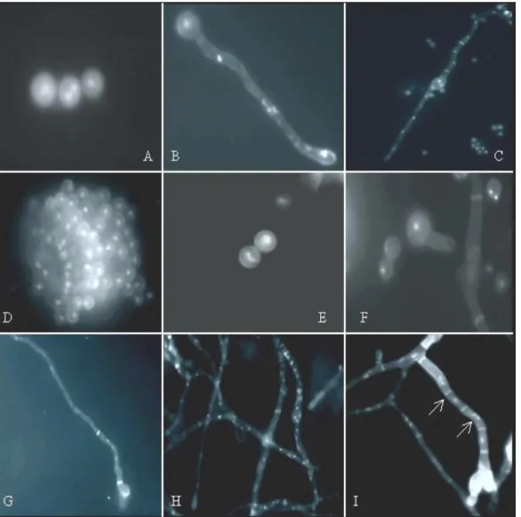
using DAPI and Calcofluor staining. Figure 1 shows the wild
type (MSE) and V103 germlings at different stages of
development. In the V103 strain, germ tubes are shorter, but
appear to establish a normal growth polarity. In contrast to the
wild type, nuclei in the mutant strain are randomly distributed
along the germ tube, suggesting a disruption in nuclear
distribution and positioning. The first septum in the wild type
germling forms shortly after the third nuclear division. This
septum is deposited at the basal end of the longer germ tube
(22). In this regard, the V103 mutant was similar to the wild
type, but approximately 40% of the germlings had a septum at
the basal end of the shorter germ tube, suggesting a
dysfunction in the mechanisms that coordinate polar growth
and septation. The V103 mutation generates phenotypes similar
to those observed in several previously classified mutations
that affect the cell duplication cycle. For instance, similar
phenotypes were observed in blocked in mitosis (bim) mutants
(12, 13). The bim mutation affects a wide range of cell cycle
functions (8). Mutants identified as defective in septation also
have some characteristics in common with the V103 mutant,
such as increased numbers of nuclei per hyphal compartment,
the occurrence of mitotic catastrophe and abnormal growth
polarity (12). V103 showed aberrant morphology similar to
that of the sep mutant, resulting in growth polarity
abnormalities and deposition of the actin ring during
cytokinesis. In turn, this may lead to an increased chromosome
mitotic index (1). In addition, analysis of hyp phenotype
suggests that wild type hypA promotes tip growth and restrains
growth of basal cells (7, 20).
Figure 1. DAPI and Calcofluor
staining (Agar Complete Medium). A–
D: wild-type strain at 2-4, 6-8, 12-16
and 32 hours respectively. E–I: the
V103 deteriorated variant at 2-4, 6-8,
12-16, 20-24 and 32 hours
respectively. H: Defective growth of
In both the MSE and V103 strains of A. nidulans,
conidiation began with formation of the aerial hyphae that
constitute the conidiophore stalk. The stalk arises as a vertical
outgrowth from a hyphal compartment known as the foot cell.
The stalk tip swells to form the conidiophore vesicle. Numerous
small outgrowths termed metulae develop from this vesicle.
Metulae give rise to sporogenous cells, termed phialides. One
phialide forms at the tip of each metulae and additional phialides
appear below and to the sides of the first phialide. The phialides
begin to form asexual spores termed conidia that accumulate in
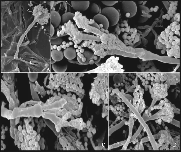
chains at the ends of the phialides, as shown in Figure 2. (10, 16).
The metulae and phialides observed in V103 were long and
indistinct, presented secondary conidiophores, multiple tiers of
sterigmata (metulae and phialides) and conidiophore stalks with
many septa were apparent. In the wild type, usually a single
vesicle is formed followed by a single tier of metulae and a single
tier of phialides. In contrast, in V103’s the conidiophore stalks
ramified and formed secondary conidiophores (Figure 2).
Figure 2. SEM showing conidia (c), metulae (m), phialides (p), stalk (s), foot-cell (cf) and hyphae (h) of wild-type(A) and V103 (B, C
and D) strains of A. nidulans. In B and C, metulae and phialides were long, indistinct and showed secondary conidiophores. In D,
Conidiophore stalks ramify. Colonies were grown at 37 oC in Agar Complete Medium for 48 hours . Magnification from A to D:
Cytological characterization of the cell division cycle,
nuclear movement and developmental morphogenesis of the
asexual development A. nidulans indicates that a complex
regulatory system is at work. This system is mainly characterized
by transcriptional control mechanisms leading to differential
expression of structural genes required for conidiophore
formation. We reported, cytologically, that the pattern of cell
growth, nuclear division, and cytokinesis changes dramatically
during formation of the different conidiophores cells. It has been
proposed that the cell cycle and the developmental program may
interact (11). Ye et al. (23, 24) have shown that the main cell
cycle regulators NIMXcdc2 and NIMA are upregulated on mRNA
and kinase activity levels in a brlA-dependent manner. Schier et
al. (19) have isolated the cyclin pclA developmental gene
required for the fast, repetitive cell divisions of the phialides,
which subsequently lead to the long conidial chains of the
conidiophore. This gene mediates events of the cell cycle and
developmental morphogenesis
This paper represents the first analysis of the development of
a spontaneously obtained mutant, starting from an inserted
segment, shown the need the molecular analyses to deepen the
knowledge and importance among the interactions of the cell
cycle with the regulator genes of the conidiogeneses.
ACKNOWLEDGMENTS
We are grateful to Drs. E.W. Kitajima and B. Leite for
permission to use the Scanning Electron Microscope (SEM) at
NAP/MEPA - ESALQ/USP. We also wish to thank Drs. M.L.R.
de Aguiar-Perecin, M. Mondin and S.C.M. Molina for sharing
their Fluorescence Microscopy equipment with us.
REFERENCES
1. Adams, T.H.; Wieser, J.K.; Yu, J.H. (1998). Asexual sporulation in Aspergillus nidulans. Microb. Molec. Biol. Rev., 62: 35-54.
2. Azevedo, J.L.; Roper, J.A. (1970). Mitotic non-conformity in Aspergillus nidulans: successive and transposable genetic changes. Genet. Res., 16: 79-93.
3. Azevedo, J.L. (1975). Altered instability due to genetic change in a duplication strain of Aspergillus nidulans. Genet. Res., 26: 55-61.
4. Enos, A. P.; Morris, N.R. (1990). Mutation of a gene that encodes a kinesin-like protein blocks nuclear division in A. nidulans. Cell, 60: 1019-1027. 5. Fischer, R. (1999). Nuclear movement in filamentous fungi. FEMS Microb.
Rev., 23: 39-68.
6. Harris, S.D.; Hamer, L.; Sharpless, K.E.; Hamer, J.E. (1997). The Aspergillus nidulans sepA gene encodes an FH ½ protein involved in cytokinesis and the maintenance of cellular polatity. EMBO J., 16: 3474-3483.
7. Kaminskyj, S.G.W. (2000). Septum position is marked at the tip of Aspergillus nidulans hyphae. Fung Genet. Biol., 31: 105-113.
8. Lies, C.M.; Cheng, J.; James, S.W.; Morris, N.R.; O’Connell, M.J.; Mirabito, P.M. (1998). BIMAAPC3, a component of the Aspergillus anaphase promoting comples/cyclosome, is required for a G2 checkpoint blocking entry into mitosis in the absence of NIMA function. J. Cell Sci., 111: 1453-1465.
9. McCully, K.S.; Forbes, E. (1965). The use of -fluorphenilalanine with “master” strain of Aspergillus nidulans form assigning gene of to linkage groups. Genet. Res., 6: 352-359.
10. Mims, C.W.; Richardson, E.A.; Timberlake, W.E. (1988). Ultrastructural analysis of conidiophore development in fungus Aspergillus nidulans using freezer-substitution. Protoplasma, 144: 132-141.
11. Mirabito, P.M.; Osmani, S.A. (1994). Interactions between developmental program and cell cycle regulation of Aspergillus nidulans. Semin. Dev. Biol.; 5: 139-145.
12. Morris, N.R. (1976). A temperature-sensitive mutant of Aspergillus nidulans reversibly blocked in nuclear division. Exp. Cell Res., 98: 204-210. 13. Morris, N.R.; Xiang, X.; Beckwith, S.M. (1995). Nuclear Migration
advances in fungi. Trends in Cell Biol., 5: 278-282.
14. Nga, B.H.; Roper, J.A. (1968). Quantitative intrachromosomal changes arising at mitosis in Aspergillus nidulans. Genet., 5: 193-209.
15. Nga, B.H.; Roper, J.A. (1969). A system generation spontaneous in intrachromosomal changes at mitosis in Aspergillus nidulans. Heredity, 21: 530-531.
16. Oliver, P.T. (1972). Conidiophore and spore developmental in Aspergillus nidulans. J. Gen. Microb,. 73: 45-54.
17. Pontecorvo, G. (1953). The Genetics of Aspergillus nidulans. Adv. Genet. 5, 141-238.
18. Queiroz, M.V.; Azevedo, J.L. (1998). Characterization of an Aspergillus nidulans mutant with abnormal distribution of nuclei in hyphae, metulae, phialides and conidia. FEMS Microbiol. Lett., 166: 49-55.
19. Schier, N.; Liese, R.; Fischer, R. (2001). A Pcl-like cyclin of Aspergillus nidulans is transcriptionally activated by developmental regulators and is involved in sporulation. Mol. Cell. Biol., 21: 4075-4088.
21. Timberlake, W.E. (1990). Molecular Genetics of Aspergillus nidulans. Annu. Rev. Genet., 24: 5-36.
22. Wolkow, T.D.; Harris, S.D.; Hamer, J.E. (1996). Cytokinesis in Aspergillus nidulans is controlled by cells size, nuclear positioning and mitosis. J. Cell Sci., 109: 2179-2188.
23. Ye, X.; Osmani, A.H.; Osmani, S.A.; Xin, M.; Morris, N.R. (1995). nudF, a
nuclear migration gene in Aspergillus nidulans, is similar to the humam LIS-1 gene required for neural migration. Mol. Biol. Cell, 6: 297-310.