Maria Judite da Silva Barreto
outubro de 2013
Current overview of the role of short-chain
fatty acids in prevention or therapy of
colorectal carcinoma
UMinho|20 13 Maria Judit e da Silva Barr et o Current o ver vie w of t he role of short-chain fatty acids in pre
vention or t
herap
y of colorect
Maria Judite da Silva Barreto
outubro de 2013
Dissertação de Mestrado
Mestrado em Ciências - Formação Contínua de Professores
Área de Especialização em Biologia e Geologia
Current overview of the role of short-chain
fatty acids in prevention or therapy of
colorectal carcinoma
Trabalho realizado sob a orientação da
Professora Doutora Ana Arminda Lopes Preto Almeida
e da
Professora Doutora Maria Manuela Sansonetty
Gonçalves Côrte-Real
Nome: Maria Judite da Silva Barreto
Endereço eletrónico: [email protected]
Telefone: 926528401
Número de Bilhete de Identidade: 10355417
Título da dissertação: Current overview of the role of short-chain fatty acids in prevention or therapy of colorectal carcinoma
Orientadoras: Doutora Ana Arminda Lopes Preto Almeida e Professora Doutora Maria Manuela Sansonetty Gonçalves Côrte-Real
Ano de conclusão: 2013
Designação do Mestrado: Mestrado em Ciências - Formação Contínua de Professores, Área de Especialização em Biologia e Geologia
É AUTORIZADA A REPRODUÇÃO PARCIAL DESTA TESE, APENAS PARA EFEITOS DE INVESTIGAÇÃO, MEDIANTE DECLARAÇÃO ESCRITA DO INTERESSADO, QUE A TAL SE COMPROMETE.
Universidade do Minho, 28 de outubro de 2013
ii À Professora Doutora Teresa Almeida por me ter permitido conhecer pessoas extraordinárias!
À Professora Doutora Ana Preto e à Professora Doutora Manuela Côrte-Real, pelas constantes palavras de incentivo, pelo apoio que sempre me deram, pelo otimismo e por todos os sorrisos com que sempre me receberam.
À Suellen e à Lisandra, pelo seu espírito solidário e por terem sido colaboradoras fundamentais na construção deste trabalho.
iii Colorectal carcinoma (CRC) is a common malignancy in the western world and is also regarded as one of the most preventable cancers. It is generally recognized that the type of diet is an important risk factor for CRC and tumor behavior. Interest in the short-chain fatty acids (SCFA) production by propionibacteria from dairy diet, on the human organism has increased rapidly in the last ten years due to the fact that gastrointestinal functions and beneficial effects are associated with these acids. SCFAs, namely butyrate, propionate and acetate are the major products of the propionibacteria fermentation and metabolism of undigested dietary fibers in the human large intestine. These SCFA have been reported as anti-proliferative and anti-neoplastic agents that induce differentiation, growth arrest and apoptosis in CRC cells lines. It is known that SCFAs protect against development of CRC and therefore, in this monographic review we focus on new aspects of cellular functions of SCFAs as a nutraceuticals in the prevention and/or treatment of CRC. In this context we also aimed at developing a simple, feasible and easily implemented protocols for undergraduate students in secondary school labs using protocols with baker´s yeast cells to illustrate the effect of acetic acid on two key cellular biological processes, namely on cell cycle and cell death.
iv O carcinoma colorretal (CCR) é um tumor maligno comum no mundo ocidental, mas é também considerado um dos cancros mais evitáveis. É geralmente reconhecido que o tipo de dieta é um importante fator de risco para o desenvolvimento e o comportamento deste tipo de cancro. O interesse nos ácidos gordos de cadeia curta (AGCC), produzidos no organismo humano pelas propionibacterias presentes nos lacticínios, cresceu rapidamente nos últimos dez anos, uma vez que várias funções gastrointestinais e efeitos benéficos estão associados a estes ácidos. Os AGCC, nomeadamente o butirato, o propionato e o acetato, são os principais produtos resultantes da fermentação das propionibacterias e do metabolismo das fibras não digeridas no intestino grosso humano. Estes AGCC têm sido referidos como sendo agentes anti-proliferativos e anti-neoplásicos, induzindo a diferenciação, a paragem do crescimento e a apoptose em linhas celulares de CRC. Sabe-se que os AGCC protegem contra o desenvolvimento do CRC e, portanto, nesta revisão monográfica concentramo-nos em novos aspetos das funções celulares dos AGCC como nutracêuticos na prevenção e / ou tratamento do CRC. Neste contexto, foi também nosso objectivo desenvolver um protocolo simples e de fácil implementação para estudantes do ensino secundário, recorrendo à utilização de fermento de padeiro, de forma a ilustrar o efeito do ácido acético em dois processos biológicos celulares chave, nomeadamente, no ciclo celular e na morte celular.
v
Abstract ... iii
Resumo ... iv
Contents ... v
List of abbreviations ... vii
Chapter 1 – Bibliographic review ... 1
1. Hallmarks of cancer: the role of apoptosis ... 2
2. Colorectal carcinoma ... 5
2.1. Types of colorectal carcinoma ... 6
2.2. Colorectal carcinoma: associated genetic alterations... 7
2.3. Classical chemotherapy for colorectal carcinoma ... 9
3. Nutraceuticals in colorectal carcinoma prevention or therapy ... 10
3.1. Prebiotics ... 11
3.2. Probiotics ... 13
3.2.1. Propionibacteria ... 15
3.3. Symbiotics ... 16
4. The association between diet, SCFA and colorectal carcinoma risk ... 18
5. Colonic SCFA concentrations and carcinogenesis ... 20
6. Effects of SCFA in CRC cells... 21
6.1. Butyrate ... 23
6.2. Propionate ... 31
6.3. Acetate ... 32
6.4. Combined effects of SCFA ... 38
7. Colorectal Carcinoma – SCFA prevention and/or therapy? ... 38
8. Conclusions ... 39
Chapter 2 – Design of a protocols for undergraduated students to illustrate the effect of the SCFA acetic acid on yeast cell cycle and cell death ... 41
1. Insights from Sacharomyces cerevisiae on the role of acetate in CRC cells - a practical application ... 42
2. Experimental Procedures ... 45
vi
2.4. Protocol for the analysis of cell cycle ... 47
2.4.1. Experimental Procedure ... 47
2. 5. Protocol for the analysis of cell death ... 48
2.5.1. Experimental Procedure ... 48
3. Results and discussion ... 49
3.1. Effect of acetic acid on cell cycle progression ... 49
3.2. Effect of acetic acid on cell death ... 53
4. Final conclusions ... 55
vii APC – Adenomatous polyposis coli
Bak – Bcl-2-antagonist/killer Bax – Bcl-2-associated X protein
Bcl-2 – B-cell lymphoma 2 gene regulator of apoptosis Bcl-W– Bcl-2-associated pro-survival protein
BRAF – v-RAF murine sarcoma viral oncogene homolog B Cat D – Cathepsin D
CD95(Fas/APO-1) – Cell surface receptor of the TNF receptor family CRC – Colorectal carcinoma
DISC – Death-inducing signaling complex DNA – Deoxyribonucleic acid
EGFR – Epidermal growth factor receptor
EPIC – European Prospective Investigation into Cancer and Nutrition FFA2 –Free fatty acid receptor 2
GSTs – Glutathione S-transferases HATs - Histone acetyltransferases HDACs – Histone deacetylases LAB – Lactic acid bacteria
MAPK – Mitogen-activated protein kinase Muc1/3/4 – Membrane-bound mucins NRP-1 – Isoform-specific receptor for VEGF
p21 (WAF1/CIP1) – Cyclin-dependent kinase inhibitor 1 pRb – Retinoblastoma protein
ROS – Reactive oxygen species SCFA – Short chain fatty acids TNF – Tumor Necrosis Factor
TRAIL – TNF-related apoptosis-inducing ligand VEGF – Vascular endothelial growth factor
1
Chapter 1 – Bibliographic review
2
1. Hallmarks of cancer: the role of apoptosis
Cancer is a very heterogeneous disease, developing in different tissue types and displaying great genetic diversity. Every day our body produces potentially malignant cells. These, however, should not be considered cancerous, we only should name it cancer when there is clinical manifestation or evidence of increased cell proliferation and capability of metastasize in the organism (Simões, 2010).
There are several evidences which indicate that tumourigenesis in humans is a multistep process and that these steps reflect genetic alterations that drive the progressive transformation of normal human cells into highly malignant derivatives (Hanahan & Weinberg, 2000). However, recent insights suggest that the underlying etiology and progression of the disease can be reduced to two events, mutations that give rise to excessive proliferation and a compensatory disruption of survival signaling pathways that ensures the persistence of these hyperproliferative cells (Green & Evan, 2002).
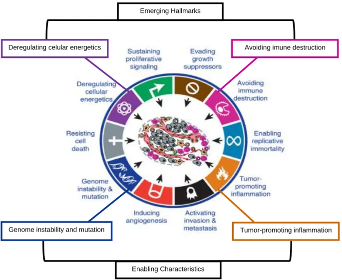
Hanahan and Weinberg (2000) proposed that six capabilities acquired during tumor development are shared in common by most and perhaps all types of human tumors. These six essential alterations in cell physiology that collectively dictate malignant growth are: self-sufficiency in growth signals, insensitivity to growth-inhibitory (antigrowth) signals, evasion of proto growth-inhibitory (antigrowth) signals, evasion of programmed cell death (apoptosis), limitless replicative potential, sustained angiogenesis, and tissue invasion and metastasis (Hanahan & Weinberg, 2000). Two enabling characteristics crucial to the acquisition of the six hallmark capabilities are the two new emerging hallmark capabilities, the metabolic alterations and signaling interactions of the tumor microenvironment crucial to cancer phenotypes (Figure 1) (Hanahan & Weinberg, 2011).
3
Figure 1 - Emerging Hallmarks and Enabling Characteristics (Adapted from Hanahan & Weinberg, 2011).
Normal tissues carefully control the production and release of growth-promoting signals that instruct entry into and progression through the cell growth-and-division cycle, thereby ensuring a homeostasis of cell number and thus maintenance of normal tissue architecture and function. Cancer cells, by deregulating these signals, become “masters of their own destinies” (Claesson et al., 2012). The ability of tumor cell populations to expand is determined not only by the rate of cell proliferation but also by the rate of cell death, being programmed cell death – apoptosis – the major source of this death. The evidence come mainly from studies in mouse models and cultured cells, as well as from analyses of human cancer tissue, that showed that cancer cells acquired resistance toward apoptosis which is a hallmark of most and perhaps all types of cancer (Hanahan & Weinberg, 2000).
Apoptosis is a genetically predetermined mechanism that may be elicited by several molecular pathways and organisms can trigger this process to remove unwanted and potentially
Avoiding imune destruction
Genome instability and mutation Tumor-promoting inflammation
Deregulating celular energetics
Emerging Hallmarks
4 dangerous cells. The activation of apoptotic genes culminate in morphological and biochemical changes leading to chromatin condensation followed by nuclear condensation, DNA fragmentation and packing of nuclear fragments into multiple membrane-enclosed apoptotic bodies (Ramos, Rabelo, Duarte, Gazzinelli, & Alvarez-Leite, 2002). Apoptosis can be also regulated via the action of several oncogenes and subsequently oncoproteins (Salakou et al., 2007).
There are two different apoptosis pathways, called the extrinsic and intrinsic pathways. In the extrinsic pathway (also known as “death receptor pathway”), apoptosis is activated by the cell-surface death receptors CD95 (Apo-1 or Fas)/TRAIL/tumor necrosis factor (TNF) receptor 1 family proteins which are located on the plasma membrane, and directly activates the caspase cascade via the recruitment of the “initiator” caspase-8 within a death-inducing signaling complex (DISC). The intrinsic pathway (also called “mitochondrial pathway”), leads to the release of cytochrome c from the damaged mitochondrion, which then binds to the adaptor molecule APAF-1 and an inactive “initiator” caspase, procaspase 9, within a multiprotein complex called the apoptosome. This leads to the activation of caspase 9, which then triggers a cascade of caspases activation (caspases 3 and 7) resulting in the morphological and biochemical changes associated with apoptosis (Tzifi et al., 2012). Caspase activated cell death is regulated by genes of the Bcl-2 family, for instance by the pro- and anti-apoptotic genes, Bax and Bcl-2, respectively (Fauser, Prisciandaro, Cummins, & Howarth, 2011; Jin & El-Deiry, 2005). Currently, the intrinsic apoptotic pathway is more widely implicated as a barrier to cancer pathogenesis. Both apoptotic pathways are targets for cancer treatment (Shao, Gao, Marks, & Jiang, 2004).
5
Figure 2 - Schematic representation of apoptotic pathways.
2. Colorectal carcinoma
Colorectal carcinoma (CRC) is the third most commonly diagnosed cancer in males and the second in females, with over 1.2 million cancer cases in the world, accounting for 9.8% of total cancer cases, according to data from GLOBOCAN project (2008). In Europe, the incidence of CRC is 229.229 cases, accounting for 13.5% of total cancer cases. In Portugal, the CRC is the second most prevalent cancer (after prostate cancer) with an incidence number of 3951 cases and an incidence rate of 16.5% of total cancer cases (Globocan project, 2008, http://globocan.iarc.fr/). Cancer prevention is an essential component of cancer control strategies because about 40% of all cancer deaths can be prevented (World Health Organization, 2008, http://www.who.int/en/).
Studies reported that dietary patterns, lifestyle, physical inactivity and obesity increased CRC risks, especially in genetically predisposed populations (Doll & Peto, 1981; Potter, 1999; Qin et al., 2010). CRC is thus causally related to both genes and environment. Environment is a risk factor that may cause mutations and initiate cancer or enhance growth by genetic and epigenetic mechanisms (Ferguson, 1999; Simões, 2010) .
6
2.1. Types of colorectal carcinoma
About 75% of patients with CRC have sporadic disease with no apparent evidence of having inherited the disorder. The remaining 25% of patients have a family history of CRC that suggests a hereditary contribution, common exposures among family members, or a combination of both. Genetic mutations have been identified as the cause of inherited cancer risk in some colon cancer-prone families; these mutations are estimated to account for only 5% to 6% of CRC cases overall. It is likely that other undiscovered genes and genetic factors contribute to the development of familial CRC in conjunction with nongenetic risk factors (National Cancer
Institute, 2013, http://www.cancer.gov/cancertopics/pdq/genetics/colorectal). Among the
non-sporadic CRC cases, 5 to 15% can be attributed to the following hereditary CRC syndromes: Lynch syndrome (also hereditary nonpolyposis CRC or HNPCC), familial adenomatous polyposis (FAP), and MUTYH-associated polyposis (MAP) (Castells, Castellvi-Bel, & Balaguer, 2009).
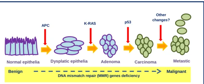
Two-thirds of CRCs are located in the rectum, rectosigmoid, or sigmoid colon with the other third distributed in the remainder of the colon. Adenocarcinomas are the most common type of CRC and have its origin in intestinal epithelial cells. The progression from adenoma to carcinoma occurs by the sequential accumulation of genetic changes, which is the more accepted model for understanding carcinogenesis process (presented in Figure 3) (Bruckner et al. 2000) Available from: http://www.ncbi.nlm.nih.gov/books/NBK20861/; Cancer Research UK).
Adenocarcinomas start in the intestinal gland cells in the lining of the colon wall. There are one or two rare types of adenocarcinoma of the colon and rectum, called mucinous tumors and signet ring tumors and these terms refer to how the cells look under the microscope. Only about 1 to 2% of colorectal cancers are the signet ring type. Squamous cells cancers are the skin like cells that make up the bowel lining together with the gland cells. Carcinoid is an unusual type of slow growing tumor called a neuroendocrine tumor. Between 4 to 17% of every carcinoid tumors diagnosed start in the rectum and between 2 to 7% out of every carcinoid tumors diagnosed begin in the large bowel. Sarcomas are cancers of the supporting cells of the body, such as bone or muscle. Most sarcomas found in the colon or rectum are leiomyosarcomas, which account for less than 2% of colorectal carcinomas and have a high chance of metastasizing. Lymphomas are cancers of the lymphatic system. Only about 1 in 100 cancers
7 diagnosed in the colon or rectum (1%) are lymphomas (Cancer Research UK, 2013, http://www.cancerresearchuk.org/cancer-help/type/bowel-cancer/about/types-of-bowel-cancer).
2.2. Colorectal carcinoma: associated genetic alterations
The model of cell transformation and progression from adenoma to colorectal carcinoma is based on the concept that progression is accompanied by the accumulation of molecular alterations in which adenomatous polyposis coli (APC), K-RAS, and p53 genes play a central role (presented in Figure 3) (Fearon & Vogelstein, 1990). A number of data indicate that the simultaneous presence of alterations of these genes is not a frequent event. In particular, K-RAS and p53 mutations rarely co-exist in the same tumor, indicating that these alterations do not represent a synergistic evolutionary pathway (Smith et al., 2002).
It was found that somatic mutations in APC gene are also found in the great majority of sporadic colorectal tumors (Miyoshi et al., 1992). APC has been proposed to function as a ‘‘gatekeeper’’ gene, regulating the entry of epithelial cells into adenoma-carcinoma progression (Kinzler & Vogelstein, 1996), maintaining low levels of -catenin in the absence of a Wnt signal, thus preventing excessive cell proliferation. Axis duplication and cell transformation are based on the activation of the canonical Wnt pathway that involves the multifunctional protein -catenin (Rao & Kuhl, 2010). A mutation of the gatekeeper leads to a permanent imbalance of cell division over cell death (Kinzler & Vogelstein, 1996).
The RAS oncogene promotes tumor formation through stimulation of cell proliferation, motility and regulation of apoptosis. Activating mutations in the RAS oncogenes (H-, N-, and K-RAS) are found in approximately 20% of all human tumors (Shaw et al., 2011) and mediates several key aspects of oncogenesis, including deregulated cell growth, evasion of apoptosis and malignant transformation, a consequence of the loss of GTPase activity (Shaw et al., 2011; Smith et al., 2002). The TP53 gene product, p53, functions as a transcription factor, exerting cell cycle control by binding to specific recognition sequences in a variety of genes including p21, Bax, and Bcl-2 in response to DNA damage or other cellular stress (A. J. Levine, 1997). About 70% of CRCs contain p53 mutations (Baker et al., 1990), rendering them susceptible to failure of apoptosis and increased accumulation of DNA damage, allowing unregulated growth (el-Deiry et al., 1993; Williams, Coxhead, & Mathers, 2003). These mutations in p53 are proposed to be relatively late events in the development of colorectal tumors, with the loss of p53-mediated
8 pathways of apoptosis considered to be an important determinant of progression from adenoma to malignant tumor (Smith et al., 2002).
Figure 3 - Genetic changes associated with colorectal tumorigenesis (Adapted from Kinzler & Vogelstein, 1996;
Smith et al., 2002).
BRAF is one of the RAF genes involved in the important RAS/RAF/MEK/MAP kinase intracellular signaling pathway, which regulates different physiological processes including cell growth, differentiation, and apoptosis (Calistri et al., 2005; Fang & Richardson, 2005). BRAF-activating gene mutations have been detected in many tumor types (Davies et al., 2002). However, Calistri et al. (2005) observed a very low frequency of mutations in their series of sporadic colorectal carcinoma, and no evident association with other specific gene mutations, suggesting that BRAF could represent an alternative pathway to both p53 and K-RAS genes, mainly in hereditary CRC characterized by microsatellite instability (MSI) (Calistri et al., 2005).
It is thought that at least 50% of colorectal cancers have a deregulation of the MAPK pathway (Fang & Richardson, 2005). Two major oncogenes are implicated in sporadic colorectal carcinogenesis, KRAS and BRAF mutations, which are alternative (Oliveira et al., 2007). Mutations on KRAS and BRAF genes are frequently found in malignant and pre-malignant colorectal lesions (Rajagopalan et al., 2002). Mutations of the KRAS proto-oncogene are an early event in development of CRCs (Bos et al., 1987). KRAS oncogene is mutated in 21% of all human sporadic cancers, including in about 30% of CRCs cases (Fang & Richardson, 2005; Oliveira et al., 2007), and BRAF mutations are found in 20% of all human cancers, including in about 10% CRCs cases (Ahlquist et al., 2008; Oliveira et al., 2007; Velho, Corso, Oliveira, & Seruca, 2010).
Normal epithelia Dysplatic epithelia Adenoma Carcinoma Metastic cancer
APC K-RAS p53
Benign Malignant Other
changes?
9
2.3. Classical chemotherapy for colorectal carcinoma
According to Sobrinho Simões (2010), there will never be possible for medicine to cure advanced cancers. However, there will have a percentage of cancers that are always curable, those who are localized “in situ” when diagnosed early that can be removed by surgical techniques that have evolved in the past years. Currently, over 50% of cancers can be cured by surgery and the other 50% have no cure; medicine only can keep them in a state of equilibrium with the immune defenses. In these cases cancer may be regarded as a chronic disease: the cancer exists, but the disease does not manifest or at least it is mitigated. Oddly enough, cancer is a disease extremely inefficient, hence the hope of turning it into a chronic disease (Simões, 2010).
The development of CRC often follows a defined pattern. The entire process frequently takes a long period, like decades, before a malignant tumor is finally formed, and is thought to develop in a multistep process, known as the “adenoma-carcinoma sequence” (Vogelstein et al., 1988). Because colon epithelia are directly exposed to dietary compounds, elimination of precancerous or cancerous cells by nutritional or chemopreventive interventions, or both, represents an approach to the lowering of the incidence of colon cancer (Cai et al., 2006; Dove-Edwin & Thomas, 2001) .
The current therapies are those in which a protein modification is identified in cancer. All modern drugs fall into one of two groups: either are small molecules which inhibit proteins action, which enter into the cell, or antibodies which are fixed on the cell surface, blocking, for example, receptors that are greatly increased in some cancer cells. These receptors stop working and the cell ceases to have the incentive to proliferate (Simões, 2010). In the last two decades several advances were achieved in the treatment of CRC. With more effective drugs, improved surgery, better radiotherapy and a strong randomized clinical trials evidence base, patients now have a higher chance of cure and, when cure is not achievable, they survive longer with their disease (Braun & Seymour, 2011). The optimum treatment strategy for patients with CRC depends on a large number of factors, such as age, performance status, the presence of other disorders or diseases and the treatment setting (adjuvant versus palliative versus neoadjuvant) (Braun & Seymour, 2011). The main types of treatment that can be used for colon and rectal carcinoma are surgery, radiation therapy, chemotherapy and targeted therapy. Surgery is the only curative treatment for CRC. Depending on the stage of the cancer, two or more of these
10 types of treatment may be combined at the same time or used after one another (American
Cancer Society, 2013,
http://www.cancer.org/cancer/colonandrectumcancer/detailedguide/colorectal-cancer-treating-chemotherapy).
In recent years new different approaches to nutritional treatment have been used to correct the deficits observed in patients with colorectal cancer (de Oliveira & Aarestrup, 2012), such as the use of nutraceuticals.
3. Nutraceuticals in colorectal carcinoma prevention or therapy
In the last years several products have been commercialized in the form of pharmaceutical products, such as pills, tablets, solutions, etc., incorporating food extracts or phytochemical‐enriched extracts to which a beneficial physiological function has been directly or indirectly attributed (Palthur, 2010). This variety of products cannot be truly classified as “food” or “pharmaceutical”, so a combined term between nutrients and pharmaceuticals, “nutraceuticals”, has been coined to designate them (Espin, Garcia-Conesa, & Tomas-Barberan, 2007).
According to Stephen Defelice (1995) “a nutraceutical is any substance that is a food or part of a food and provides medical or health benefits, including the prevention and treatment of disease. Such products may range from isolated nutrients, dietary supplements and specific diets to genetically engineered designer foods, herbal products, and processed foods such as cereals, soups and beverages” (DeFelice, 1995). However, there is often confusion in the use of this terminology as there is a slight difference between the functional foods and nutraceuticals. When food is being cooked or prepared using "scientific intelligence" with or without knowledge of how or why it is being used, the food is called "functional food". Functional food provides the body with the required amount of vitamins, fats, proteins, carbohydrates, etc. needed for its healthy survival. When functional food helps in the prevention and/or treatment of disease(s) and/or disorder(s) it is called a nutraceutical (Pandey, 2010).
The nutraceuticals revolution began in the early 1980s, sparked off when the actual or potential clinical benefits of calcium, fiber and fish oil were supported by clinical studies published in distinguished medical journals, and when physicians began to educate their colleagues and consumers about these substances via the media (DeFelice, 1995). Within
11 European Medicines law, a nutraceutical can be defined as a medicine for two reasons: it can be used for the prevention, treatment or cure of a condition or disease or it can be administered with a view to restoring, correcting or modifying physiological functions in human beings (Richardson, 1996). However, no specific regulation exists in Europe to control nutraceuticals (Espin et al., 2007). The majority of the definitions indicate the health benefits of nutraceuticals and among these health benefits are prevention and treatment of diseases.
The major impact of eating habits on the prevalence of CRC has triggered efforts to design an optimal diet and/or to create food supplements specifically reducing the risk of cancer. Already in 1989 Fuller checked the growing interest in the use of live microbial agents for health maintenance and disease prevention or treatment (Fuller, 1989).
3.1. Prebiotics
A prebiotic was first defined by Gibson and Roberfroid as ‘‘a nondigestible food ingredient that beneficially affects the host by selectively stimulating the growth and/or activity of one or a limited number of bacteria in the colon, and thus improves host health’’ (G. R. Gibson & Roberfroid, 1995). However many food components, especially many food oligosaccharides and polysaccharides (including dietary fiber), have been claimed to have prebiotic activity without due consideration to the criteria required.
In practical terms, prebiotics are short-chain carbohydrates (SCCs) that are nondigestible by human enzymes and that have been called resistant SCCs. They are sometimes referred to as nondigestible oligosaccharides (NDOs). Nonetheless, NDOs are not strictly oligosaccharides and their nondigestibility is largely assumed but not always proved (Cummings, Macfarlane, & Englyst, 2001). An oligosaccharide, according to the International Union of Pure and Applied Chemistry Joint Commission on Biochemical Nomenclature (IUPAC-IUB JCBN) definition, is a molecule containing a small number (2 to about 10) of monosaccharide residues connected by glycosidic linkages ("IUPAC-IUB Joint Commission on Biochemical Nomenclature (JCBN). Abbreviated terminology of oligosaccharide chains. Recommendations 1980," 1982). Some of the carbohydrates that are named prebiotics often fall outside this definition because several of them have a degree of polymerization (DP) higher than 10 (Cummings et al., 2001). Not all dietary carbohydrates are prebiotics, and clear criteria need to be established for classifying a food ingredient as a prebiotic. These criteria are, by Gibson and Roberfroid, (1) to resist gastric
12 acidity, hydrolysis by mammalian enzymes and gastrointestinal absorption; (2) to be fermented by the intestinal microflora; (3) to stimulate selectively the growth and/or activity of intestinal bacteria associated with health and wellbeing (G. R. Gibson, Probert, Loo, Rastall, & Roberfroid, 2004).
In 2004 the definition was updated when prebiotics were defined as “selectively fermented ingredients that allow specific changes, both in the composition and/or activity in the gastrointestinal microflora that confer benefits upon host wellbeing and health” (G. R. Gibson et al., 2004). The definition considers microflora changes in the whole gastrointestinal tract and extrapolates the definition into other areas that may benefit from a selective targeting of bifidobacteria and lactobacilli (Bellei G., 2012). Any dietary material that is non-digestible and enters the large intestine is a candidate prebiotic. This includes polysaccharide-type carbohydrates such as resistant starch and dietary fiber, as well as proteins and lipids. However, current prebiotics are confined to non-digestible oligosaccharides, many of which seem to confer the degree of fermentation selectivity that is required (regarding bifidobacteria)(G. G. Gibson et al., 2010)
This prebiotics compounds have been shown to be a source of SCFA both in vitro and in vivo. Pan et al (2009) demonstrated, using a rat model, that the intake of selected prebiotic oligosaccharides improved concentrations of feacal SCFA, including butyrate (Pan et al., 2009). By producing a greater concentration of butyrate, the preferred energy source for colonocytes (Scheppach, 1994), a trophic effect may result within the gastrointestinal tract (Pan et al., 2009). Hence, prebiotics can have an impact on gut health in general, and are believed to play an important role in the prevention of CRC, as it has been highlighted by a Fotiadis and co-workers review (Fotiadis, Stoidis, Spyropoulos, & Zografos, 2008). For example, Bindels et al. (2012) proposed that propionate production by propionibacteria could be one of the gut microbial functions responsible for the anti-tumor effect of prebiotic nutrients (Bindels et al., 2012) .
Among the established prebiotics, inulin-type fructans, present in foods, have been studied widely in the setting of CRC (Pool-Zobel, 2005) and have been demonstrated to elevate the levels of bifidobacteria and to increase SCFA concentrations in the colon (Bouhnik et al., 1999). Various studies have shown that these prebiotics prevent chemically induced preneoplastic lesions, aberrant crypt foci (ACF) and tumors in the colon of rats and mice (Reddy, Hamid, & Rao, 1997; Verghese, Rao, Chawan, & Shackelford, 2002).
13
3.2. Probiotics
The term probiotic was first used in 1965 in contrast to the word antibiotic and defined as “substances secreted by one microorganism, which stimulates the growth of another” (Schrezenmeir & de Vrese, 2001). These probiotics are nonpathogenic micro-organisms that, when ingested, exert a positive influence on the health or physiology of the host (Fuller, 1989). They can influence intestinal physiology either directly or indirectly through regulation of the endogenous microflora. This complex multicellular entity plays an important role in maintaining homeostasis in the body (Macfarlane & Macfarlane, 2012).
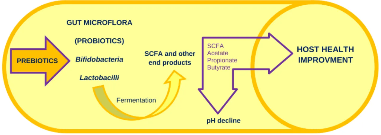
The mammalian intestinal tract contains a complex, dynamic, and diverse microbial community dominated by nonpathologic bacteria or “good bacteria” (Teitelbaum & Walker, 2002). The vast majority of bacteria in the human body reside in the large intestine, where the slow transit time, availability of nutrients, anaerobic conditions and pH are favorable for microbial growth (G. G. Gibson et al., 2010). Colonic microorganisms have ample opportunity to degrade available substrates, which may be derived from either the diet or by endogenous secretions (Bergman, 1990; Cummings & Macfarlane, 1991; Miller & Wolin, 1979). Bacterial fermentation involves a variety of reactions and metabolic processes in the anaerobic microbial breakdown of organic matter, yielding metabolizable energy for microbial growth and maintenance and other metabolic end products for use by the host (Cummings & Macfarlane, 1991; Wong, de Souza, Kendall, Emam, & Jenkins, 2006). In terms of end products, a variety of different metabolites arise, including short-chain fatty acids (SCFA) such as acetate, propionate and butyrate, (G. R. Gibson, Willems, Reading, & Collins, 1996). Thus, carbohydrate fermentation generally leads to health promoting SCFA production (Figure 4).
Each person has a distinct and highly variable microbiota, but a conserved set of gut colonizers (the core gut microbiota) and genes (the core microbiome) are shared among individuals (Qin et al., 2010; Turnbaugh et al., 2009) and may be required for the correct functioning of the gut (Tremaroli & Backhed, 2012). The human gut microflora differs in composition between infants and adults at various stages of life. Initial bacterial colonization of the human intestine begins at birth and on weaning Bifidobacteria decrease and a more “adult” profile of bacteria are present. In healthy elderly people, a decrease in the number of Bacteroides and Bifidobacteria has been reported (Roy, Kien, Bouthillier, & Levy, 2006). It is important that older people ingest sufficient amounts of dietary fiber to obtain the required amount of SCFA in
14 the gut lumen. Dietary supplements with defined food ingredients that promote particular components of the microbiota may also prove useful for maintaining health in older people (Claesson et al., 2012; Macfarlane & Macfarlane, 2012) .
When probiotics are ingested, they are able to resist the physicochemical conditions prevailing in the digestive tract. The strains most frequently used as probiotics consist mostly of strains of Lactobacillus, Bifidobacterium and Streptococcus, bacterial types which have been used for centuries in the production of fermented dairy products (Fooks & Gibson, 2002; Heyman & Menard, 2002). Because of all their benefits, probiotics represent an emerging therapeutic option. There is accumulating evidence describing the capacity for probiotic strains to prevent CRC, and in some cases, treat established tumor (Geier, Butler, & Howarth, 2006).
The predominant species used as probiotic agents belong to the group of lactic acid bacteria (LAB). Due to their long history of safe use in foods, most species of LAB are considered as commensal microorganisms with no pathogenic potential (Chukeatirote, 2003). Within the LAB group, the genus Lactobacillus is the most widely encountered for probiotics (Chukeatirote, 2003). L. acidophilus NCFM strain exhibits ability to reduce levels of free amines in the intestine, leading to a low risk of colon carcinoma (Goldin & Gorbach, 1984). However, also here the controverse is present, for example, Shahani et al. (1980) observed that consumption of large quantities of fermented milk products containing Lactobacillus or Bifidobacteria were associated with a lower incidence of CRC (Shahani & Ayebo, 1980) although, other studies suggested that consumption of fermented dairy products had little influence or no protection (Kampman, Goldbohm, van den Brandt, & van 't Veer, 1994).
The mechanisms by which probiotics may inhibit CRC are not yet fully characterized. The production of SCFAs is one key mechanism by which probiotics and prebiotics may impart beneficial effects (Figure 4). Mattar et al. (2002) demonstrated that the addition of the probiotic Lactobacillus casei GG (LGG) to the Caco-2 cells induced MUC-2 expression (responsible for the production of mucins of the intestine mucus layer) that correlated with LGG dosage. They surmise that LGG may bind to specific receptor sites on the enterocyte and stimulate the up-regulation of MUC-2, resulting in increased inhibition of bacterial translocation (Mattar et al., 2002). Ohkawara et al. (2005) investigated the bacterial strain Butyrivibrio fibrisolvens MDT-1 in the context of CRC treatment as it produces high amounts of butyrate, a SCFA with well-known apoptotic ability in cancer cells. In the 1,2-dimethylhydrazine-induced mouse model of colon carcinoma, administration of Butyrivibrio fibrisolvens MDT-1 led to a significant decrease in ACF,
15 and the number of mice with an increased proportion of aberrant crypts per foci was also reduced, indicating an inhibited progression of tumor development. This suggests that MDT-1 may be a potential new probiotic with the ability to reduce the incidence of CRC (Ohkawara et al., 2005).
The genetic manipulation of probiotics is another area of research in the treatment of CRC, which can be a method to deliver important anti-neoplastic factors to the colon. Steidler et al. (2000) demonstrated that Lactococcus lactis genetically-engineered to produce the anti-inflammatory cytokine IL-10, reduced colonic inflammation in the dextran sulphate sodium model of colitis (Steidler et al., 2000). This study highlighted the potential for probiotics to be used as a delivery system for anti-inflammatory or anti-tumorigenic substances which could help in the prevention or treatment of CRC (Geier et al., 2006). This is an important advance in the field of treatment of CRC, as a probiotic strain could potentially be engineered to produce other cytokines. Castagliuolo et al. (2005) used a nonpathogenic strain of E. coli designed to deliver TGF- genes to the colonic mucosa that successfully demonstrated to reduce the severity of experimental colitis in mice (Castagliuolo et al., 2005). This strategy could also be beneficial in the prevention of CRC, since TGF- could be administered as an anti-proliferative factor, thus suppressing tumor development (Geier et al., 2006). The use of this technology provides a mean by which probiotic strains can be tailored to deliver a wide array of therapeutic genes or factors including other anti-proliferative factors, or pro-apoptotic factors including anti-inflammatory cytokines (Geier et al., 2006).
3.2.1. Propionibacteria
Among different dietary bacteria, the Propionibacterium form a genus, which is found in specific dairy products (Mantere-Alhonen, 1995). Several experiments have shown that propionibacteria also possess probiotic characteristics when used alone or together with lactic acid and/or bifidobacteria (Mantere-Alhonen, 1995). Propionibacteria possess a peculiar fermentative metabolism which leads to the production of carbon dioxide and SCFA, such as propionate and acetate (Lan, Lagadic-Gossmann, Lemaire, Brenner, & Jan, 2007). In the last decades, several studies showed that the SCFA, namely acetate, propionate and butyrate, induces apoptosis in CRC cells but not in normal cells (Tang, Chen, Jiang, & Nie; Sakata 1987; Sauer, Richter, & Pool-Zobel, 2007). These results will be discussed later in this work.
16 The first time that the probiotic and growth promoting effect of pure propionibacteria was observed was in a study made with piglets by Mantere-Alhonen, in 1982 (Mantere-Alhonen, 1982). It was a rather large test material, thus the positive results of the feeding experiments should be considered as statistically significant, and they prove the ability of propionibacteria to act as probiotics (Mantere-Alhonen, 1995). In 2002 Jan and Balzacq proposed that propionibacteria could constitute probiotics efficient in digestive cancer prophylaxis via their ability to produce SCFA which induced apoptosis (Jan et al., 2002). Propionibacteria can survive in the human intestine and was found to induce apoptosis in colorectal cancer cells but not in normal cells, at least in part, due to their specific property to produce propionate and acetate (Jan et al., 2002).
Immerseel et al. (2010) suggested, that an ideal probiotic would be a colonizing bacterium that combines systemic anti-inflammatory and immunoregulatory effects with delivery of high butyrate levels at the site of action and that can be ingested in a stable form, such as spores (Van Immerseel et al., 2010). These studies indicate that SCFA delivery via probiotic ingestion may be an exciting new prevention/treatment option for CRC (Geier et al., 2006).
The possibility of using live propionibacteria in diet as preventive anti-cancer agents remains to be determined first by in vivo relevance of SCFA-based therapeutic strategy, and second by efficiently deliver SCFA to cancer cells (Lan et al., 2007).
3.3. Symbiotics
When prebiotics and probiotics are administered simultaneously, the combination is termed symbiotics. Symbiotics have been proposed as a new preventive and therapeutic option.
The mechanisms by which pro-, pre- and symbiotics may inhibit colon carcinoma are beginning to be understood (Fotiadis et al., 2008). The prebiotic in the symbiotic mixture improves the survival of the probiotic bacteria and stimulates the activity of the host’s endogenous bacteria (Mugambi, Musekiwa, Lombard, Young, & Blaauw, 2012). Consumption of probiotics and prebiotics together can increase the beneficial effects of each, since the stimulus of probiotic strains leads to selection of ideal symbiotic pairs (de Oliveira & Aarestrup, 2012).
A human study has investigated the effect of an oligofrutose and inulin mixture together, a product called ‘Synergy’ that combines short-chain oligofrutose and long-chain inulin with Lactobacillus rhamnosus GG and Bifidobacterium lactis Bb-12 on biomarkers of cancer (Rafter et
17 al., 2007). The study involved a twelve week double blind placebo-controlled trial in patients with cancer and polypectomised individuals. The symbiotic intervention resulted in significant alterations in the composition of the colonic bacterial ecosystem, which presumably have consequences for the metabolic activity of this organ. Colorectal cell proliferation and genotoxicity were significantly reduced, and the intestinal barrier function increased (Rafter et al., 2007).
The most important conclusion from Brady et al. (2000) is that in animals it appears to be a synergistic effect of consumption of probiotic bacteria and prebiotics such as fructoligosaccharides on the attenuation of the development of CRC. The effect is often not large, but it could be beneficial, in combination with other ways to reduce risk (Brady et al., 2000). For example, in vitro studies only comprising prebiotics, the increase in acetic acid was reported to be between two and six times higher as compared to controls and for butyric acid the highest concentrations observed were four times higher as compared to control (van Zanten et al., 2012). These increases in concentrations of acetic and butyric acids were however lower than the increases observed for all symbiotic combinations investigated in van Zanten et al. (2012) study, where concentrations were three to eight times higher for both acetic and butyric acids as compared to control. These findings emphasize that a synergistic effect may be obtained when combining the prebiotic with the probiotic strains (van Zanten et al., 2012).
Figure 4 - General process of colonic fermentation by symbiotics. Prebiotics compounds are food source for probiotic microorganisms, which produce SCFA that contribute to host health improvement (Adapted from Huazano-García & López, 2013). PREBIOTICS GUT MICROFLORA (PROBIOTICS) Bifidobacteria Lactobacilli Fermentation
SCFA and other end products SCFA Acetate Propionate Butyrate HOST HEALTH IMPROVMENT pH decline
18
4. The association between diet, SCFA and colorectal carcinoma risk
Although there is a strong genetic component in the development of colorectal adenomas or carcinomas, it is generally accepted that environmental factors including diet and lifestyle have a major impact on risk (Azcarate-Peril, Sikes, & Bruno-Barcena, 2011). Diet and nutrition are estimated to explain as much as 30-50% of the worldwide incidence of CRC and a high intake of calories, fats, red meat and low consumption of fruits and vegetables are associated with the risk of CRC development (Cervi, 2005; Chan & Giovannucci, 2010). It is believed that a proper diet can prevent three to four million new cases per year (Garófolo, 2004).
Dietary fiber has been consumed for centuries and has been recognized as having health benefits. The consumption of foods rich in this dietary component such as fresh vegetables and fruits, whole grains and nuts is associated with gastrointestinal benefits, such as increasing stool bulk and improving laxation (Schneeman, 1999). Despite all the benefits, Scharlau et al. (2009) referred that human studies are not showing that fruit and vegetable intakes are associated with a reduced cancer incidence in general and in particular with reduced CRC risk (Fuchs et al., 1999; Scharlau et al., 2009). For example, Terry et al. (2001) results do not support the hypothesis that high consumption of cereal fiber decreases the risk of colon or rectal carcinoma, even based on a much broader range of cereal fiber intake than had been examined in previous cohort studies (Terry et al., 2001). However, it is also likely that the frequency of fruit and vegetables consumption that is adequate to decrease cancer risk, taking into account other health consequences, probably varies with individual factors and, perhaps, with other cofactors in the population, such as multivitamin use and whether foods are fortified with other micronutrients. In contrast, the role of diet in cancer development is strongly supported by epidemiological studies, in particular in the case of cancers of the digestive tract (Vano, Rodrigues, & Schneider, 2009). Topping and Clifton (2001) observed that some studies showed that native East Africans, consuming a diet high in unrefined cereals, were at lower risk of CRC, diverticular disease, and constipation than Europeans who ate a diet low in such foods (Topping & Clifton, 2001). Another study by Bamia et al. (2013) allowed to conclude that adherence to Mediterranean diet may be associated with lower CRC risk (Bamia et al., 2013) . The traditional Mediterranean diet is characterized by high intakes of vegetables, fruit/nuts, fish, cereals and legumes, moderate alcohol consumption (particularly wine during meals), low to-moderate consumption of dairy products (mainly cheese and yogurt) and low consumption of meat/meat
19 products (Trichopoulou & Lagiou, 1997) . The main source of lipids is olive oil consumed in large quantities, and it is the main source of monounsaturated fatty acids in Mediterranean populations
(Bamia et al., 2013). It has been suggested that up to 25% of colorectal carcinomas could be
prevented by shifting to a Mediterranean diet (Trichopoulou, Lagiou, Kuper, & Trichopoulos, 2000).
Regarding diet, resistant starch, a type of dietary fiber, has been hypothesized to have specific anti-cancer properties since this form of fiber is preferentially fermented by microflora into potentially beneficial SCFA in the colon (Chan & Giovannucci, 2010). These SCFA produced from undigested dietary fibers in the human large intestine, have been extensively reported as antitumor agents that induce differentiation, growth arrest and apoptosis in colon carcinoma cells (Tang, Chen, Jiang, & Nie, 2011a). The most important role of SCFA in colonic physiology is
their trophic effect on the intestinal epithelium. Sakata reported that the presence of SCFA in rat colon stimulates mucosal proliferation (Sakata, 1987). In human, SCFAs production from inulin-type fructan can increase the metabolic activity, pointing to trophic effects for normal colonocytes (Sauer, Richter, & Pool-Zobel, 2007).
SCFA are organic fatty acids with 1 to 6 carbon atoms, increasing from acetic (C2:0), propionic (C3:0), butyric (C4:0), valeric (C5:0) and caproic (C6:0) acids (Fauser et al., 2011). These intermediate carboxylic acids at the physiological pH predominate in their dissociated form acetate, propionate, butyrate, valerate and caproate are the principal anions which arise from bacterial fermentation of polysaccharide, oligosaccharide, protein, peptide, and glycoprotein precursors in the colon (Bergman, 1990; Cummings & Macfarlane, 1991; Miller & Wolin, 1979). Among these, however, butyrate, acetate, and propionate have been mainly emphasized. In particular, butyrate was addressed to be more beneficial for promoting colonic health and more effective for stimulating the proliferation of intestinal mucosal cells than acetate and propionate (Sakata, 1987). These SCFA, especially butyrate, are recognized for their potential to act on secondary chemoprevention by slowing growth and activating apoptosis in CRC cells. Additionally, SCFA can also act on primary prevention by activation of different drug metabolizing enzymes. This can reduce the burden of carcinogens and, therefore, decrease the number of mutations, reducing cancer risk (Scharlau et al., 2009).
Results from Takashi Sakata (1987) indicated that the stimulatory effect of SCFA on intestinal epithelial cell proliferation in vivo is substantial and highly reproducible, and that the effect of SCFA persists sufficiently long to be of nutritional significance (Sakata, 1987). When the
20 concentrations of SCFA used were compared with the lumen concentrations measured by Yang et al. (1970), it was clear that physiological doses of acetate had a trophic effect on colonic epithelium, and butyrate had a trophic effect on both jejunal and colonic epithelium. In contrast, propionate was effective only at superphysiological doses (Sakata, 1987; Yang et al., 1970).
Several benefits can be obtained when the ingestion of dietary fibers are higher: increases in SCFA result in decreased pH, which indirectly influences the composition of the colonic microflora (eg, reduces potentially pathogenic clostridia when pH is more acidic), decreases solubility of bile acids, increases absorption of minerals (indirectly), and reduces the ammonia absorption by the protonic dissociation of ammonia and other amines (Wong et al., 2006).
Because different types of dietary fiber produce varying amounts of the specific SCFA (Cummings, 1981), it is likely that the exact composition of fiber within the colonic lumen may determine its cellular effects, including its possible beneficial role in the prevention and/or treatment of colon cancer (McIntyre, Gibson, & Young, 1993).
5. Colonic SCFA concentrations and carcinogenesis
SCFA constitute approximately two-thirds of the colonic anion concentration (70-130 mmol/l), however, the rate and amount of SCFA produced depends on the species and amounts of microbiota present in the colon, the substrate source and gut transit time (Macfarlane & Macfarlane, 2012; Mortensen & Clausen, 1996). Total SCFA and local differences in SCFA concentration along the intestinal track are implicated in diseases of the colon, especially in cancer and gastrointestinal disorders, where disease often occurs distally. Therefore, increased SCFA production and a greater delivery of SCFA distally may have a role in preventing these diseases (Wong et al., 2006).
In vivo, the study of SCFA is more difficult and relies mostly on determination of the concentrations in feces. The three main SCFA, butyrate, propionate and acetate, can be found in the gut in considerably high concentrations. These concentrations range from 40–80mM, 10– 25mM and 10–20mM for acetate, propionate and butyrate, respectively (Alles et al., 1999; Jenkins et al., 1999; Topping & Clifton, 2001). The relative molar proportions range from 50– 65% for acetate, from 10–25% for propionate and from 10–25% for butyrate, depending on the fiber consumed (Alles et al., 1999; Jenkins et al., 1999; Topping & Clifton, 2001). Nonetheless,
21 the in vitro production of total colonic SCFA is difficult to determine because more than 95% of the SCFAs are rapidly absorbed (Roy et al., 2006) and metabolized by the host (Cook & Sellin, 1998; Topping & Clifton, 2001).
SCFA absorption rates are the same in all the regions of the colon, but as the concentrations of fermentation substrates are highest in the cecum and ascending colon, the concentration of SCFA decreases from the proximal to the distal colon (Roy et al., 2006). In the gut, butyrate is the major energy source for colonocytes (Ahmad et al., 2000), propionate is largely taken up by the liver and acetate enters the peripheral circulation to be metabolized by peripheral tissues (Wong et al., 2006).
6. Effects of SCFA in CRC cells
A variety of biological effects of SCFA have been reported, and there is a vast number of experimental works showing new aspects of these molecules.
Although most studies focus on SCFA-induced apoptosis, Tang and co-workers demonstrated for the first time that butyrate and propionate are able to induce autophagy in human colon cancer cells (Tang et al., 2011a). Autophagy is an evolutionarily conserved catabolic process in which the cytoplasmic contents and organelles are transferred into double membrane vesicles, called autophagosomes (Glick, Barth, & Macleod, 2010). Autophagosome ultimately fuses with a lysosome, where its contents are broken down by degradative enzymes and subsequently recycled (Glick et al., 2010). This mechanism of type II Programed Cell Death (Tang et al., 2011a), is responsible for the turnover of intracellular long-lived proteins and damaged organelles during cellular homeostasis. Autophagy plays also important roles in tissue development, differentiation and remodeling (B. Levine & Klionsky, 2004) and has been implicated in tumor
development. In Tang et al. (2011a) in vitro study, human colon carcinoma cells were treated
with propionate and butyrate at concentrations (1–3 mM) below their IC50 (half maximal inhibitory concentration) value toward the cancer cells and instead of inducing apoptosis, these SCFA induced extensive morphological alterations characteristics of autophagy. Consequently, the induced autophagy may provide tumor cells with an alternative energy supply to allow for adaptive protein synthesis and help overcome mitochondria defects causing a cellular energy crisis. Autophagic degradation of defective mitochondria could retard the occurrence of apoptosis by circumventing the release of proapoptotic factors such as cytochrome c from the mitochondria
22 and the activation of the apoptotic caspase cascade. The results presented in their study suggest that induced autophagy by SCFAs would increase the resistance and flexibility of colon carcinoma toward an adverse microenvironment and compromise the efficacy of SCFAs themselves in colon carcinoma prevention (Tang et al., 2011a).
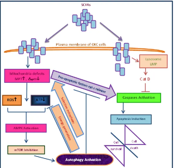
Therefore, SCFA may have opposing effects either inducing autophagy and hence increasing cell resistance, or inhibiting the proliferation of cancer cells through induction of apoptosis. In line of this latter effect we have recently demonstrated that lysosomal membrane permebilization (LMP) and the release of Cat-D is important in regulation of apoptosis by acetate (Marques et al., 2013) (Figure 5). An extensive revision was performed concerning the precise role of SCFA: butyrate, propionate and acetate in colorectal carcinoma and all data was compiled in Table I, and will be described in brief.
23
Figure 5 - Schematic representation of the proposed role of SCFAs in orchestrating two opposing cellular events: induction of autophagy which increases tumor resistance and contributes to its development or induction of apoptosis which increases tumor sensitivity and promotes elimination of colon cancer cells. MPT, mitochondrial
membrane permeability transition; Δψm, mitochondrial membrane potential; ROS, reactive oxygen species
(Adaptated from Marques et al., 2013; Tang, Chen, Jiang, & Nie, 2011b).
6.1. Butyrate
Of all SCFA butyrate is the most extensively studied because of its role in the prevention of CRC, inducing a variety of changes within the nucleus as a consequence of inhibition of cell differentiation, promotion of cell-cycle arrest and apoptosis of transformed colonocytes (Heerdt, Houston, & Augenlicht, 1997). Hinnebusch et al. (2002) demonstrated that apoptosis levels in human colon carcinoma cells were higher for treatment with butyrate than with other SCFA (Hinnebusch et al., 2002). On the other hand, Marques et al. (2013) showed that acetate
24 induces high levels of apoptosis in CRC cells (Marques et al., 2013). Butyrate presents the same effects in other types of malignant cells both in vitro and in vivo (Table 1).
Butyrate is a major energy source for colonic epithelial cells in vivo, accounting for about 70-90% of total energy consumption (Cook & Sellin, 1998; G. R. Gibson & Roberfroid, 1995; Scheppach, 1994), while most other cell types use glucose (Donohoe et al., 2011), and is thought to stimulate cell proliferation (Scheppach et al., 1992). Cells that metabolize butyrate at a higher rate are likely to be less susceptible to its apoptosis-inducing effects, which may explain why normal colonocytes are unaffected by the very high levels of this SCFA in the distal colon (Medina et al., 1997). However, an important conclusion from Singh et al. (1997) study was that the response of colonic epithelial cells to butyrate may depend in part on the other energy sources available to the epithelium: in conditions of low energy availability, butyrate could stimulate growth, but in the presence of high levels of alternative high energy sources such as glucose, butyrate could switch from a growth stimulator to a growth inhibitor and/or an inducer of apoptosis, depending upon factors such as the level of exposure and the intracellular milieu (Sengupta, Muir, & Gibson, 2006; Singh et al., 1997). Nevertheless, differentiation of colonocytes, either induced by butyrate or by other conditions, is reversible at early times of exposure. It has been hypothesized that less differentiated transformed cells are more sensitive to the apoptotic effects of butyrate than differentiated colonocytes (Matthews, Howarth, & Butler, 2006; Scharlau et al., 2009). Thus, if a continuous in vivo exposure to high concentrations of butyrate is not attained, the irreversible commitment to differentiation might not occur and cells could return to their normal malignant phenotype and regain the capacity to proliferate (Cai et al., 2006).
Boren and et al. (2003) observed that differences exist in the metabolism of butyrate between a butyrate-sensitive (HT29) and a butyrate-resistant (MIA) cell types. Incubation of MIA cells with butyrate had no effect on glucose utilization and was not associated with cellular differentiation (Boren et al., 2003). It was, therefore, proposed that a cell will undergo apoptosis or differentiation depending on the ability of that cell to metabolize butyrate. However, cancer cells that are capable of metabolize butyrate have been identified (Lopez de Silanes et al., 2004) and they are protected against its role as an apoptotic inducer. This finding suggests that the colonic butyrate-rich microenvironment may play a role in the selection of more aggressive colon carcinoma cells that maintain the ability to use butyrate as a carbon and energy source (Serpa et al., 2010). Additionally, several studies showed that butyrate-resistant cancer cells may be
25 selected and give rise to more aggressive cancers (Chai, Evdokiou, Young, & Zalewski, 2000; Lopez de Silanes et al., 2004; Mariadason, Corner, & Augenlicht, 2000).
Other studies propose a down regulation of butyrate transporters, such as MCT1, in human colon carcinoma tissue (Lambert, Wood, Ellis, & Shirazi-Beechey, 2002; H. Li et al., 2003) which results in a reduced uptake and metabolism of butyrate in the colonocytes (Hamer et al., 2008). A decline in the abundance of MCT1 in the membrane of colonic epithelial cells, and hence in butyrate uptake, would in turn reduce the availability of intracellular butyrate as a source of energy and as an important regulator of cellular homeostasis. In 2009 Thangaraju et al. showed that butyrate also elicits effects in colon cells extracellularly by serving as a ligand for GPR109A (Thangaraju et al., 2009), a receptor in the lumen facing apical membrane of colonic epithelial cells that binds butyrate with low affinity (Clarke et al., 2012). The expression of GPR109A is silenced in colon carcinoma by DNA methylation whereas its re-expression by butyrate exposure in cancer cells results in apoptosis without the involvement of histone deacetylases (HDAC) (Thangaraju et al., 2009). Butyrate was also able to activate the receptor with an EC50 (concentration necessary for half-maximal activation of the receptor) of ≈1.6 mmol/L. Although the levels of butyrate in circulation are too low (≈5 mol/L) to activate the receptor, butyrate is present at high levels (≈20 mmol/L) in colonic lumen (Hamer et al., 2008). If GPR109A is expressed in the lumen-facing apical membrane of colonocytes, it might suggest that the ability of butyrate to prevent cancer and inflammation in the colon may also be mediated extracellularly via the receptor without entering into cells (Thangaraju et al., 2009). Still regarding to transporters, the gene of the SMCT1 transporter is highly methylated in colon adenomas of African Americans, who are more likely to die from colon carcinoma than others, which points to its potential use as a marker for early detection (Brim et al., 2011). Re-expression of SMCT1 in the presence of butyrate results in colon carcinoma cellular apoptosis (Brim et al., 2011). Thangaraju (2008) observed that the induction of apoptosis in SW480 colon cancer cells by SMCT1/butyrate was associated with upregulation of pro-apoptotic genes (p53, Bax, Bad, Bak, FAS ligand, FAS receptor, TRAIL, and TRAIL receptors) and downregulation of anti-apoptotic genes (Bcl-2, Bcl-W, BclxL, Bfl-1, and survivin) (Thangaraju et al., 2008). Butyrate causes cell death in colon carcinoma cells by two independent but complementary mechanisms: one through GPR109A independent of HDACs and other through SMCT1-mediated entry of butyrate into cells with subsequent inhibition of HDACs (Thangaraju et al., 2009). By acting as an HDAC inhibitor, butyrate leads to histone hyperacetylation (Cousens, Gallwitz, & Alberts, 1979) and
26 enhances the accessibility of transcription factors to the DNA, causing biological effects in colon carcinoma cells (Wong et al., 2006), including cell death (Shao et al., 2004).
For Donohoe and co-workers (2012) butyrate inhibits the growth of cancerous colonocytes because it is metabolized inefficiently due to the Warburg effect, described as a high degree of aerobic glycolysis. Consequently, butyrate accumulates in the nucleus, acts as an HDAC inhibitor and causes cell death (Shao et al., 2004; Wong et al., 2006). Robert Li and Cong Jun Li (2006) confirmed the accumulation of acetylated histone 3 (H3) due to butyrate treatment (R. W. Li & Li, 2006). In addition to this function, butyrate increases histone acetylation through conversion to acetyl-CoA and stimulating histone acetyltransferases (HATs) (Barshishat, Polak-Charcon, & Schwartz) activity (R. W. Li & Li, 2006). However, the metabolic state of the cell influences intranuclear butyrate and acetyl-CoA levels and determines whether butyrate functions to inhibit HDACs or stimulate HATs to epigenetically regulate the expression of different target genes (Donohoe et al., 2012). Deregulation in the expression or activity of HATs and HDACs may lead to alterations in gene expression profiles, associated with the reactivation or silencing of genes critical for cancer progression, differentiation and apoptosis (Iacomino, Tecce, Grimaldi, Tosto, & Russo, 2001; Marchion & Munster, 2007; Sambucetti et al., 1999). For example, hyperacetylation of histones can support chromatin opening and induction of p21 (WAF1/CIP1) gene expression (Davie, 2003), and increased histone acetylation in the promoter of p21WAF1 gene has been reported after butyrate treatment. Activation of tumor-suppressor genes, like p21WAF1, is claimed to be the major cause of growth arrest and/or apoptosis. On the other hand, down regulation of p21WAF1 expression in colorectal carcinomas is associated with histone hypoacetylation of total chromatin (Lu et al., 2008). It is possible, that nucleosome conformations are altered due to histone H3 hypoacetylation, and that the access to chromatin of transcriptional regulatory proteins may be reduced in colorectal carcinomas (Lu et al., 2008). It has also been reported that butyrate can induce a hypermethylation of DNA (de Haan, Gevers, & Parker, 1986), contributing to repression of the transcription of a specific region of DNA. Still regarding to DNA molecules, alkaline phosphatase (ALP) is a hydrolase isoenzyme responsible for removing phosphate groups nucleotides, and their levels are frequently elevated in patients with metastatic CRC (Saif, Alexander, & Wicox, 2005). ALP activity and the dipeptidyl peptidase-IV have been used as markers of colonocyte differentiation in many studies with Caco-2 and HT29 colon carcinoma cells and were increased during culture of malignant cells in the presence of butyrate (Whitehead, Young, & Bhathal, 1986). Regarding transcription, it has been demonstrated that
27 transcriptional attenuation triggered by butyrate plays an important role in the down regulation of two key genes that regulate colonic cell maturation and transformation – c-myc and cyclin D1, and therefore eliminate their increased steady state levels, which might be expected from the elevation in Wnt signaling (Cai et al., 2006; Maier et al., 2009). Scharlau et al. (2009) also observed that butyrate treatment enhanced mRNA gene expression, protein expression and enzyme activity of GSTs, and other stress response genes in human primary colon cells, LT97 cells and HT29 cells (Scharlau et al., 2009). In response to DNA damage, p53 is activated and turns on the transcription of one of its downstream genes, p21WAF1 (Archer, Meng, Shei, & Hodin, 1998; el-Deiry et al., 1993). For Harper et al. (1993) the p21WAF1 product binds to cyclin complexes and inhibits the function of cyclin-dependent kinases (Harper et al., 1993). Nakano et al (1997) found that butyrate markedly induces p21WAF1protein and causes G1-phase arrest. They also observed that butyrate can strongly activate the WAF1/CIP1 promoter, and that the two p53-binding sites are not required for the transcriptional activation by butyrate (Nakano et al., 1997). By inhibiting the HDAC activity recruited to the p21WAF1 promoter by Sp1 or Sp3, butyrate induces the expression of p21WAF1 and thereby stops cell proliferation (Davie, 2003). Nakano et al (1997) suggested that butyrate-induced growth arrest in human colon carcinoma cells is due to the p53-independent activation of p21 promoter mediated through specific Sp1 sites in the promoter region (Nakano et al., 1997). Siavoshian et al. (2000) showed that exposure to butyrate resulted in arrest in the G1 phase of the cell cycle, and that was associated with p21WAF1 induction at the protein and mRNA level and overexpression of cyclin D3 (Siavoshian et al., 2000). Regarding cell cycle progression, Robert Li and Cong Jun Li (2006) observed that MDBK cells (Madin-Darby bovine kidney epithelial cells) were arrested at the G1/S boundary and DNA replication was blocked after butyrate treatment (R. W. Li & Li, 2006). A different study showed that after 6 hours following treatment of CRC cells with 5 mM of butyrate, the percentage of cells in G1 phase of the cell cycle increased by 40% (Maier et al., 2009). Another gene that is regulated by p53 and that could influence the decision to commit to an apoptotic pathway is Bax, which belongs to Bcl-2 family. It is known that members of the Bcl-Bcl-2 family play important roles in regulating apoptosis by functioning as promoters (e.g. Bax, Bak, and Bok) or inhibitors (e.g. Bcl-XL, Bcl-w, A1, and Mcl-1) of cell death of transformed cells (Carpinelli et al., 2012; Gewies, 2003; Tzifi et al., 2012). It is also known that overexpression of Bcl-2 can block p53-mediated apoptosis (A. J. Levine, 1997). Bax binds to Bcl-2 and antagonizes its ability to block apoptosis so a p53-dependent Bax synthesis could tip the scales toward apoptosis (A. J. Levine, 1997). Hague et al. (1996)