FACULDADE DE CIÊNCIAS E TECNOLOGIA
PHYTOCHEMICAL STUDIES AND BIOLOGICAL
ACTIVITY OF CARNIVOROUS PLANTS FROM
THE MEDITERRANEAN REGION
(Tese para a obtenção do grau de doutor no ramo de Ciências Biotecnológicas, especialidade de Biotecnologia Vegetal)
T
OMÁSG
REVENSTUKF
AROFACULDADE DE CIÊNCIAS E TECNOLOGIA
PHYTOCHEMICAL STUDIES AND BIOLOGICAL
ACTIVITY OF CARNIVOROUS PLANTS FROM
THE MEDITERRANEAN REGION
(Tese para a obtenção do grau de doutor no ramo de Ciências Biotecnológicas, especialidade de Biotecnologia Vegetal)
T
OMÁSG
REVENSTUKOrientador: Prof. Doutora Anabela Romano
F
ARODECLARATION
This thesis contains results that were, or will be published in collaboration with A Romano, S Gonçalves, J Vervoort, JJJ van der Hooft, P de Waard, C Quintas, N Gaspar, AL Escapa, N Coelho, S Almeida, JMF Nogueira and N Neng.
I hereby declare that this thesis is a presentation of original research work. Wherever contributions of others are involved, every effort is made to indicate this clearly, with due reference to the literature, and acknowledgement of collaborative research and discussions.
Faro, September 2010
I would like to express my sincere gratitude and appreciation to my supervisor, Professor Dr. Anabela Romano, for offering me the chance to apply as a PhD student at the Plant Biotechnology Laboratory. I am very grateful for her help with outlining this research project, guidance, encouragement, good spirits and for the special bond that was created and strengthened throughout the past years, but also for taking the time for the thorough revision of this thesis.
I am very grateful to Dr. Sandra Gonçalves, my “unofficial” co-supervisor, without whose support, motivation and encouragement, I would possibly not have considered a career following this direction. I am very grateful for her help with planning and guidance of great part of the experimental work, for the proof-reading, for solving countless setbacks and for giving her opinion on so many occasions.
I would like to thank Professor Dr. Jacques Vervoort for welcoming me at the Wageningen NMR Centre (Wageningen University), where it was possible to work with state-of-the-art LC-MS and LC-NMR equipment and learn the basics of NMR fundaments and interpretation of spectra for structure elucidation. I am thankful for his revision of Chapter 3 of this thesis and would like to add that his expertise and understanding in this field was inspiring and added considerably to my graduate experience. I am very grateful to Justin van der Hooft for the training and help in the operation of the analytical equipment, interpretation of results, for his many comments and suggestions, but mostly for his patience in helping a rookie in NMR analysis. I would also like to thank Dr. Pieter de Waard for assistance in NMR experiments.
I am also grateful to Professors Drs. Célia Quintas and Nelma Gaspar (Laboratory of Microbiology, ISE, Algarve University) for their help with the antimicrobial activity assays.
I am grateful to Professor Dr. Gabriela Bernardo-Gil for receiving me at the Supercritical Extraction Laboratory (IST, Technical University of Lisbon) and for putting the supercritical extraction equipment at our disposal. I would also like to thank Paula Pereira and Dr. Maria João Cebola for their assistance and guidance with the experiments.
I would also like to thank Professor Dr. José Manuel Florêncio Nogueira and Nuno Neng of the Laboratory of Chromatography and Capillary Electrophoresis (FC,
in Chapter 5.
Also, I would like to thank Natacha Coelho and Sara Almeida for their help with the bioassay experiments, and Ana Luísa Escapa and once more Natacha Coelho for helping with the development of micropropagation protocols. I would also like to thank my lab colleagues and Rosália Almeida, our lab technician, for their help and kind disposition.
I am very grateful to Dr. Miguel Porto for providing information on the location of natural populations of D. intermedia, D. rotundifolia and to Professor Dr. Henrique Pereira (Peneda-Gerês National Park, ICNB) and Mr. António Rebelo who very kindly provided seeds of P. vulgaris. I am also grateful to Dr. Jorge Jesus for collecting P. lusitanica seeds.
I greatly appreciate the financial support of the Portuguese Foundation for Science and Technology by funding the PhD fellowship (FCT, Grant SFRH/BD/31777/2006) and the European Community activity Large-Scale Facility Wageningen NMR Center (FP6-2004-026164 (2006–2009), which supported the research activities held at Wageningen, the Netherlands.
I would also like to thank my parents for their loving support and for creating an environment in which following this path seemed so natural, and my dear friends, for their company, high spirits and for not reminding me of work in their presence!
And last but not least Alexandra, not necessarily for coming along at the right time, but for the very special person she is, and for the incredible amount of patience she had with me during the redaction of this manuscript.
δ chemical shift
1D one-dimensional
2D two-dimensional
AAPH 2,2’-azobis-2-methyl-propanimidamide dihydrochloride
ABTS 2,2’-azinobis(3-ethylbenzothiazoline-6-sulfonic acid
ABTS•+ ABTS radical cation
amu atomic mass units
ANOVA analysis of variance
AOC antioxidant capacity
ATCC American type culture collection
AUC area under curve
BA 6-benzyladenine
CFU colony forming units
COSY correlation spectroscopy
DAD diode array detector (or diode array detection)
DMSO dimethyl sulfoxide
DQF double-quantum-filtered
EI electron impact
ESI electrospray ionization
F-C Folin-Ciocalteu
g, mg, µg gram, milligram, microgram
GAE gallic acid equivalent
h, min, s hour, minute, second
HAT hydrogen atom transfer
HMBC heteronuclear multiple bond coherence
HPLC high performance liquid chromatography
HSQC heteronuclear single quantum coherence
HTS high throughput screening
Hz, kHz, MHz, GHz Hertz, kiloHertz, megaHertz, gigaHertz
i.d. internal diameter
IAA indole-3-acetic acid
IBA indole-3-butyric acid
INADEQUATE Incredible Natural Abundance Double Quantum Transfer
Spectroscopy
IZD inhibition zone diamters
K Kelvin
Kin kinetin
L, mL, µL liter, milliliter, microliter
cm, mm, nm centimeter, millimeter, nanometer
m/z mass to charge ratio
MAE microwave-assisted extraction
mAU milli absorbance units
MDR multidrug resistance pumps
MHA Mueller Hinton agar
MHB Mueller Hinton broth
MIC minimum inhibitory concentration
MPa megaPascal
MRSA methicillin-resistant Staphylococcus aureus
MS mass spectrometer (or mass spectrometry)
MS Murashige and Skoog culture medium
NAA 2-naphthaleneacetic acid
NCCLS national committee for clinical laboratory standards
NMR nuclear magnetic resonance
NOESY nuclear-Overhauser-effect spectroscopy
ºC degree Celsius
ORAC oxygen radical absorption capacity
P statistical probability
PCA plate count agar
PGR plant growth regulator
ppm part per milion
ROESY rotational frame nuclear Overhauser effect spectroscopy
ROS reactive oxygen species
rpm revolutions per minute
S/N signal to noise ratio
SE standard error
SET single electron transfer
SFE supercritical fluid extraction
SPE solid phase extraction
SPSS statistical package for the social sciences
TE trolox equivalent
TEAC trolox equivalent antioxidant capacity
TOCSY total-correlation spectroscopy
TOF time of flight
tR retention time
UAE ultrasound assisted extraction
UV ultraviolet
v/v volume per volume
w/v weight per volume
In this thesis several studies were conducted with four carnivorous plant species which occur on Portuguese territory: Pinguicula lusitanica, Pinguicula vulgaris, Drosera intermedia and Drosera rotundifolia. Most habitats of these plants are threatened and natural populations are scarce, therefore micropropagation protocols were developed to produce biomass for the subsequent studies. Efficient micropropagation protocols were developed for P. lusitanica and D. intermedia enabling large scale biomass production, while protocols for the other two species have still to be optimized (in Chapter 2). The in vitro established cultures represent active germplasm collections of Portuguese natural populations and contribute therefore for their conservation. In Chapter 3 extracts prepared from micropropagated plant material were analyzed using state of the art HPLC-ESI-MS and HPLC-SPE-NMR equipment which enabled the identification of the major secondary metabolites produced by P. lusitanica and D. intermedia, directly from essentially crude extracts. The metabolites identified in P. lusitanica belong to the iridoid glucosides and caffeoyl phenylethanoid glycosides and D. intermedia was shown to produce mainly flavonoid glucosides, ellagic acid derivatives and the naphthoquinone plumbagin. The evaluation of the biological activities of these extracts, compiled in Chapter 4, showed that the methanol extract of P. lusitanica has considerable antioxidant activity and that the n-hexane extract of D. intermedia has high antimicrobial potential. In Chapter 5 a method for the extraction of plumbagin from micropropagated D. intermedia plants was optimized and its potential as an alternative for bioprospection evaluated. It was shown that the commercial exploitation of plumbagin from D. intermedia cultures might be viable and that UAE with n-hexane followed by an SPE purification step is an efficient procedure for obtaining large quantities of high purity plumbagin. It is hoped that this study represents an enrichment of the knowledge on these plants and contributes to their conservation and valorisation.
Keywords: Pinguicula; Drosera; micropropagation; conservation; hyphenated
analytical techniques; antioxidant activity; antimicrobial activity; bioprospection; plumbagin.
Nesta tese apresentam-se resultados de diversos estudos realizados em quatro espécies de plantas carnívoras que ocorrem naturalmente em Portugal continental: Pinguicula vulgaris, Pinguicula lusitanica, Drosera rotundifolia e Drosera intermedia. As plantas carnívoras mantêm todas as características de qualquer outro ser vivo do reino vegetal: são plantas verdes onde ocorre fotossíntese, contudo estas plantas desenvolveram a capacidade única de capturar e digerir pequenas presas, pertencentes essencialmente ao grupo dos artrópodes. A maior parte das plantas carnívoras terrestres ocorre em turfeiras ou pântanos, onde persistem condições desfavoráveis constantes. Nestes habitats os solos encontram-se frequentemente submersos ou saturados em água e são de natureza ácida e relativamente pobres em relação ao teor de nutrientes disponíveis. Tendo em conta que o hábito carnívoro nas plantas surgiu em várias famílias distintas de forma independente, crê-se que representa uma adaptação aos factores de stress típicos destes habitats.
As plantas carnívoras despertaram o interesse dos biólogos desde longa data, devido à sua morfologia peculiar e aos seus hábitos carnívoros, no entanto, poucas espécies têm sido estudadas em relação aos metabolitos secundários que produzem e às suas potenciais aplicações farmacológicas. As plantas são organismos sésseis desprovidos de sistema imunitário e portanto desenvolveram estratégias alternativas que envolvem a produção de compostos orgânicos bioactivos capazes de dissuadir ataques de herbívoros ou infecções por parte de microorganismos. Milhares de anos de evolução resultaram na imensa diversidade de metabolitos secundários produzida actualmente pelas plantas. Apesar do desenvolvimento das técnicas de modelação molecular e síntese química, as plantas continuam a ser uma fonte importante de novas drogas e estruturas químicas, fornecendo pistas importantes para o tratamento de varias doenças. Estima-se que aproximadamente um quarto das drogas actualmente em uso clínico tenham sido isoladas directamente ou derivadas de fitoquímicos. É de salientar que as estruturas químicas provenientes de plantas para além de serem usadas directamente podem servir de precursores para novos medicamentos por processos de modelação química. Deste modo, o estudo da composição química de extractos preparados a partir de plantas,
âmbito da identificação de substâncias bioactivas com interesse farmacológico.
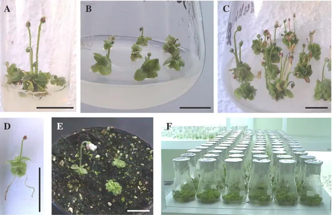
O estado de conservação dos habitats das espécies estudadas neste trabalho é precário e as populações naturais não suportam a colheita de exemplares para a realização de um trabalho de investigação deste âmbito. Desta forma, no Capítulo 2 descrevem-se os protocolos de micropropagação desenvolvidos para as espécies em estudo, garantindo a produção de biomassa para os estudos subsequentes. A aplicação de técnicas de cultura in vitro é muito importante para alcançar os objectivos deste trabalho porque permite a obtenção, de forma rápida, de grandes quantidades de material a partir de quantidades reduzidas de tecido vegetal inicial. As culturas das quatro espécies foram iniciadas a partir de rebentos provenientes de germinantes produzidos in vitro de forma a evitar a recolha de exemplares do campo, reduzir a probabilidade de contaminações e manter uma diversidade genética elevada nas culturas estabelecidas. Em geral obtiveram-se percentagens de germinação relativamente baixas, pelo que poderá ser interessante testar o efeito de algumas técnicas de estratificação. No entanto, os germinantes obtidos demonstraram elevada capacidade de proliferação e permitiram avaliar os parâmetros de crescimento das espécies em vários meios de cultura.
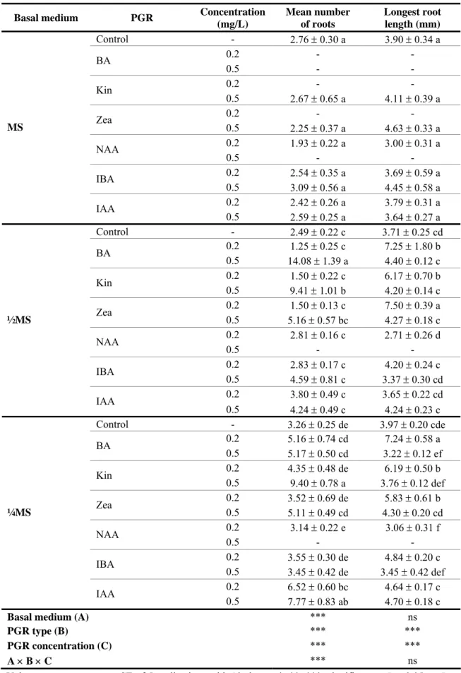
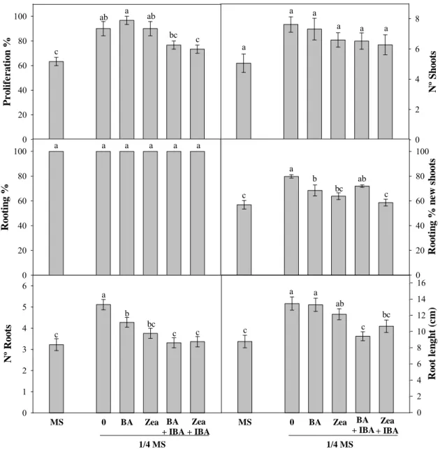
O efeito da concentração do meio MS sem reguladores de crescimento foi testado em todas as espécies tendo-se verificado, de uma forma geral, que as culturas têm uma preferência para meios de cultura com baixas concentrações de macronutrientes, o que vai de encontro com as condições naturais em que as plantas carnívoras prosperam. No caso da espécie P. lusitanica testaram-se meios de cultura com três concentrações de meio basal (MS total, ½MS e ¼MS) suplementados com citocininas (BA, Kin e Zea) ou auxinas (NAA, IBA, IAA) a duas concentrações (0.2 e 0.5 mg/mL) e verificou-se que na grande maioria dos casos as culturas produziam simultaneamente novos rebentos e raízes, independentemente da composição do meio em reguladores de crescimento. Desta forma determinou-se que o protocolo de micropropagação para esta espécie dispensa uma fase de enraizamento adicional e que plântulas enraizadas podem ser produzidas num único passo. As culturas de P. lusitanica responderam melhor em meio MS suplementado com citocininas na concentração mais alta (0.5 mg/mL), obtendo-se cerca de 26 novos rebentos por explantado inicial e percentagens de enraizamento
é pouco comum e deve ser investigado. No entanto, as culturas também mostraram capacidade de proliferação e enraizamento elevadas em meios sem reguladores de crescimento, o que pode indicar níveis endógenos elevados de citocininas e auxinas. Meios de cultura suplementados simultaneamente com citocininas e auxinas não promoveram a proliferação de rebentos.
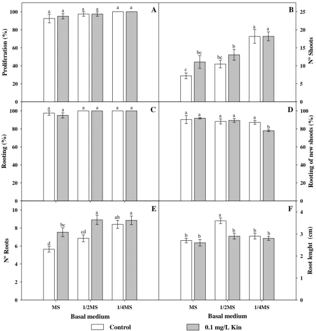
O desenvolvimento de um protocolo de micropropagação para a espécie P. vulgaris foi uma tarefa mais complicada devido à fragilidade e pouca viabilidade dos rebentos produzidos in vitro. Os rebentos mostraram-se muito susceptíveis ao passo de individualização no início de cada ensaio e portanto a quantidade de meios testada para esta espécie foi inferior. Em primeiro lugar foi testado o efeito da concentração de meio basal na capacidade de resposta das culturas usando as concentrações de MS total e ¼MS e posteriormente testou-se o efeito da adição de 0.1 mg/mL das citocininas BA e Zea individualmente, ou em combinação com a auxina IBA a 0.01 mg/mL, ao meio basal mais adequado. Os resultados demonstraram que o meio MS total influenciou negativamente o crescimento das culturas de P. vulgaris e que não se verificaram diferenças entre o controlo e meio de cultura suplementado com citocininas, em termos de rebentos produzidos ou percentagens de enraizamento. No entanto, do ponto de vista morfológico, os rebentos produzidos em meio de cultura sem reguladores de crescimento eram mais vigorosos enquanto os rebentos produzidos em meios suplementados com BA ou Zea mostraram por vezes sinais de necrose e reduzido desenvolvimento. Tal como no caso de P. lusitanica, a combinação de citocininas e auxinas não promoveu a proliferação das culturas de P. vulgaris. Desta forma, dos meios de cultura testados, o mais indicado para a micropropagação de P. vulgaris é o meio ¼MS sem reguladores de crescimento. No entanto, este protocolo deverá ser optimizado uma vez que a baixa viabilidade das culturas produzidas não permite produção de biomassa em larga escala.
Ao contrário das outras espécies estudadas, as percentagens de germinação de D. intermedia foram bastante elevadas. Curiosamente, registaram-se percentagens de germinação superiores no ensaio controlo do que no tratamento de estratificação a frio, ao contrário do que tinha sido observado para outras plantas do mesmo género. Na espécie D. intermedia foram testados meios com as concentrações de meio basal ¼MS,
A resposta de D. intermedia foi semelhante à de P. vulgaris na medida em que a redução da concentração de macronutrientes melhorou a proliferação dos rebentos e que a adição de Kin não induziu diferenças significativas na resposta das culturas. Quando cultivadas em meio ¼MS sem reguladores de crescimento, as culturas de D. intermedia produziram em media 15.8 rebentos ao fim de 8 semanas e em todos os casos os explantados iniciais formaram raízes, bem como mais de 80% dos novos rebentos formados. Como seria difícil melhorar qualquer um dos parâmetros de crescimento não foram testados outros meios. Enquanto os protocolos de micropropagação de P. lusitanica, P. vulgaris e D. intermedia foram desenvolvidos pela primeira vez neste trabalho, a cultura in vitro da espécie D. rotundifolia já tinha sido descrita previamente. No entanto, nestes estudos em vez de sementes, foram usados explantados recolhidos de exemplares de campo como material de partida. Para esta espécie testaram-se apenas dois meios, nomeadamente ¼MS e MS total. Apesar de se ter obtido um número de rebentos razoável nos dois meios testados, os rebentos eram de tamanho reduzido, muitas vezes difíceis de contabilizar e demasiado pequenos para iniciar novas culturas. Esta dificuldade foi também referida por outros autores, pelo que deve ser considerada uma fase de alongamento e uma fase de indução de raízes de forma a desenvolver um protocolo eficiente.
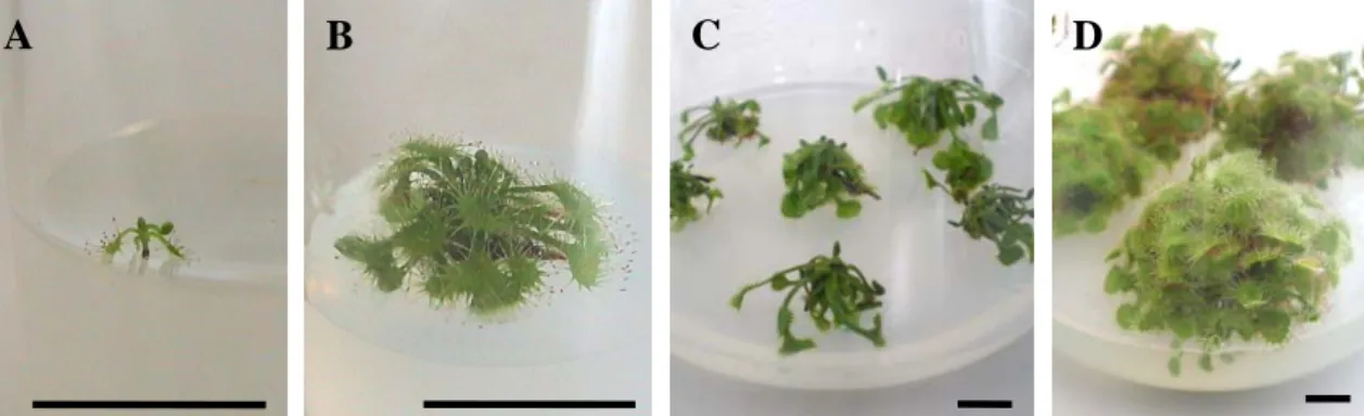
As plântulas micropropagadas de P. lusitanica, P. vulgaris e D. intermedia com sistemas radiculares bem desenvolvidos foram aclimatizadas com sucesso às condições ex vitro apresentando um desenvolvimento normal, sem aparentes anomalias morfológicas e com folhas funcionais capazes de capturar insectos, podendo ser usadas em programas de reintrodução em populações naturais. Em suma, foram desenvolvidos protocolos de micropropagação eficientes para P. lusitanica e D. intermedia, que permitiram a produção de material suficiente para as fases subsequentes deste trabalho. Os protocolos de micropropagação de P. vulgaris e D. rotundifolia têm que ser optimizados, no entanto, as culturas estabelecidas in vitro representam uma colecção activa de germoplasma que se pode tornar valiosa na eventualidade de extinção das populações.
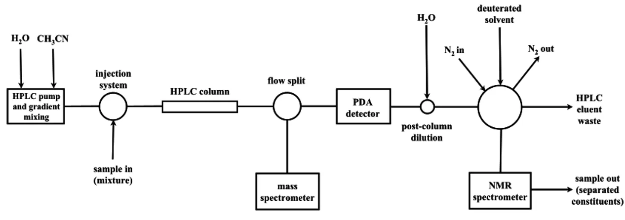
secundários identificados bem como a sua significância taxonómica. Em projectos de descoberta de produtos naturais bioactivos é vantajoso realizar uma caracterização química numa fase precoce do trabalho para evitar o estudo alargado de compostos já conhecidos e caracterizados. Para o efeito recorreu-se às técnicas hifenadas HPLC-ESI-MS e HPLC-SPE-NMR. A espectroscopia de massa é uma ferramenta extremamente útil na identificação de compostos em amostras naturais, que para além da sua elevada sensibilidade, permite obter a massa molecular com grande precisão e portanto inferir a fórmula molecular, e em alguns casos até informação estrutural por análise do padrão de fragmentação. Em termos de informação estrutural a técnica de NMR é a mais valiosa, dando informações sobre distâncias intramoleculares entre átomos e grupos funcionais e acerca da orientação espacial de substituintes em torno de centros quirais, permitindo assim determinar a estrutura completa de uma molécula bem como relações de estereoisomerismo. O maior impedimento para o uso mais alargado da técnica de NMR, para além do custo elevado, está relacionado com a relativa baixa sensibilidade. No entanto, para além da miniaturização das células de fluxo, bobinas arrefecidas a temperaturas criogénicas e campos magnéticos cada vez mais potentes, o acoplamento de uma unidade automatizada entre o HPLC e o equipamento de NMR capaz de extrair e concentrar picos cromatográficos em cartuchos de SPE, tem contribuído muito para o ganho de sensibilidade. Desta forma, a utilização destas técnicas avançadas permitiram identificar os compostos maioritários directamente a partir de extractos sem ter que recorrer ao isolamento por técnicas de cromatografia preparativa para obter informação estrutural.
A espécie P. lusitanica não foi previamente estudada do ponto de vista bioquímico, tornando impossível a utilização de padrões. No entanto, explorando as potencialidades destas técnicas hifenadas foi possível a identificação dos seus metabolitos maioritários que pertencem a dois grupos de compostos naturais: iridóides e feniletanóides glicosídicos. A partir do extracto metanólico de P. lusitanica identificaram-se os iridóides ácido mussaenosídico, globularina e scutellarioside II e os feniletanóides acteoside, R/S campneoside I e R/S campeneoside II. Por análise das intensidades dos sinais dos protões anoméricos, determinou-se de forma aproximada que os compostos presentes em maior quantidade são o ácido mussaenosídico e acteoside. O extracto metanólico continha também um composto cuja estrutura foi impossível de determinar
seus sinais encontravam-se sobrepostos no espectro de NMR. No entanto, os dados preliminares indicam que o composto poderá não ter sido identificado previamente, podendo revelar-se interessante proceder ao seu isolamento.
No caso da espécie D. intermedia identificaram-se vários flavonóides glicosídicos (galactopiranosídeo, glucopiranosídeo, quercetina-3-O-(2’’-O-galoil)-galactopiranosídeo), glucopiranosídeo e miricetina-3-O-(2’’-O-galoil)-galactopiranosídeo) e derivativos de ácido elágico (ácido elágico, 3-O- acido metil-elágico, ácido 3,3'-di-O-metil-elágico e ácido 3,3'-di-O-metil-elágico 4-O-glucopiranosídeo), para além de uma naftoquinona que poderá possivelmente ser hidroplumbagina di-1,4-O-glucopiranosídeo. O extracto aquoso de D. intermedia não foi investigado e o extracto hexânico era constituído exclusivamente por um composto, que foi analisado directamente por NMR e identificado como a naftoquinona plumbagina. Grande parte dos metabolitos secundários identificados foram atribuídos pela primeira vez a D. intermedia, fazendo com que este trabalho represente também uma contribuição para a melhor compreensão da bioquímica desta espécie. Do posto de vista taxonómico, o perfil de metabolitos secundários obtido para P. lusitanica e D. intermedia corroboram estudos realizados previamente noutras espécies das famílias Lentibulariaceae e Droseraceae, respectivamente.
Sendo impraticável testar um grande número de actividades biológicas em simultâneo neste trabalho, como ponto de partida decidiu-se avaliar a actividade antioxidante e antimicrobiana dos extractos preparados a partir de P. lusitanica e D. intermedia. A determinação da actividade antioxidante de um extracto é um dado importante na medida em que pode indicar potenciais actividades contra outros alvos biológicos. A avaliação da actividade antimicrobiana é igualmente importante devido à necessidade urgente de encontrar novas fontes de agentes anti-sépticos em resposta ao desenvolvimento de resistências múltiplas aos antibióticos em uso clínico. Quando possível, tentaram estabelecer-se relações de estrutura-actividade entre os compostos identificados previamente nos extractos e os resultados obtidos nos ensaios. Os resultados mostraram que o extracto com maior actividade antioxidante foi o extracto metanólico de P. lusitanica, possivelmente devido a um dos seus compostos
reacções em cadeia. O facto de terem sido comprovadas actividades contra vários alvos biológicos e ser considerada não tóxica, torna esta molécula interessante para estudos posteriores. O extracto metanólico de D. intermedia também apresentou actividade antioxidante considerável, que pode ser explicada pela combinação de flavonóides e derivativos de ácido elágico presente no extracto.
Os extractos de D. intermedia foram mais eficazes nos ensaios de actividade antimicrobiana, especialmente o extracto hexânico, que inibiu o crescimento de todas as estirpes incluídas no painel de microorganismos seleccionados, à excepção de P. aureginosa. Esta actividade foi atribuída ao composto maioritário do extracto, plumbagina, para o qual já tinha sido comprovado elevada actividade antimicrobiana. A actividade desta naftoquinona parece estar relacionada com a sua capacidade de se ligar covalentemente a biomoléculas tornando-as inactivas, mas também com a sua capacidade de produzir radicais livres em sistemas biológicos. Porém, a possível pequena margem de segurança da plumbagina tornam a sua aplicação farmacológica incerta. O extracto metanólico de D. intermedia também inibiu o crescimento de grande parte dos microorganismos testados incluindo, curiosamente, a estirpe multirresistente P. aeruginosa. Como os antibióticos activos contra esta estirpe são escassos seria interessante estudar em maior detalhe o mecanismo subadjacente. Por sua vez, o extracto de P. lusitanica mostrou possuir reduzida actividade antimicrobiana. Apesar deste estudo não ter identificado potenciais candidatos para o desenvolvimento de novos fármacos é importante a continuação de programas com o intuito de escrutinar aplicações de extractos vegetais contra alvos biológicos, tendo em conta o enorme contributo que o Reino das plantas tem dado à medicina moderna.
As análises efectuadas no Capítulo 3 demonstraram que era possível obter, de uma forma relativamente simples, quantidades significativas de plumbagina de elevada pureza a partir de material micropropagado de D. intermedia. Desta forma, aliado ao valor comercial desta naftoquinona, decidiu-se avaliar o potencial de prospecção de plumbagina a partir de culturas in vitro de D. intermedia. A produção de biomassa foi monitorizada ao longo do tempo para determinar o período de crescimento máximo e o material micropropagado foi extraído por várias metodologias: maceração com agitação, extracção Soxhlet, extracção assistida por ultrasons (UAE) e extracção com fluidos
biomassa ser bastante elevada, as quantidades de plumbagina produzidas por D. intermedia são superiores em relação à actual fonte de prospecção de plumbagina, as plantas do género Plumbago. A comparação dos métodos de extracção levou a concluir que a melhor alternativa para a extracção de plumbagina é aplicar ultrasons à matriz vegetal colocada no solvente de extracção (n-hexano). Este procedimento traz vantagens em relação ao tempo de operação e permite obter rendimentos de extracção superiores, assim como maiores concentrações de produto, um factor importante para os passos subsequentes de purificação. Em alternativa, o material vegetal pode também ser extraído com fluidos supercríticos. Apesar de esta metodologia ter produzido resultados inferiores à técnica de UAE, os rendimentos foram consideráveis para uma primeira abordagem. A possibilidade de evitar o uso de solventes orgânicos nocivos é uma grande vantagem do ponto de vista ambiental. Na segunda parte do desenvolvimento do processo de extracção de plumbagina avaliou-se a potencialidade de usar colunas de SPE para concentrar e purificar os extractos. Os resultados demonstraram que usando esta abordagem é possível remover grande parte das impurezas co-extraídas num único passo com pequenas perdas de produto. Aplicando a purificação por SPE ao extracto obtido por UAE é possível produzir plumbagina em grandes quantidades com uma pureza final próxima dos 100%.
Neste trabalho pretendeu-se estudar a composição química e avaliar as propriedades biológicas de extactos preparados a partir de algumas espécies de plantas carnívoras que existem em Portugal. Para tal foi imperativo o desenvolvimento de técnicas de micropropagação que permitiram também optimizar um processo de bioprospecção de um metabolito secundário de valor. Espera-se que este trabalho tenha, de forma geral, contribuído para a conservação e valorização das espécies estudadas.
Palavras-chave: Pinguicula; Drosera; micropropagação; conservação; técnicas
analíticas hifenadas; actividade antioxidante; actividade antimicrobiana; bioprospecção; plumbagina.
Papers in international scientific periodicals with referees
Grevenstuk T, Coelho N, Gonçalves S, Romano A. 2010. In vitro propagation of Drosera intermedia in a single step. Biologia Plantarum 54: 391-394.
Grevenstuk T, van der Hooft JJJ, Vervoort J, de Waard P, Romano A. 2009. Iridoid and caffeoyl phenylethanoid glycosides of the endangered carnivorous plant Pinguicula lusitanica (Lentibulariaceae). Biochemical Systematics and Ecology 37: 285-289. Grevenstuk T, Gonçalves S, Coelho N, Romano A. 2009. Evaluation of the antioxidant
and antimicrobial properties of in vitro cultured Drosera intermedia extracts. Natural Product Communications 4: 1063-1068.
Gonçalves S, Escapa AL, Grevenstuk T, Romano A. 2008. An efficient in vitro propagation system for Pinguicula lusitanica, a rare insectivorous plant. Plant cell, tissue and organ culture 95: 239-243.
Two publications concerning results that are presented in Chapter 3 (chemical investigation of D. intermedia) and Chapter 5 are in preparation.
Patents
Grevenstuk T, Romano A. 2010. A new and effective method for the bioprospection of plumbagin from Drosera spp. Provisional patent, process number: 105221 (Filed 27/07/2010).
Conferences
Grevenstuk T, Gonçalves S, Romano A. 2010. Development of a sustainable method for the bioprospection of plumbagin from D. intermedia. 28th International Horticultural Congress. 22-27 August Lisbon, Portugal, 2: Pp 75.
Grevenstuk T, Domingos T, Gonçalves S, Quintas C, Romano A. 2009. Antimicotic potency of Drosera intermedia extracts on fungi and yeasts causing biodeterioration on food commodities. 3rd International Conference on Environmental, Industrial and
260.
Grevenstuk T, van der Hooft JJJ, Vervoort J, Gonçalves S, Romano A. 2009. Identification of antimicrobial agents from Drosera intermedia using HPLC-MS/HPLC-SPE-NMR. 57th International Congress and Annual Meeting of the Society for Medicinal Plant and Natural Product Research. 16-20 August Geneva, Switzerland. Planta Medica 75: Pp 912.
Grevenstuk T, Gonçalves S, Almeida S, Coelho N, Quintas C, Gaspar MN, Romano A. 2009. Antioxidant and antimicrobial activities of Drosera intermedia. International Congress of Aromatic and medicinal Plants. 26-28 March Marrakech, Morocco, BA126 Pp 111.
Grevenstuk T, Gonçalves S, Xavier C, Alberício F, van der Hooft JJJ, Vervoort J, Romano A. 2008. Antiproliferative, cytotoxic and antioxidant capacity of methanolic extracts of Pinguicula lusitanica. International PSE Symposium on Natural Products in Cancer Therapy. 23-26 September Naples, Italy, Pp P18.
Grevenstuk T, van der Hooft JJJ, Vervoort J, Romano A. 2008. Chemical Investigation of Pinguicula lusitanica by HPLC-MS and HPLC-SPE-NMR. 2008. 7th Joint Meeting of GA, AFERP, ASP, PSE & SIF, Natural Products with Pharmaceutical, Nutraceutical, Cosmetic and Agrochemical interest. 3-8 August Athens, Greece. Planta Medica 74: Pp 1101.
Grevenstuk T, Gonçalves S, Nogueira JMF, Romano A. 2007. Chemical characterization of in vitro cultures of Pinguicula lusitanica (L.) by GC-MS. Congresso Nacional Micro’07-Biotec’07-XXXIII JPG. 29 November - 2 December, Lisboa, Portugal, Pp 113.
Grevenstuk T, Escapa AL, Gonçalves S, Romano A. 2007. Simultaneous multiplication and rooting of Pinguicula lusitanica cultures. 3rd International Symposium on Acclimatization and Establishment of Micropropagated Plants. 12-15 September, Faro, Portugal, Pp 186.
Declaration………...…………....…...… i Acknowledgements……….………..…... iii List of Abbreviations…….………..……..…..…... v Abstract……….………...….…….. vii
Resumo……….... ix
Publications prepared from this thesis………..………... xvii
CHAPTER 1: General Introduction………... 3
1.1. Current status of drug discovery from natural products……… 3
1.2. Drug discovery from plants………... 4
1.2.1. The biogenetic significance of secondary metabolites………... 5
1.2.2. The importance of biotechnological approaches………. 6
1.2.3. Strategies for drug discovery………... 7
1.3. Plant description, taxonomy and biology………. 9
1.3.1. The trait of carnivory in plants………... 9
1.3.2. The genus Pinguicula………... 11
1.3.2.1. Taxonomy and geographical distribution………... 11
1.3.2.2. Biology, morphology and ecology………. 12
1.3.2.3. P. vulgaris……….. 13 1.3.2.4. P. lusitanica………... 14 1.3.3. The genus Drosera……….. 16
1.3.3.1. Taxonomy and geographical distribution………... 16
1.3.3.2. Biology, morphology and ecology………. 17
1.3.3.3. Drosera rotundifolia………... 18 1.3.3.4. Drosera intermedia………... 19
1.4. Objectives………. 20
1.5. References………. 21
CHAPTER 2: Micropropagation of P. vulgaris, P. lusitanica,
D. rotundifolia and D. intermedia………. 27
2.1. Introduction……….. 29
2.1.1. The importance of micropropagation for the conservation
of carnivorous plants……….……....
29
2.1.2. Advantages of micropropagation……….…... 30
2.1.3. Development of micropropagation protocols……….…. 30
2.1.4. Objectives……….…... 34
2.2. Experimental……….… 35
2.2.1. Seed collection, seed germination and establishment of cultures…….…... 35
2.2.2. Proliferation and rooting………..… 36
2.2.3. Plantlet acclimatization……….…... 37
2.2.4. Statistical analysis………..….. 37
2.3. Results and discussion………..…. 38
2.3.1. P. lusitanica……….… 38 2.3.2. P. vulgaris……….... 44 2.3.3. D. intermedia……….….. 48 2.3.4. D. rotundifolia……….… 52 2.4. Conclusions……….….. 55 2.5. References……….… 56
and Drosera intermedia……….. 61
3.1. Introduction………... 63
3.1.1. Phytochemical characterization of extracts………. 63
3.1.2. Phytochemical data……….. 63
3.1.3. Collection of plant material and sample preparation………... 63
3.1.4. Analytical techniques……….……. 65
3.1.5. Separation techniques……….……. 66
3.1.5.1. High Performance Liquid Chromatography (HPLC)………. 66
3.1.6. Hyphenated techniques……… 67 3.1.6.1. HPLC-UV………... 67 3.1.6.2. HPLC-MS………... 68 3.1.6.3. HPLC-NMR……… 69 3.1.6.4. HPLC-SPE-NMR………... 72 3.1.7. Objectives………. 74 3.2. Experimental………. 75
3.2.1. Plant material and sample preparation………. 75
3.2.2. HPLC-MS and HPLC-SPE-NMR measurements……… 76
3.2.2.1. General experimental setup……… 76
3.2.2.2. HPLC-MS and HPLC-SPE-NMR experiments……….. 77
3.2.3.2.1. HPLC-(DAD) gradient optimization………. 77
3.2.3.3.2. HPLC-ESI-MSexperiments………... 78
3.2.3.3.3. HPLC-SPE-NMR experiments………. 78
3.2.3.3.4. Direct NMR analysis………. 79
3.3. Results and Discussion………. 80
3.3.1. P. lusitanica... 80
3.3.1.1. HPLC gradient optimization………... 80
3.3.1.2. HPLC-ESI-MS………..…………. 82
3.3.1.3. HPLC-SPE-NMR………... 83
3.3.1.3.1. SPE trapping procedure………. 83
3.3.1.3.2. HPLC-SPE-NMR………. 85
3.3.1.3.2.1. Iridoid glucosides……… 85
3.3.1.3.2.2. Phenylethanoid glycosides……….. 93
3.3.1.4. Biological and taxonomical relevance……… 97
3.3.2. D. intermedia………... 99
3.3.2.1. HPLC gradient optimization and SPE trappings……… 99
3.3.2.2. Direct NMR analysis of the n-hexane extract……… 103
3.3.2.3. HPLC-SPE-NMR analysis of the methanol extract of D. intermedia… 105
3.3.2.3.1. Flavonoid glucosides………. 106
3.3.2.3.2. Ellagic acid derivatives………. 110
3.3.2.3.3. Naphthoquinone glycosides……….. 114
3.3.3. Biological and taxonomical importance... 115
3.4. Conclusions………... 119
4.1. Introduction……….….…. 129
4.1.1. Antioxidant activity……….….… 129
4.1.1.1. Reactive oxygen species and their biological importance……….….… 129
4.1.1.2. Methods for the determination of antioxidant capacity………….….… 130
4.1.1.2.1. ORAC assay………..…… 131
4.1.1.2.2. TEAC assay……….….…. 132
4.1.1.2.3. F-C assay……….…….…. 133
4.1.2. Antimicrobial activity……….………. 134
4.1.2.1. The issue of antibiotic resistance………..…….. 134
4.1.2.2. Antibacterial and antifungal assays………..…….. 137
4.1.2.2.1. Agar diffusion methods……….……….……... 137
4.1.2.2.2. Dilution methods
(Minimal inhibitory concentration determination)……….……..…. 138
4.1.2.3. General considerations on antimicrobial assays………..….. 138
4.1.3. Objectives………..…….. 139
4.2. Experimental……….…… 140
4.2.1. Plant material and sample preparation………..…... 140
4.2.2. Antioxidant activity……….….…… 140
4.2.2.1. Oxygen radical absorbance capacity (ORAC) assay………..…… 140
4.2.2.2. Trolox equivalent antioxidant capacity (TEAC) assay…………..……. 141
4.2.2.3. Folin-Ciocalteau (F-C) assay………..…… 142
4.2.3. Antimicrobial activity……….…. 143
4.2.3.1. Microorganisms………..… 143
4.2.3.2. Agar disc diffusion assay……….... 143
4.2.3.3. Minimum inhibitory concentration (MIC) determination……….. 144
4.2.4. Statistical analysis……….... 144
4.3. Results and discussion………..…. 145
4.3.1. Antioxidant capacity……….... 145
4.3.2. Antimicrobial activity……….. 150
4.3.2.1. D. intermedia………..…… 150 4.3.2.2. P. lusitanica………..….. 157
4.3.3. Evaluation of antimicrobial assays……….…. 157
4.3.4. Potential of D. intermedia metabolites as antimicrobial agents………..…. 158
4.4. Conclusions………... 160
4.5. References……….… 162
CHAPTER 5: Method development for the bioprospection
of plumbagin from micropropagated D. intermedia………..…….. 171
5.1. Introduction……….…….. 173
5.1.1. Biocompound extraction from plants………..……. 173
5.1.2. The naphthoquinone plumbagin………..……. 174
5.1.2.1 Importance of plumbagin………..…... 174
5.1.2.2. Occurrence and biological significance of plumbagin………..…. 176
5.1.2.3. Chemical and physical characterization of plumbagin………..…. 177
5.1.2.4. The exploitation of plumbagin………... 177
5.1.3. Methods for plant secondary metabolite extraction………..…... 179
5.1.3.1. Solvent extraction………..…. 179
5.1.3.5. Supercritical fluid extraction (SFE)……….... 180
5.1.3.5.1. SFE Operation………... 182
5.1.3.5.2. Operation parameters in SFE……….... 183
5.1.3.5.2.1. Plant matrix………..……… 183
5.1.3.5.2.2. Effect of pressure and temperature……….……. 184
5.1.3.5.2.3. Extraction time and flow rate………..……. 184
5.1.3.5.3. SFE of plumbagin………..…… 185
5.1.4. Evaluation of extraction efficiency………..… 185
5.1.5. Solid Phase Extraction (SPE) procedure……..……….... 185
5.1.6. Objectives………..….. 187
5.2. Experimental………. 188
5.2.1. Biomass production………..… 188
5.2.2. Plant material extraction……….…. 188
5.2.2.1. Solvent extraction………..…. 188
5.2.2.2. Supercritical fluid extraction (SFE)……….... 189
5.2.2.2.1. General experimental setup………... 189
5.2.2.2.2. SFE Operation………... 190
5.2.3. Sample treatment………..… 191
5.2.4. SPE procedure………..… 191
5.2.5. Plumbagin quantification………... 192
5.2.6. Statistical analysis……… 193
5.3. Results and Discussion………..…… 194
5.3.1. Evaluation of biomass production………..….. 194
5.3.2. Evaluation of extraction methods……….... 195
5.3.3. D. intermedia as a source of plumbagin……….. 199
5.3.4. Evaluation of the SPE purification procedure……….. 201
5.4. Conclusions..………... 206
5.5. References………. 207
C
HAPTER
1
______________________________________________________________________
G
ENERAL
I
NTRODUCTION
1.1. Current status of drug discovery from natural products
Although the modern pharmaceutical industry was born from natural product research, synthetic approaches to drug discovery have become standard. The role of natural products in drug discovery has recently been diminished by the advent of structure activity-guided organic synthesis, combinatorial chemistry, and computational (in silico) drug design (Schmidt et al., 2008). This trend is in great part due to the increased compatibility of these synthetic approaches with high throughput screening (HTS) methods, whereby a large number of samples (up to 100000 in 24 h) can be screened for a single activity using molecular targets (Bindseil et al., 2001; Gurib-Fakim, 2006; Schmidt et al., 2008). Natural extracts, being comprised of a complex mixture of compounds are difficult to implement in HTS platforms (Bindseil, 2001). However, this modern approach has led to a decline in new drug development in the past two decades (Butler, 2004; Rishton, 2008).
Comparative analysis of structural diversity in natural product mixtures and combinatorial libraries suggests that nature still has an edge over synthetic chemistry. Despite the fact that combinatorial libraries use superior elemental diversity, this does not compensate for the overall molecular complexity, scaffold variety, stereochemical richness, ring system diversity, and carbohydrate constituents of natural product libraries (Lee and Schneider, 2001; Feher and Schmidt, 2003; Newman, 2008). It is generally believed that the complexity of plant-produced secondary metabolites and the vast number of natural products will constitute a resource beyond the capacity of current synthetic chemistry for a long time (Koch et al., 2005). In addition, natural products, characterized as small-molecule secondary metabolites that originate from terrestrial and marine plants, microorganisms and animals, tend to present more structurally diverse ‘‘drug-like’’ and ‘‘biologically friendly’’ molecular qualities than pure synthetic compounds at random, and are an important source of novel lead structures for the synthetic and combinatorial chemistry aspects of drug discovery (Bindseil et al., 2001; Vuorelaa et al., 2004; Pan et al., 2010). This is because the importance of natural product molecules to medicine lies not only in their pharmacological effects but also in their role as template molecules for the production of new drug substances. Morphine from the opium poppy, for example, which continues to be used as a highly effective analgesic for the relief of terminal pain, has also served as a template molecule for the
design of numerous drugs including analgesics such as pethidine and pentazocine and the cough suppressant dextromethorphan (Philipson, 1994). An organic chemist considering the structure of morphine would be quick to point out that such a molecule would never have been conceived of by medicinal chemists engaged in a rational drug design program for pain. Without morphine as a small molecule tool for pharmacology and without its unique chemical structure for inspiration, drug discovery scientists might never have developed an analogously effective therapy for pain (Rishton, 2008). Industrial funding for natural product-based drug discovery has been declining (Bindseil et al., 2001), yet the percentage of natural product-derived small molecule patents has remained relatively unchanged and there has also been a steady introduction of new natural product and natural product-derived drugs (Butler, 2004; Koehn and Carter, 2005). Between 2000 and 2003 a total of 15 drugs were launched which included new drug types such as the antimalarial arteether (Graul, 2001), the antifungal caspofungin (Graul, 2002), the anti-Alzheimer’s drug galantamine (Heinrich and Teoh, 2004) and the antibacterial lipopeptide daptomycin (Frantz, 2004). On the contrary, while the investment in R&D and clinical development using current drug discovery approaches has skyrocketed, the output of newly launched drugs has fallen (Butler, 2004). Surprisingly, to date, there has been only one drug approved by the US Food and Drug Administration (sorafenib for renal carcinoma in 2005) resulting from high-throughput screening of combinatorial chemistry libraries followed by the optimization of hits (Newman, 2008). This way, it can be stated that the major achievements of natural product research of the past decades have clearly demonstrated that natural products represent an unparalleled reservoir of molecular diversity to drug discovery and development, and are complementary to combinatorial libraries (Pieters and Vlietink, 2005).
1.2. Drug discovery from plants
Plants continue to serve as a valuable source of therapeutic compounds because of their vast biosynthetic capacity. It is estimated that plant-derived natural products represent more than 25% of all drugs in clinical use in the world (Rates, 2000; Gurib-Fakim, 2006). Examples of important drugs obtained from plants are digoxin from Digitalis
Catharanthus roseus, atropine from Atropa belladonna and morphine and codeine from Papaver somniferum (Rates, 2000). However, the potential of higher plants as a source for new drugs is still largely unexplored. Among the estimated 250000-500000 plant species, only a small percentage has been investigated phytochemically and the fraction submitted to biological or pharmacological screening is even smaller (Hamburger and Hostettmann, 1991).
The wide molecular diversity of metabolites throughout the plant kingdom represents an extremely rich biogenic resource for the discovery of novel drugs and for developing innovative drugs. Not only do plant species yield raw material for useful compounds but knowledge on their biochemistry also provides pointers for rational drug development (Phillipson, 2007). Plant constituents have a key position in the advancement of knowledge on biological activity because bioactive plant compounds are themselves products of metabolism, and hence function in life processes in a similar way to
compounds that operate in humans and animals (Gurib-Fakim, 2006).Most of the plant
compounds that have been found to be medicinally useful and interesting tend to be secondary metabolites.
1.2.1. The biogenetic significance of secondary metabolites
A typical character of plants is the production and storage of usually complex mixtures of secondary metabolites. Although the function of most is unknown, and only limited numbers of secondary metabolites have been studied in detail in terms of physiology, biochemistry and ecology, it is safe to assume that secondary metabolites are not functionless waste products (as suggested earlier in the 20th century), but important for the plants in an ecological context (Wink, 2008).
Despite the uses that mankind may have for secondary metabolites, they are compounds that have important functions in the organism that produces them (Macías et al., 2007). Ever since their existence, plants had to cope with infectious diseases and animals which tried to feed on them, and although being obvious, it is important to note that plants cannot run away when challenged by a herbivore nor do they have an elaborate
immune system to fight off a microbial infection.As a common defence measure, plants
poison herbivores and which can inhibit growth and development of bacteria, fungi and even viruses (Wink, 2008). Some of the defense compounds are constitutive (phytoanticipins) while others can be induced under stress conditions (phytoalexins) and are synthesized de novo when a plant is challenged by bacteria, fungi or viruses (Macías et al., 2007). Because plants have to compete with other plants for light, water and nutrients, secondary metabolites often also serve as mediators in plant-plant interactions, termed as allelopathy. During evolution, secondary metabolites were apparently optimized in such a way that they did not only exhibit defensive but also additional non-defence functions: some have additional physiological and ecological functions (for example, as nitrogen storage compounds or UV protectants) or serve as signal compounds to attract pollinating or seed dispersing animals and can mediate the interactions between symbiotic bacteria and their plant hosts (Wink, 2008).
The metabolic system of a plant may be regarded as being constituted of regulated processes within which biochemical conversions and mass transfer take place. The metabolic performance of living organisms can be distinguished into primary metabolism and secondary metabolism. Primary metabolism is associated with fundamental life processes common to all plants. It comprises processes such as photosynthesis, pentose cycle, glycolysis, the citric acid cycle, electron transport, phosphorylation and energy regulation and management. Secondary metabolites are therefore termed as group of compounds that are not directly involved in the normal growth, development or reproduction of organisms. Primary and secondary metabolisms are interconnected in the sense that the biosynthesis of accumulating secondary metabolites can be traced back to ubiquitous primary metabolites. However, in contrast to primary metabolites, secondary metabolites represent features that can be expressed in terms of ecological, taxonomic and biochemical differentiation and diversity
(Gurib-Fakim, 2006). Secondary metabolites are often restricted to a narrow set of species
within a phylogenetic group and can therefore provide a basis for chemosystematics.
1.2.2. The importance of biotechnological approaches
Many higher plants which are used as sources of pharmaceuticals and are of value in drug discovery are rare or threatened with extinction (Phillipson, 1994). In addition,
harvested from nature. Quantitative considerations regarding the average yield of active compounds and the amount of starting crude plant material required for the discovery, development and launch of a new drug on the market emphasize the urgency of using alternative procedures by which it can be obtained: 50 kg of raw material are necessary to provide 500 mg of pure compound for bioassays, toxicology, and in vivo evaluation; and full pre-clinical and clinical studies can require 2 kg of pure compounds obtained from 200 ton of raw material (Rates, 2000).
Placlitaxel is a good example of the application of biotechnological strategies to this field. Placlitaxel is one of the most important natural product-derived antitumor agents
found in the recent past and was initially isolated from Taxus brevifolia.However, the
biggest obstacle to its clinical use was obtaining the material, considering that in order to produce 2.5 kg of taxol, 27000 tons of T. brevifolia bark were required and 12000 trees had to be cut down. Due to the high demand, this species of Taxus would soon be extinct if no alternative source could be developed (Hamburger and Hostettmann, 1991). The antitumour agent contains 11 chiral centres with 2048 possible diastereoisomeric forms so its synthesis de novo on a commercial scale appears to be unlikely (Phillipson, 1994). Currently the drug is produced by plant cell fermentation and Taxus trees are no longer used in the process. Plant biotechnology offers the possibility of improved production methods of cultivated medicinal plants as well as alternative approaches to the production of natural products for the preparation of pharmaceuticals (For further details see Sections 2.1.2 and 5.1.1.).
1.2.3. Strategies for drug discovery
Different approaches to drug discovery using higher plants can be distinguished; however, all have the final goal of isolating new bioactive products or lead structures with novel structures and novel mechanisms of action. In all cases plants can be either selected randomly or as a follow-up of bioactivity reports or ethnomedical uses (Cos et al., 2006). The most common approach and the one accessible to most laboratories consists on performing biological assays using essentially crude extracts and selecting the most promising extracts for chemical analysis with the intent of trying to identify the active compounds. The entire extract can be chemically characterized and the active components can be identified by confirming the activity with commercial standards of
the identified compounds. However, following this approach the generated new knowledge is limited as only the activity of known compounds is determined, although possibly in a new bioactivity context. Alternatively, following the approach of bioactivity-guided fractionation, the plant extracts can be sequentially fractionated and the active components can be identified by subjecting each fraction to bioassay (Verpoorte, 1989). The compound or compounds of the active fraction can then be purified by chromatographic methods and structurally characterized by spectroscopic methods. However, this approach also presents some limitations. Bioactivity-guided fractionation may exclude compounds with relevant pharmacological activities when the effect is not caused by a single compound, but rather by a combination of compounds, as a result of pharmacodynamic synergism. A good example of this is Panax ginseng in which the whole plant or its saponin fractions are more active than the isolated compounds (Hamburger and Hostettmann, 1991). In addition, when only one activity is considered in pharmacological screens the possibility exists of missing out compounds with interesting activities for which the assay does not test for. Catharanthus roseus was initially studied for its anti-diabetic activity described in folk medicine, but was then shown to produce the powerful anti-tumour compounds, vincristine and vinblastine (Rates, 2000). Another issue concerns the possibility of isolating already known and characterized compounds after the laborious and time consuming efforts to isolate and determine the structure of the active compound.
Another strategy consists on performing a chemical screening prior to biological assays and submitting only new or structurally interesting compounds to bioassays. The process of rapidly indentifying known compounds is known as dereplication and ensures that novelty is brought into the isolation process and that no time is wasted on re-investigating existing and known molecules (Sprogøe et al., 2007). The high sensitivity and efficiency of current hyphenated techniques such as UV, HPLC-MS and HPLC-NMR allow for the rapid identification of known compounds in an early stage of the procedure, and identification of enough of an unknown structure to prioritise or conduct an isolation (Wolfender et al.; 2003, Butler, 2004). This can mean either a full identification of a natural product after only partial purification, or partial identification to the level of a family of known compounds after which the most promising lead-structures are selected for further investigation.
A more radical approach consists on generating libraries of pure natural products which are compatible with HTS and can be tested against a large number of molecular targets in a reduced time (Bindseil et al., 2001). Based on the principle that natural products offer structural diversity that is not rivalled by the creativity or synthetic ingenuity of synthetic chemists, it is suggested that the most important paradigm shift for natural product chemistry is the general change from activity-guided extract screening to pure-compound screening, which implies activity-independent pure-compound isolation and characterization. Despite being obvious that pure compound isolation includes significant investments before screening, the overall process from screening to a validated lead is much faster, as well as significantly less expensive, when pure natural-compound libraries are used as basic raw materials, and not crude extracts. Resulting from a collaboration between the pharmaceutical companies Aventis Pharma AG (Vitry sur Seine, France) and AnalytiCon Discovery (Berlin, Germany), a library of 4000 pure natural products was generated and in most cases, the natural product libraries showed superior hits to in-house combinatorial libraries (Bindseil et al., 2001). This confirms the potential of natural products in modern drug discovery and it is believed that this strategy will be responsible for the revival of natural product research.
1.3. Plant description, taxonomy and biology
1.3.1. The trait of carnivory in plants
Carnivorous plants have acquired the unique ability to capture prey and to absorb nutrients from the captured animals. The majority of terrestrial carnivorous plants grow in bog and fen soils, where they endure persistent unfavourable conditions, i.e., the soils are usually wet or waterlogged, mostly acidic, and poor in available mineral nutrients (Adamec et al., 2005). The multiple, independent evolution of carnivory in diverse plant families suggests that it is an adaptation to the stress factors typical of these habitats (Givnish et al., 1984; Ellison and Gotelli, 2001; Adlassnig et al., 2005). Givnish et al. (1984) proposed a cost-benefit model that predicts that carnivory is adaptive only in nutrient-poor environments that are well lit and moist, because the photosynthetic costs to carnivory are thought to exceed the benefits in either shady or dry habitats. Thus, in these habitats carnivory confers an important competitive advantage in the ability to
obtain nutrients without an overwhelming cost to photosynthesis (Ellison and Gotelli, 2001).
The prey of carnivorous plants range from unicellular organisms to small mammals, although the most common prey are insects (Darnowski et al., 2006), and therefore these plants are often referred as insectivorous plants. In order to be carnivorous, a plant must attract, trap and digest prey, followed by nutrient absorption. To attract prey the plants secret nectar or exhale a sweet odor or exhibit bright colors (Adlassnig et al. 2005). A variety of active and passive mechanisms exist for trapping prey. A well-known example of an active mechanim is that of the Venus flytrap, Dionaea muscipula (Droseraceae), which possesses modified leaves, the lobes of which close on prey when trigger hairs are touched. A passive example is found in the pitcher-plants (Sarracenia spp. and Heliamphora spp; Sarraceniaceae) which present pitfall traps filled with water or digestive fluid.
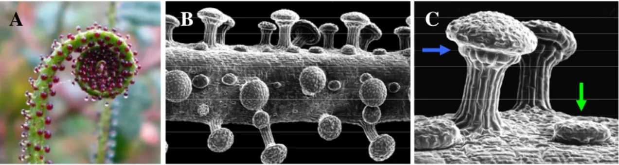
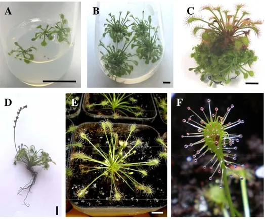
Drosera spp. (Droseraceae) use traps generically termed as fly-paper traps, which are partly passive in their action, consisting of highly specialized leaves bearing two types of glands, stalked glands which attract insects by their distinctive red coloured head and by their mucilage secretions rich in carbohydrates, and sessile glands which produce digestive enzymes (Figure 1.1.1). Rost and Schauer (1977) found that the mucilage of Drosera capensis is composed of a 4% aqueous solution of a complex polysaccharide containing xylose, mannose, galactose, and glucoronic acid with no protein and a pH around 5, at which value the viscosity is maximal. When prey adheres to a tentacle and struggles, there is an initial rapid movement of the individual tentacle, followed by the slower inflection of the leaf lamina itself ensuring that the prey is held. Leaves are induced by the presence of trapped insects to secrete enzymes for digestion of the prey (Matusikova et al., 2005). One to two weeks after prey has been caught, the leaf opens and the blackened remains, mostly chitinous pieces of legs and wings, fall off or blow away, allowing for the leaf to make repeated captures (Crowder et al., 1990). Other genera of carnivorous plants that use the same basic mechanism include Byblis (Bylidaceae), Drosophyllum (Drosophyllaceae), Pinguicula (Lentibulariaceae) and Triphyophyllum (Dioncophyllaceae) (Darnowski et al., 2006).
A
B C
Figure 1.1.1 - Example of specialized glands of a carnivorous plant using the fly-paper mechanism
(Drosophyllum lusitanicum): leaf extremity bearing distinctive red coloured stalked glands (A); Scanning electron microscopy of leaf (B) and detail of stalked (blue arrow) and sessile gland (green arrow) (C). (Photographs by S Gonçalves, used with permission).
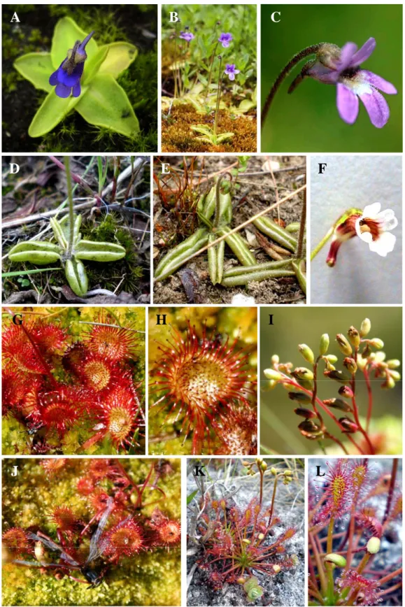
A considerable number of these carnivorous plant species are included in the pharmacopeias (Blumenthal et al., 1998), however, very few have been studied from a chemical and pharmacological perspective. In Portugal, three genera of terrestrial carnivorous plants are represented, the genus Drosera (D. rotundifolia and D. intermedia), Pinguicula (P. vulgaris and P. lusitanica) and Drosophyllum (D. lusitanicum). D. lusitanicum has been previously studied at the Plant Biotechnology Lab and besides the development of a micropropagation protocol (Gonçalves and Romano, 2005), extracts prepared from this species were investigated for their biological activities (Gonçalves et al., 2009) and chemical composition (Grevenstuk et al., 2008), and the encouraging results obtained incentivized the study of other carnivorous plant species. The distribution in Portugal of the plant species under study in this thesis are depicted in Figure 1.1.2.
1.3.2. The genus Pinguicula
1.3.2.1. Taxonomy and geographical distribution
Pinguicula is one of the three genera that together with Genlisea and Utricularia compose the Lentibulariaceae family. The genus Pinguicula consists of some 85 currently accepted species (Cieslak et al., 2005) which are present on all continents except Australia and in Africa is limited to the extreme north-west of the continent (Heslop-Harrison, 2004). The greatest concentration of Pinguicula spp. is in the humid mountainous regions of Central America and South America, where they probably originated as it is the centre of diversity. In Europe, 12 species were described by
Casper (1962) and nine species occur on the Iberian Peninsula, of which five are endemics (Blanca et al., 1999). The representatives of the genus in the Iberian Peninsula belong to the three subgenera into which it has been subdivided: subgenus Pinguicula, Isoloba and Micranthus. The Lentibulariaceae family is placed in the Lamiales order which has been supported by cladistic analysis (APG, 1998).
◦
▪
▪
•
P. lusitanica P. vulgaris D. rotundifolia D. intermedia•
◦
▪
▪
•
P. lusitanica P. vulgaris D. rotundifolia D. intermedia•
Figure 1.1.2 - Distribution of P. lusitanica, P. vulgaris (Blanca et al., 1999), D. rotundifolia and D.
intermedia (Crowder et al., 1990) in continental Portugal.
1.3.2.2. Biology, morphology and ecology
The Pinguicula spp., commonly known as butterworts, are herbaceous, relatively short-lived perennials (although occasionally behaving as annuals) and of rosette habit in active growth, while some overwinter as resting buds (hibernacula). As well as sexual reproduction by seed, many reproduce vegetatively by means of bulbils or buds which later take root. Unlike members of the other two genera in the family (Genlisea and Utricularia), all species of Pinguicula bear true roots, which are generally fibrous, tufted, and ephemeral. The leaves, which in most species lie appressed to the ground, are occasionally heterophyllous and the later formed ones may be larger and semi-erect (whereby the plant can tolerate more shaded conditions). The leaves are adapted for insectivory and bear stalked and sessile glands on the upper surfaces. The stalked glands carry permanent mucilaginous droplets giving the characteristic greasy feel and its
However, unlike the Drosera spp., the leaves of most Pinguicula spp. are sessile and therefore the capture system is entirely passive (Blanca et al., 1999). As with almost all carnivorous plants, the flowers of the butterworts are held far above the rest of the plant by a long stalk, in order to reduce the probability of trapping potential pollinators.
Pinguicula plants are restricted to nutrient-poor habitats, such as bogs and swamps, which remain sunny and moist at least during the growing season (Blanca et al., 1999). Despite the rarity of such sites in the Mediterranean ecosystems, many Pinguicula species are known from the Mediterranean basin (Casper, 1962). In these regions, where plant growth is greatly limited by water availability, suitable habitats for the Pinguicula species are scattered. Populations of the same species are often separated from one another by large distances and the isolation of the populations might have played an important role in the speciation processes. Since the current aridity of the Mediterranean basin has made small, isolated populations vulnerable to extinction, there is an urgent need to ensure the conservation of these species (Zamora et al., 1996).
1.3.2.3. P. vulgaris
Pinguicula vulgaris (L.) Linneaus, or the common butterwort, is a perennial plant consisting of a rosette of 4-7 leaves lying close to the ground, shallowly anchored by a tuft of fine, fibrous roots and overwinters as a hibernaculum (Figure 1.1.3 A). The leaves are bright, yellowish-green, and fleshy in texture with the margin somewhat involute and the upper surface covered with stalked glands. Each plant can produce 1-8 scapes in succession in the growing season which increases in length as the fruit develops (Figure 1.1.3 B). P. vulgaris produces violet, solitary, bisexual and zygomorphic flowers (Figure 1.1.3 C). Normally the flower assumes a horizontal posture at anthesis, but is occasionally held more or less erect (Heslop-Harrison, 2005). Besides seed formation, P. vulgaris also reproduces vegetatively by means of buds formed in the axils of the last foliage leaves of the season (Blanca et al., 1999). Plants produce flowers, after being grown from seed or bulbils, usually in their third year, i.e. after their second season of vegetative growth. Flowering and subsequent seed set are usually then of annual occurrence, if conditions are favourable. Seedling establishment in the wild is precarious because the tiny seed size provides negligible food reserves, and suitable wet sites free from competition by other species are rare. Although small in
number, the axillary buds probably provide an effective method of reproduction because these can draw on relatively large starch reserves stored in the bud scales (Heslop-Harrison, 2005).
P. vulgaris has a northern circumpolar distribution. The species is widespread in the northern and upland parts of Europe, extending into Corsica, Italy and Macedonia and across Siberia into north Asia but it thins out eastwards to Ukraine. Its most northerly limit is on the east coast of Greenland and southwards into central Spain and north Portugal. In North America it extends from Alaska in the north, as far south in the USA as northern New York State, the southern limit being roughly equivalent to that in Europe (Heslop-Harrison, 2005). It occurs mainly in seepage channels in the less acid parts of bogs, mires, calcareous fens and flushes, wet heaths and on wet rocks and seems to be indifferent to soil type (Blanca et al., 1999). A high humidity requirement during the growing season limits the number of suitable habitats available for the species, and it can survive only some degree of desiccation as a hibernaculum. Leaf extracts of Pinguicula spp. were found by early herbalists to be effective in giving spasmodic relief in cases of whooping cough, asthma, tuberculosis and spasms of intestinal pain (Christen, 1961; Hegnauer, 1966; in Heslop-Harrison, 2005). Biochemical data indicates that P. vulgaris produces iridoid glucosides (Damtoft et al., 1985; 1994; Marco et al., 1985; Section 3.1.2).
1.3.2.4. P. lusitanica
Pinguicula lusitanica (L.) is a small herbaceous plant consisting of a horizontal rosette of 5-12 leaves, lying close to the ground, shallowly anchored by a tuft of fine, fibrous roots (Figure 1.1.3 D). Unlike most species of Pinguicula in Europe, P. lusitanica belongs to the subgenus Isoloba and its morphological, vegetative and floral characteristics are closer to the species centred in the Gulf of Mexico, rather than to the other European ones (Casper, 1962). The leaves are oval or oblong-oval and sometimes the margins are inrolled exposing little of the lamina which is pale green with dark red veins and covered with stalked red-headed glands on the upper surface (Heslop-Harrison, 2005). In general, P. lusitanica plants flower successively over a period of months from May until August, but it can vary significantly according to climate as