Integrin alpha5beta1
and resistance to cancer therapies
Role of endocytosis in resistance to gefitinib
Departamento de Ciências
Biomédicas e Medicina
Departamento de Ciências
Biomédicas e Medicina
2016/2017
Integrin alpha5beta1
and resistance to cancer therapies
Role of endocytosis in resistance to gefitinib
Mestrado em:
Oncobiologia - Mecanismos Moleculares do Cancro
Trabalho efetuado sob a orientação de:
Professor Maxime Lehmann
Doutora Bibiana Ferreira
i
Titulo do trabalho:
“Integrin alpha5beta1 and resistance to cancer therapies: Role of endocytosis in resistance to gefitinib ”
Declaro ser a autora deste trabalho, que é original e inédito. Autores e trabalhos consultados estão devidamente citados no texto e constam da listagem de referências incluída.
Copyright Elisabete Cruz da Silva
________________________________________________
A Universidade do Algarve tem o direito, perpétuo e sem limites geográficos, de arquivar e publicitar este trabalho através de exemplares impressos reproduzidos em papel ou de forma digital, ou por qualquer outro meio conhecido ou que venha a ser inventado, de o divulgar através de repositórios científicos e de admitir a sua cópia e distribuição com objetivos educacionais ou de investigação, não comerciais, desde que seja dado crédito ao autor e editor.
ii “Education is not the filling of a pail but the lighting of a fire”
by William Butler Yeats
First, I would like to thank the greatest opportunity given by Professor Monique Dontenwill by accepting me into her team and allowing me to live this experience. To Professor Maxime Lehmann for all the time spent teaching me, for giving me the joy of sharing his knowledge, for giving me support in all moments and for being inspiring full.
To the whole team for their help and support mainly to Fanny Noulet. Also to the collaborators Romain Vauchelles and Oleksandr Glushonkov for their help in the experiments.
To all the Professors at the Master in Oncobiology - Molecular Mechanisms of Cancer for their support and knowledge given, specially to Doutora Bibiana Ferreira and Professor Doutor Alvaro Tavares that were my support in the university.
“The greatest gift of life is friendship, and I have received it” by Hubert H. Humphrey
From the oldest to the newest, thank you to all my friends, especially to Vanessa Henriques, Bastien Lambert, Marie-Cecile Mercier and Mickaële Hémono.
“Family is not an important thing, it is everything” by Michael J. Fox
For all the love, support and affection I would like to thank to my family. Because even with the distance, without them nothing would be possible!
iii Glioblastoma is an aggressive, invasive and resistant brain cancer. Despite the promisor role of EGFR, targeted therapies failed without giving insights about predictive factors. EGFR endocytosis and trafficking have been reported to influenciate therapy resistance. Integrin is known as a regulator of EGFR oncogenic activity during tumor progression by affecting its trafficking. Previous results from the team in cell evasion showed that α5 integrin depletion sensitizes cells to gefitinib treatment. For that, my main objective was to determine if the endocytic pathway is involved in the integrin-mediated resistance to gefitinib.
Endocytosis involvement on gefitinib treatment was evaluated by dynamin inhibition. Dynamin is GTPase involved in the fission of endocytic vesicles. There were performed cellular evasion and EGFR internalization assays under gefitinib treatment with or without dynamin chemical inhibitiors (dynasore and dyngo4a). We showed that endocytosis is involved in the gefitinib-mediated inhibition of U87 cell evasion regardless the α5 integrin expression level. Gefitinib induces ligand-bound EGFR internalization, being this impaired with the addiction of dynasore. EGFR and α5β1 integrin distribution were evaluated using immunofluorescence confocal microscopy. Gefitinib was shown to induce internalization of both receptors, being them founded co-localized inside of early endosomal vesicles. Gefitinib induced EGFR internalization occurs in U87 and others glioma cell lines independently of α5 level.
We postulate that α5β1 integrin may impact on EGFR trafficking and function during membrane trafficking after endocytosis confering resistance towards gefitinib treatment. There was already described a regulatory role of integrin in EGFR trafficking to promote carcinoma cell invasion, for that this role of integrin should be further evaluated.
In conclusion, endocytosis plays a relevant role in gefitinib treatment. EGFR trafficking gains here a strong evidence for its role in therapy resistance. Modulators of this
endosomal trafficking, such as α5β1 integrin can predict responsiviness towards EGFR targeted therapies.
iv
Graphical abstract: Dynamin inhibition enhances gefitinib resistance. Gefitinib
treatment induces EGFR internalization independently of integrin expression level. α5 integrin depletion sensitizes cells to gefitinib treatment, since there is no evasion under these conditions. When endocytosis is impaired, cells become resistant (with a more evading behaviour) to gefitinib independently of α5 level. α5β1 integrin may impact on EGFR trafficking and function during membrane trafficking after endocytosis confering resistance towards gefitinib treatment.
v Glioblastoma é um dos tumores cerebrais mais agressivos, sendo extremamente invasivo e resistente à terapia. Na sua progressão ocorrem diversas alterações génicas, sendo uma delas a amplificação e/ou mutação do gene erbB 1.
O gene erbB 1 codifica o receptor tirosina cinase EGFR, um receptor de sinalização que está envolvido em muitos processos celulares Incluindo a migração, constituindo um importante alvo terapêutico no tratamento do glioblastoma. No entanto, as terapias dirigidas não têm sido bem sucedidas em ensaios clínicos. De momento, não existem quaisquer marcadores predictivos que determinem o carácter responsivo de um paciente a este tipo de terapia. Em outros tipos de cancro, já se encontram descritos diversos mecanismos de resistência, sendo um deles a cooperação entre EGFR e outros receptores, como por exemplo com as integrinas.
As integrinas são receptores de adesão celular. Em particular, a integrina α5β1 desempenha um papel importante na progressão do glioma, sendo um descrito alvo terapêutico. Esta integrina encontra-se sobreexpressa em estadios mais avançados da progressão do glioma, estando associada a um pior prognóstico e a um carácter mais invasivo e resistente do tumor. As integrinas e receptores de factores de crescimento como o EGFR cooperam em diversos níveis. A regulação do trafego intracelular já se encontra descrita como um mecanismo de resistência a terapias.
Devido ao interesse do grupo no papel da integrina α5β1 na progressão tumoral do glioblastoma, foi previamente geneticamente manipulada uma das mais comuns linhas celulares de glioblastoma (U87). Esta manipulação teve como objectivo a sobre-expressão e a sub-sobre-expressão da integrina α5. Estas linhas celulares foram tratadas com gefitinib, um inibidor tirosina cinase especifico para o receptor EGFR, e foi avaliada a evasão celular. A evasão celular foi avaliada através da migração de células de um pequeno agregado celular (esferoide). As células com níveis acrescidos de integrina α5 demonstraram um comportamento resistente após o tratamento com gefitinib. Foi também verificada uma internalização do receptor EGFR após tratamento através da técnica de imunofluorescência.
Perante estes resultados os objectivos do meu trabalho foram a verificação do papel da endocitose no tratamento com gefitinib e a importância da integrina α5β1 neste fenótipo.
vi é composta por três isoformas homólogas mas com diferentes padrões de expressão. A dinamina 1 encontra-se apenas expressa em neurónios, a dinamina 2 tem uma expressão ubíqua e a dinamina 3 encontra-se expressa no cérebro e nos testículos. A dinamina está envolvida na fissão de vesículas endociticas, sendo esta função dependente da sua actividade como GTPase. Após a ligação de GTP, a dinamina polimeraliza em torno do pescoço da vesícula. A hidrólise do GTP altera a conformação da dinamina, levando a fissão da vesícula pela geração de forças. A inibição da dinamina foi provocada por dois inibidores químicos diferentes. Dynasore é um inibidor não competitivo da actividade GTPase de ambas dinamina 1 e 2. Dynasore interfere com a actividade catalítica da proteína. Dyngo-4a é um inibidor mais potente, embora seja mais selectivo para dinamina 1 do que para dinamina 2. Dyngo-4a inibe a atividade GTPase ao ligar-se ao domínio G responsável pela ligação e hidrólise ao GTP.
A evasão celular foi avaliada com o tratamento com gefitinib e com ou sem inibição das dinaminas. Observou-se uma diminuição da evasão celular após tratamento com gefitinib, verificando-se novamente um carácter mais resistente por parte das células que sobre-expressam integrina α5. A inibição da dinamina reverte completamente o efeito negativo do gefitinib em relação à evasão celular, independentemente do nivel da integrina α5. Observou-se ainda um aumento da evasão celular com os inibidores da dinamina mas apenas na presença de gefitinib.
O efeito do gefitinib e dos inibidores da dinamina na internalização do ligando EGF foi avaliado recorrendo ao uso de EGF acopolado com fluorforo. Pode verificar-se que o tratamento com gefitinib aumenta a internalização do ligando, sendo este aumento inibido com a inibição da dinamina. Este fenótipo é integrina α5 independente semelhante ao estudo da evasão celular.
Para se averiguar a importância da endocitose no tratamento com gefitinib, imunofluorescência foi efetuada com anticorpos anti-EGFR e anti-EEA1. EEA1 é um marcador de endosomo inicial, sendo este a primeira estrutura vesicular após endocitose/internalização. Foi demonstrado que, após tratamento com gefitinib, EGFR é internalizado e encontra-se em proximidade de EEA1. Esta co-localização aumenta com o tempo de incubação com gefitinib e é integrina α5 independente. Isto demonstra a importância da endocitose neste tratamento.
vii integrina β1. Foi demonstrado que após tratamento, EGFR é internalizado juntamente com as integrinas. Devido à limitada resolução da microscopia confocal, esta co-localização foi confirmada por microscopia de super resolução, mais propriamente microscopia estocástica de reconstrução optica. Deste modo, confirmou-se a proximidade destas duas proteínas dentro de estruturas endomembranares.
Para verificar se este fenómeno de endocitose ocorre transversalmente em diferentes linhas celulares de glioma foi realizada imunofluorescência usando anticorpos anti-EGFR e anti-integrina β1. Verificou-se que o EGFR foi internalizado em todas as linhas celulares, e que este fenótipo é independente do nível de expressão de integrina α5, visto que as linhas celulares que apresentam um fenótipo mais marcante são as que quase não expressam esta proteína. É de reforçar que nestas linhas celulares apenas foi averiguado a occorrência da internalização de EGFR e integrina β1. Para averiguar o carácter resistente ou sensitivo destas células ao tratamento com gefitinib é necessário futuramente realizar estudos de evasão celular. Além do mais, foram utilizadas as condições de tratamento de gefitinib usadas na linha celular U87. Não foram efetuados nenhuns estudos de toxicidade ou efetividades do gefitinib, de modo a determinar as condições ótimas de concentração e duração de tratamento.
Os resultados obtidos sugerem que a migração é independente da endocitose do receptor EGFR, visto que quando a endocitose do receptor é impedida a migração celular é aumentada. A internalização do receptor EGFR após tratamento com gefitinib aparenta ser uma forma de diminuir a sinalização do receptor. A influência da integrina α5 na resistência ao tratamento com gefitinib deverá ser independente da endocitose, provavelmente tendo um papel após a internalização do receptor Alguns estudos mostraram que as alterações no tráfego dos receptores bem como a promoção da sua reciclagem por parte das integrinas funcionam como promotores de tumorigénese e potenciam a progressão tumoral. No caso do glioblastoma ainda não se encontram descritas alterações no tráfego de receptores como promotores de progressão tumoral. Deste modo, este trabalho é um inovador ao demonstrar a importância da endocitose e do trafégo intracelular na resistência à terapia em glioblastoma.
viii Acknowledgements ... ii Abstract ... iii Resumo ... v Contents... viii Figures Index ... x Abbreviations ... xi 1. Introduction ... 1 1.1 Glioblastoma ... 1 Epidemiology ... 1
Risk factors for CNS tumors ... 1
Symptoms of GBM ... 1
Glioblastoma classifications and characterization ... 2
Glioblastoma Invasive behavior ... 6
Glioblastoma Treatment ... 7
Predictive and prognostic factors of GBM ... 9
Cancer initiating tumor cells ... 9
Effect of brain tumor in BBB permeability ... 10
Pre-clinical models of glioblastoma ... 10
1.2 EGFR... 13
EGFR and Glioblastoma ... 13
HER family ... 13
EGFR signaling pathway ... 15
Endocytic pathway of EGFR ... 17
Therapies against EGFR ... 19
Antibodies ... 19
Tyrosine kinase inhibitors ... 19
Secondary effect of EGFR therapies ... 22
Resistance to EGFR-targeted therapies ... 22
1.3 Integrins ... 25
Integrins Family ... 25
Integrin expression and function ... 26
Integrins signaling pathways ... 27
ix
1.4 Objective ... 33
2. Methods ... 34
2.1 Cell culture ... 34
2.2 Formation of tumoral spheroids ... 35
2.3 Preparation of methylcellulose solution ... 37
2.4 Immunofluorescence ... 37
2.5 Stochastic Optical Reconstruction Microscopy (STORM) ... 38
2.6Immunoblot Blot ... 39
2.7EGFR uptake assay ... 41
2.8Statistical Analysis ... 41
3. Results ... 43
3.1 Dynamin inhibition reverts negative effect of gefitinib in cell evasion ... 43
3.2 Endocytosis is important in gefitinib treatment ... 51
3.3 Gefitinib induces EGF uptake and dynasore impaired this phenotype ... 55
3.4 β1 integrin and EGFR co-localized in endomembranar strutures after gefitinib treatment...59
3.5 Gefitinib-induced EGFR and β1 integrin internalization occurs in others glioma cell lines ... 63
4. Discution ... 67
5. Conclusion ... 67
6. Bibliography references ... 78
x
Figure 1.1: Magnetic resonance image from a glioblastoma tumor ... 2
Figure 1.2:Diffuse astrocytic and oligodendroglial ... 3
Figure 1.3:Molecular changes in primary brain tumor progression... 4
Figure 1.4: Gene expression of the four subtypes of glioblastoma.. ... 6
Figure 1.5: Hypothetic model of GBM progression.. ... 7
Figure 1.6: Schematic of ErbB receptor structure and its dimerization and activation ... 14
Figure 1.7: Schematic of some EGFR signaling pathways involved in glioma progression. ... 16
Figure 1.8: Schematic of clathrin-dependent EGFR internalization and consequent trafficking.. ... 20
Figure 1.9: TKI mechanism of inhibition.. ... 21
Figure 1.10: Structure of ATP and Gefitinib. ... .. 22
Figure 1.11: Family of integrins. ... 26
Figure 1.12: Mechanisms of interaction between integrins and growth factor receptors ... 30
Figure 1.13: Loss of integrin α5 expression in glioblastoma cell line (U87) sensitizes cell to EGFR-targeted therapy (gefitinib) ... 33
Figure 2.1: Schematics of spheroid evasion assay ... 37
Figure 3.1: Dynamin struture and its role in membrane fission.. ... 44
Figure 3.2: GTPase function of dynamin in clathrin-mediated endocytosis.. ... 44
Figure 3.3: Cell evasion on U87 α5+ under treatment with dynasore and gefitinib ... 46
Figure 3.4: Cell evasion on U87 α5- under treatment with dynasore and gefitinib ... 47
Figure 3.5: Cell evasion on U87 α5+ under treatment with dygo-4a and gefitinib. ... 49
Figure 3.6: Cell evasion on U87 α5- under treatment with dygo-4a and gefitinib ... 50
Figure 3.7: EGFR is localized in early endosomes in U87α5+. ... 52
Figure 3.8: EGFR is localized in early endosomes in U87α5-. ... 53
Figure 3.9: Co-localization of EGFR and EEA1 increases with the gefitinib incubation time. ... 54
Figure 3.10: Negative (4°C) and Positive (37°C) controls of EGF uptake assay ... 56
Figure 3.11: EGF uptake after treatment with dynasore and/or gefitinib ... 57
Figure 3.12: Quantification of EGF uptake after treatment with dynasore and/or gefitinib ... 58
Figure 3.13: β1 integrin and EGFR are co-internalized after gefitinib treatment ... 60
Figure 3.14: α5 integrin and EGFR are co-internalized after gefitinib treatment in U87α5+ ... 61
Figure 3.15: β1 integrin and EGFR are co-localized after gefitinib treatment ... 62
Figure 3.16: Protein expression of α5 integrin and was analyzed by immunoblotting using GAPDH as a loading control. ... 63
Figure 3.17: EGFR and β1 integrin localization in absence (Control) and presence (Gef) of 20µmol.ml -1of gefitinib………..65
Figure 3.18: EGFR and β1 integrin localization in absence (Control) and presence (Gef) of 20µmol.ml -1of gefitinib.……….66
xi
A
ANOVA – Analysis of Variance AP-2 – Adaptor protein 2
APPL1 - Adaptor protein, phosphotyrosine interacting with PH domain and leucine zipper 1
ARF - ADP Ribosylation Factors ASCL1 - Achaete-scute homolog 1 ATCC – American Type Culture Collection ATP – Adenosine triphosphate
B
BBB – Blood-brain barrier
bFGF - Basic fibroblast growth factor BSA – Bovine serum albumin BTC- Betacellulin
C
CD - Cluster of differentiation CDK4 - Cyclin-dependent kinase 4
CDKN2A - Cyclin Dependent Kinase Inhibitor 2A CHI3L1 - Chitinase 3 Like 1
CNS – Central Nervous System CR - Cysteine residue CYP – Cytochromo P450
D
DAPI - 4',6-diamidino-2-phenylindole DCX – Doublecortin
DLL3- Delta Like Canonical Notch Ligand 3 DMSO - Dimethyl-sulfoxide
DNA - Deoxyribonucleic acid
DPBS - Dulbecco's phosphate buffered saline DRP1 - Dynamin-related protein 1
E
ECM - Extracellular matrix
EDTA - Ethylenediamine tetraacetic acid EEA1 - Early Endosome Antigen 1 EGFR – Epidermal growth factor receptor EMEM - Eagle's Minimum Essential Medium EPGN - Epithelial Mitogen
EPS - Epidermal Growth Factor Receptor Pathway Substrate
EREG – Epiregulin Gene
ERK - Extracellular signal-regulated kinases
ESCRT - Endosomal sorting complexes required for transport
F
FAK- Focal adhesion kinase FDA – Food and drug administration FRET - Förster resonance energy transfer
G
GABRA1 - Gamma-Aminobutyric Acid Type A Receptor Alpha1 Subunit
GBM - Glioblastoma
G-CIMP – Glioma CpG island methylator phenotype GDP – Guanosine diphosphate
GED - GTPase effector domain GFAP - Glial fibrillary acidic protein GFP – Green fluorescent protein GFR –Growth factor receptor GPCR - G protein–coupled receptors GPI - Glycosylphosphatidylinisotol GTP - Guanosine-5'-triphosphate
H
HB-EGF - Heparin-binding EGF-like growth factor HCl - Hydrochloric acid
HER - Human epidermal growth factor receptor HRP - Horseradish peroxidase
hTERT - Human telomerase reverse transcriptase
I
IC50 - Half maximal inhibitory concentration
IDH - Isocitrate dehydrogenase
IGFR - Insulin-like growth factor receptor ILK – Integrin linked kinase
J
JACOP – Just another co localization plugin
K
kDa – Kilo daltons
KRAS - Kirsten rat sarcoma viral oncogene homolog
M
mAb – Monoclonal antibody
MAP2 – Microtubule associated protein 2 MAPK - Mitogen Activated Protein Kinases MDM2 - Mouse double minute 2 homolog MEK – Mitogen-activated protein kinase kinase
MGMT - O6-methylguanine DNA methyltransferase
MIG6 - Mitogen-inducible gene-6 MMP - Matrix metalloproteinases mRNA – Messenger ribonucleic acid
MTIC-3-methyl-(triazen-1-yl)imidazole-4-carboxamide mTOR - Mammalian target of rapamycin
MTT-3-(4,5-dimethylthiazol-2-yl)-2,5-diphenyltetrazolium bromide MVE – Multi-vesicular endosome
xii
N
NEFL – Neurofilament light NF1 - Neurofibromin 1 NF-ƙb - Nuclear factor kappa B NOS – Nitric oxide synthase
NSCLC – Non-small cell lung carcinoma
O
OCT4 - Octamer-binding transcription factor 4 OLIG2 - Oligodendrocyte transcription factor
P
PI3K - Phosphatidylinositol-4,5-bisphosphate 3-kinase PBS - Phosphate buffered saline
PDGFR - Platelet-derived growth factor receptor PDX – Patient- derived xenograft
pH – Potential of hydrogen PH - Pleckstrin homology domain
PIQ - Plateforme d'Imagerie Quantitative
(http://imageriepiq.u-strasbg.fr) PLC- Phospholipase C PRD - PTS Regulation Domain
PTEN - Phosphatase and tensin homolog PTP1B - Protein-tyrosine phosphatase 1B PVDF - Polyvinylidene fluoride
R
RALT - Receptor-associated late transducer Rb1 – Retinoblastoma 1
RGD - Arginylglycylaspartic acid ROS – Reactive oxygen species rpm - Revolutions per minute RTK – Receptor tyrosine kinase
S
SDS - Sodium dodecyl sulfate SEM - Standard error of the mean
SLC12A5 - Solute Carrier Family 12 Member 5 SNP - Single nucleotide polymorphism SPRY2 - Sprouty RTK Signaling Antagonist 2 STAT – Signal transducer and activator of transcription STORM-Stochastic Optical Reconstruction Microscopy SYT1 - Synaptotagmin 1
T
TBS - Tris-buffered saline TCF4 - Transcription Factor 4
TCPTP - T cell protein tyrosine phosphatase TG - Tris-Glycine
TGF - Transforming growth factor TIC – Tumor initiating cell
TIMP - Tissue inhibitor of metalloproteinase TJ – Tight Junction
TK – Tyrosine kinase
TKI - Tyrosine kinase inhibitor TMZ - Temozolomide TNF – Tumor necrosis factor TP53 – Tumor protein p53
TRADD - Tumor necrosis factor receptor type 1-associated death domain
U
UK – United Kingdom
uPA – Urokinase- type plasminogen activator USA – United States of America
UV - Ultraviolet
V
VEGF – Vascular endothelial growth factor
W
1
1. Introduction
1.1 Glioblastoma
Epidemiology
In Europe, central nervous system (CNS) tumors are the 17th most common cancer type, with 57 100 new cases diagnosed in 2102. They are the 11th cause of cancer death, with around 45 000 deaths in 2012 (1). Glioblastoma (GBM) is the most common and most aggressive malignant tumor in CNS representing 20% of all the primary brain neoplasm. GBM are the highest-grade astrocytoma and remained essentially incurable. Despite numerous efforts, the overall 5 years survival rate does not exceed 5 years and median survival is around 15 month (2). GBM can appear anywhere in the brain, but their preferred localization is the supratentorial region, having edema surrounding the tumor (3). GBM is more prominent in men than women, having a peak of incidence between 45 and 70 years old (2).
Risk factors for CNS tumors
The risks factor associated with GBM development are ionizing radiation, decreased susceptibility to allergy and immune factors and genetic alterations. There are single nucleotide polymorphisms (SNP) that increase risk of GBM with inherited variation, in the chromosome 9p21 that contain cyclin-dependent kinase inhibitor 2B gene, and two SNPs in the regulator of telomere elongation helicase 1 (3,4).
Symptoms of GBM
The usual symptoms englobe headache, seizures, nausea, vomiting and hemiparesis. The diagnosis is made by cranial magnetic resonance imaging (fig.1.1) (3,4).
2 Figure 1.1: Magnetic resonance image from a glioblastoma tumor.
Adapted from (5)
Glioblastoma classifications and characterization
For decades, brain tumor classification was based on their histology and the microscopic similarities observed on light microscope after various coloration. This bring to the creation of groups of tumors that can be highly heterogeneous such as the astrocytoma or the oligodendrocytoma. In 2016, the World Health Organization (WHO) presented a new classification of CNS tumors based in integrated phenotypic and genotypic parameters (Fig.1.2) (6). Glioblastomas are now classified as grade IV diffuse astrocytic and oligodendroglial tumors. Glioblastomas are further segregated depending of their analysis of IDH status. IDH-wildtype GBM represent 90% of the GBM and the other 10% shared a genetic driver mutation on IDH1 and IDH2 genes. The evaluation of IDH status is made by R132H IDH1 immunohistochemistry and IDH sequencing. When IDH
3 evaluation cannot be performed, the GBM are denominated GBM NOS (not otherwise specified) (6).
Figure 1.2:Diffuse astrocytic and oligodendroglial tumours subcategorization in 2016 WHO Classification for CNS tumors. Adapted from (6)
But in this classification the subcategorization of GBM is only based in one molecular marker, the IDH status.
GBM can also be divided into de novo and secondary GBM, each characterized by different pathways (Fig.1.3). De novo GBM doesn’t have evidences of previous lesions, being 80% of all GBM and usually affects older patients (over 55 years old).
Genetically, de novo GBMs are characterized by HER1 amplification, PTEN mutations and p16 deletions. They are also IDH1 wild type and mutated in hTERT promoter. The chromosomal events underneath de novo GBM formation could be the amplification of 12q14 region, where are encoded the CDK4 and MDM2 genes, occurring then a disruption of p53 and Rb1 pathways; the homozygous deletion of 9p, where are encoded the genes
4 p16, p15 and p14 ARF; the loss of heterozygosity of 10q, where are encoded the gene PTEN. HER1 amplification is found in 40% of de novo GBM, followed by mutations that result in the constitutive activation of EGFR. PTEN mutation is found into 45% of de novo GBM, leading to a constitutive activation of PI3K/AKT pathway.
Secondary GBM affects young patients and develops from previously described low grade astrocytoma or anaplastic astrocytoma. In secondary GBM there are low grade genetic alterations, such as TP53, PDGF-A, Rb1, ATRX, and IDH1. (2,4,7,8).
Figure 1.3:Molecular changes in primary brain tumor progression. In orange there is represented the cell cycle alterations, in green the signaling pathways alterations and in blue the heterozygous alterations. Adapted from (7)
In a preclinical study, Verhaak et al described four subtypes of GBM (Proneural, Neural, Classical and Mesenchimal) based on gene expression, demonstrating a greater inter heterogeneity between patients. In figure 1.4, are represented the main genetic alterations that distinguish these subtypes. The proneural subtype is characterized by alterations of PDGFRA, points mutations in IDH1 and TP53 mutations. Focal amplification of PDGFRA is associated also with high levels of PDGFRA gene expression. Tp53 mutations were associated with loss of heterozygosity. Its signature also has proneural developmental genes (SOX, DCX, DLL3, ASCL1 and TCF4) and oligodendrocytic ones (PDGFRA, NKX2-2, OLIG2) (9). The neural subtype has expression of neuron markers
5 such as NEFL, GABRA1, SYT1 and SLC12A5 (9). Classical subtype has the typical GBM amplification of chromosome 7 and loss of the 10. Classical GBM are characterized by high expression levels of erbB1, without the presence of mutations on TP53. However, alterations in RB pathway with homozygous deletion of CDKN2A are found. In this subtype there are also expression of neural precursor markers NES, as markers from Notch and Sonic Hedgehog pathways (9). The classical subtype is more responsive to therapy (temozolomide/radiotherapy). Finally, the mesenchymal GBM are characterized by the focal hemizygous deletion at 17q11.2 leading to loss of NF1. The expression of mesenchymal markers (CHI3L1 and MET) is associated with epithelial-to-mesenchymal transition. The significant existence of necrosis and inflammation can be explained by the expression of genes from TNF and NF-Kb pathways (TRADD, RELB) (9).
Intra-tumoral heterogeneity is a hallmark of GBM. To explain this heterogeneity there are three theories: the clonal evolution, the cancer stem cell theory and interclonal cooperativity. In the clonal evolution, the initial cell suffers somatic alterations giving arise to different clones that are genetically unstable. From these clones, only the ones with the most aggressive behavior and less sensitive to therapy are favored and survive. In the cancer stem cell theory, only a group of cells have the ability to self-renew, continuous proliferation and ability to give arise to different clones. Numerous works describe that these cells are also resistant to therapy. In the interclonal cooperativity theory, the heterogeneity is due to the interactions between tumor cells and a changing microenvironment (immune cells, stromal cells and the extracellular matrix). For example, tumor evolution and self-renewing of glioblastoma cancer stem are affected by the hypoxic perivascular niche (9–11).
Intra-heterogeneity has a relevant clinical implication because it usually leads to therapy failure. So it should be important to perform the molecular analysis in distinct tumor biopsies from different parts of the tumor (10).
6 Figure 1.4: Gene expression of the four subtypes of glioblastoma. The heatmap represents three different sources of DNA, where DNA microarrays for the main genes that characterized each GBM subtype were analyzed. The results are represents in a gradient colorated scale when green means a loss of expression and red a gain of expression. Adapted from (9).
Glioblastoma Invasive behavior
GBM is a very locally invasive tumor. This migration capacity allows cells to develop tumor in the opposite hemisphere from the primary site or even multifocal GBM tumors (3).
Primary brain tumors have a unique pattern of invasion and rarely metastasize outside of the brain. Usually GBM cells, invade as single cells to almost anywhere in the brain. They infiltrate along blood vessel walls periphery, corpus callosum of neural fibers and astrocytes glia limitans externa. This invasion phenotype is not the same in tumors that metastasize to the brain, since these are more static, and when they invade it happens in group and only in short distances (13).
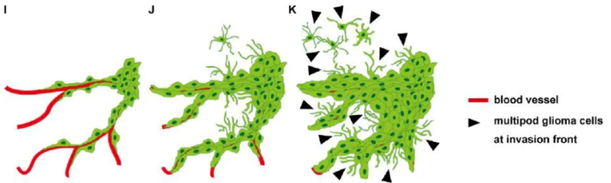
During evasion, cellular morphology changes (fig.1.5). When GBM cells migrate along blood vessels, they present a spindled shape with a single pseudopodium that extends toward the movement direction by polarization of actin polymerization. When they migrate through the brain parenchyma, they present multiple pseudopodia pointed in different directions. One of these directions will be chosen and be an invasion guide. GBM cells migrate using mesenchymal motility form that is dependent on the adhesion to the extra cellular matrix (ECM) and their remodulation. The invasion results then on
7 the combination of cell shape, position and tissue architecture, being needed PI3K signaling activation and also small GTPases. The pseudopodium interacts with ECM mainly through integrins and their focal adhesion complexes, acid hyaluronan receptor CD44. These two receptors have as main ligands proteins that are abundant in brain parenchyma as hyaluronan, collagen, fibronectin and laminin. ECM is remodeled by serine proteases, cysteine proteases and metalloproteases (MMP). In the serine proteases, the most studied is the complex urokinase-type plasminogen activator (uPA)/ uPA receptor that activates plasmin and degrades fibronectin and laminin. In the cysteine proteases, Cathepsin B is involved in laminin and collagen degradation and in GBM invasion. In GBM, the most important MMPs involved in cell invasion are MMP2 and -9. The inhibition of these MMPs leads to less migration and invasion in glioma cell lines and also in glioma cells xenografts. Tissue inhibitors of MMP (TIMP) modulates the proteases activity by forming complexes with them. Their addiction is reported to decrease cell invasion (14–18).
Invasion is a process with multiple involving factors that can be targeted in a way to treat GBM.
Figure 1.5: Hypothetic model of GBM progression. Two distinct cellular morphologies whether cells invade along the periphery of blood vessels (I) or through the brain parenchyma (K). Adapted from (19).
Glioblastoma Treatment
GBM treatment starts with surgical resection, followed by radiotherapy and chemotherapy. Until 2005, after surgical resection were performed a radiotherapy with adjuvant carmustine, a nitrosourea drug with alkylating function (4). A clinical trial (trial 22981/26981) showed that concomitant administration of temozolomide (TMZ) with radiotherapy, with adjuvant TMZ resulted in a better survival for the patient with minimal
8 levels of toxicity (20). Surgical resection is decompressive and cytoreductor, being associated with the increase of survival if there is complete resection of the tumor. To facilitate the tumor removal, a tumor fluorescence derived from 5-aminolevulinic acid is used to enhance the contrast of normal-tumor tissue (21).
The radiotherapy used in the study was a fractionated focal type, where occured a irradiation of 2 Gy/ fraction, once a day for five days/week, for a period of six weeks (total of radiation given to the patient was 60Gy) (22).
The drugs used to treat GBM must be able to cross the blood-bran barrier (BBB), so they must have a low molecular weight, high lipidic solubility and low ionization, and they also must have minimal protein binding capability (23). TMZ is an oral alkylating agent. It is a pro-drug that is spontaneous converted to the active metabolite, imidazole-4-carboxamide. It is able to methylate DNA, in N-7 or O-6 positions of guanine residues. TMZ is a small, lipophilic compound and so it is able to cross the BBB (24,25). TMZ is given concomitantly with radiotherapy for the following reasons:
- A daily administration of low doses has a greater intensity of activity without additional toxicity,
- After radiotherapy, the enzyme MGMT is activated and repairs the DNA damage. A continued administration of an alkylating agent such as TMZ depletes this enzyme.
- It was observed an in vitro synergetic effect by the concomitant use of TMZ and radiotherapy.
- TMZ was also chosen by its capability of crossing the BBB and the spontaneous conversion into the active metabolite (MTIC).
TMZ was administrated daily, all days of the week during radiotherapy, and for 5 days in the adjuvant six cycles that occurred during 4 weeks (22).
Even with treatment, the median survival rate is less than a year, between 9 to 15 months (26). After recurrence, the tumor is often different from the primary. The recurrent tumor doesn’t respond well to TMZ, and it also presents high expression levels of VEGF. In a clinical trial for recurrent GBM, combined treatment with bevacizumab (humanized IG1 monoclonal antibody for VEGF) and irinotecan (topoisomerase1 inhibitor) gives a survival rate of 7-9 months after treatment, similar to the conventional treatment (27).
9
Predictive and prognostic factors of GBM
Predictive factors/markers are used to evaluate the responsiveness to a treatment, with the objective of stratifying the patients according to the benefit or not from a specific treatment. Prognostic factors/markers are used to evaluate the overall outcome of the patients (28,29).
In GBM, there are good prognostic factors such as young age at diagnosis, cerebral location and maximal tumor resection. The Methylation status of the O6-methylguanine-DNA methyltransferase (MGMT) promotor, IDH1/2 mutation, erbB1 amplification, glioma-CpG island methylator phenotype (G-CIMP), TP53 mutation and losses of chromosomes are genetic prognostic factors. The MGMT is an enzyme that removes alkyl groups from the O6-guanine, producing a resistance to alkylating agents. After methylation of the MGMT promotor, this one is silenced and cells are incapable to repair the DNA damage and become more sensitive to TMZ. MGMT promoter methylation has then a prognostic significance. Besides this, MGMT promoter methylation also has a predictive one since it predicts tumor responsiveness to alkylating agents such as TMZ. The MGMT promoter is methylated in approximately 50% of GBM, being associated with IDH1/2 mutations (common in secondary GBM). In IDH1/2 mutations the more frequent ones are in IDH1 appearing mainly in secondary GBM. This mutation is associated with lesions with less necrosis and small areas of tumor, having then a more favorable prognosis. G-CIMP occurs in 10% of GBM, more common in secondary ones, being associated with IDH1/2 mutations (3,30). Mutations in ATRX cause alternative lengthening of telomeres, being present associated with IDH1/2 and TP53 mutations, mainly in secondary GBM. TERT mutation is most frequent in de novo GBM, being correlated with erbB1 amplification and a shorter patient survival (3,30). EGFR overexpression was associated with worse prognosis in younger patients bearing Tp53-wildtype tumors, while in older ones appears to have a better prognosis. So, TP53 mutations are not a definitive prognostic marker (3,30).
Cancer initiating tumor cells
Tumor initiating cells (TIC) are a subpopulation of cells in a tumor. These cells have some stem cell properties such as: renewing capability, unspecialized characteristics and capability to become into differentiated cells. To evaluate these stem cells properties, must be study the self-renewal properties and their capability to initiate a tumor (31,32).
10
Neural stem and progenitor cell are cell types present in the brain, expressing both CD133+. In Singhs et al study, CD133+ cells with stem cell properties in vitro were isolated from human brain tumors. CD133+ GBM cells represent a proportion between 3-30% of the tumor. These cells were capable to produce tumors in NOCID mice. These tumors resemble the human tumor in the expression of markers such as nestin, MIB-1, GFAP, MAP2. In the tumor obtained, were found CD133 positive and negative cells, and the CD133+ cells were different to MAP2+ cells. These data evidence that the CD133+ initiating cells could give arise to differentiated cells. These study showed that the hypothesis of TIC in GBM should be taken seriously, due to the fact that conventional therapies don’t kill them, having them the possibility to allow tumor progression or relapse (33). Sub-populations of TIC have high levels of SOX2, OCT4, and NANOG, all known to maintain self-renewal and cellular proliferation (30).
Effect of brain tumor in BBB permeability
Brain tumors increase BBB permeability by disruption of tight junctions (TJ) and due to increased angiogenesis. It is well known that GBM is characterized by a high level of angiogenesis. In GBM, vessels are tortuous and leaky. One growth factor responsible for angiogenesis, VEGF, is present in high levels in GBM. VEGF enhances endocytosis of VE-cadherin, and consequently there is a disruption of the endothelial barrier, increasing in this way also its permeability. Other factors produced in GBM, such as TGF-β2, caveolin-1, ROS and aquaporins, induce secretion and activation of MMPs that degrade TJ. It has been reported the loss of claudin3 and occludin in primary brain tumors that enhances disruption of BBB permeability. In GBM there are also increased levels of membrane transporters such as folate and insulin receptors that can facilitate the entry of molecules through the BBB. By another way, even with BBB permeability changed, others mechanism involved into the protection of chemical entry into the brain are intact. It has been shown that the expression of P-gp was not altered in GBM, remaining functional and limiting the brain diffusion of chemicals such as therapeutic drugs .
Pre-clinical models of glioblastoma
GBM cell culture is a useful tool to study cell processes before using tumor behavior in animal models. Usually, a cell culture has optimized conditions for proliferation and survival of the cells. The medium used supply the cells with all metabolites, growth factors and cytokines. But tumor cells in culture have unlimited oxygen and optimal pH,
11 and that is not the reality in vivo due a hypoxic microenvironment. Furthermore, in culture the cells don’t have a three-dimensional interaction with the other cells and matrix. To overcome this, there are spheroids models that mimetic it (37).
Due to selection pressure on cell culture, genetic alterations can occur and alter the genetic and phenotypic profile of the cancer cell lines in comparison to the original tumor (38). After a study between solid primary GBMs and GBM cell lines were identified 160 proteins gained and 60 proteins lost in culture, losing then the GBM heterogeneity and making difficult any comparation between in vitro and in vivo. One of this lost is EGFR overexpression (27–31).
The GBM cell lines are obtained from human brain diagnosed with GBM, astrocytoma grade IV. After the excision of the tumor, some cells are put into petri dishes. Here most of the cells die, and the ones which survived after a few passages become into an immortalized cell line. For glial cells, Pontén and Macintyre in 1968 were the first ones to optimize the culture conditions (39).
One of the usual GBM cell lines is U87. Genetically, U87 is hypodiploid human cell line, that easily forms tumors when injected in mice. These tumors have a huge vessels network (40).
The use of tumor initiating cell lines that are maintained in serum free conditions with growth factors (PDGF, bFGF, EGF) and growth as tumor spheroids are able to retain the tumor phenotype and tumor initiating capacity. But it was shown that growth of tumor initiating cells in adherent culture maintains highly pure stem cells populations (41,42).
Patient derived xenografts (PDX) are a tool to improve pre-clinical studies, since the tumor cells grow in an in vivo environment. PDX can be made using fresh tumor samples or cryopreserved ones (tissue cryopreserved at low temperature right after tumor excision). The single cell suspension can be implemented in the brain (orthotropic) or in the mice flank (heterotopic). But that was reported that heterotopic PDX do not demonstrate a local invasive profile compared to the orthotropic ones. This different can be explained by the different microenvironment in the two cases. But even in orthotropic xenograft, the murine brain microenvironment is different molecular and functionally from the human. The xenograft should be done in mice lacking immune system, since it was demonstrated that residual active immune system prevents tumor formation (43,44).
12 As we can see, GBM is a malignant and higly resistant tumor. Its biology needs to be better studied. One interesting therapeutic target in GBM is EGFR, since it is founded overexpressed in 40% of the cases. EGFR is a signaling receptor that is involved in the main signaling pathways that leads to cell migration and invasion, which are typical in GBM.
13
1.2 EGFR
EGFR and Glioblastoma
The discovery of EGFR in malignant transformation was made in the 80’s by oncogenic viruses that showed EGFR as a cellular homolog of the avian erythroblastosis virus v-erbB oncogene (45).
The EGFR signaling network is critical for tumor progression because it promotes cancer cell survival, growth and invasion. In GBM, erbB1 the gene encoding of the EGFR/ErbB1 protein is amplified is 40-60% after gene rearrangement and/or focal amplification. This amplification is often associated with mutations. These mutations can lead to ligand independent activity of the receptor, and are also reported to enhance motility and invasion by inducing genes of the extracellular matrix, metalloproteases and serine proteases. erbB1 mutations in GBM are common, and they can be N-terminal truncation (EGFRvI), deletion of exons 14 – 15 (EGFRvII), deletion of exons 25 – 27 (EGFRvIV), C-terminal truncation (EGFRvV) and C-terminal duplications and truncations. The most common mutation in GBM is EGFRvIII (occured in more than 50% of the GBM cases). EGFRvIII is a truncated protein due to loss of exons 2-7, that gives arise to a 801 base pair deletion. The amino acids 6-273 are replaced by a glycine residue, and so the resulted protein is a 145 kDa glycoprotein with constitutive, ligand-independent activation. The constitutive activation is due the reduced interaction with E3-ligase Cbl, leading to a reduced degradation of the receptor which a negative feedback regulation system. EGFRVIII is also occurs in lung, breast, ovarian and prostate cancers (37–39,41,42).
As described above, EGFR overexpression has a relevant role in GBM progression. EGFR is a signaling receptor that is involved in diverse cellular processes, like cell migration and invasion (characteristics of GBM).
HER family
EGFR belongs to a family called HER family, which has four transmembrane receptors: EGFR (HER1), ERBB2 (HER2), ERBB3 (HER3) and ERBB4 (HER4) (50).
14 The EGFR is a 170 kDa glycoprotein, with 1186 amino acids that is composed by three main domains: an extracellular ligand-binding domain (ectodomain), a hydrophobic transmembrane domain and a cytoplasmic tyrosine kinase domain (46,47,50).
Figure 1.6: Schematic of ErbB receptor structure and its dimerization and activation. A – Schematic of ErbB receptor showing the different parts: ectodomain (L1,L2 – leucine-rich domains, CR1, CR2 – cysteine rich domains), transmembrane domain, tyrosine kinase domain and c-terminal tail. B- Schematic of dimerization and receptor activation: i- receptor in inactive state, ii- ligand binds to L1 and L2 changing receptor conformation, iii- receptor dimerize through cysteine domains, creating docking sites in tyrosine kinase domain, iv- ErB2 in is inactive state has the same conformation that the rest active receptors. Adapted from (47). The ectodomain contains four subdomains: two leucine-rich subdomains (in figure 1.6 L1 and L2) and two cysteine-rich subdomains (in figure 1.6 CR1 and CR2). The leucine domains directly bind to ligand, while the cysteine domains are involved in interaction and dimerization with others receptor. The leucine domain is different between the family members, giving to them different ligand specificity.
While, the cytoplasmic domain is a highly conserved bilobed tyrosine kinase. Only HER3 does not have kinase activity. Between the two lobes there is a ATP binding site. The activation of the receptor by ligand binding (between L1 and L2) creates an extended conformation. This expose the dimerization loop present in CR1, allowing dimerization. In this moment, occurs the interaction between the N-lobe of one domain with the C-one of another, creating phosphorylated binding sites as docking sites (46,47,50).
The EGFR known ligands are EGF, TGFA/TGF-𝛼, amphiregulin, epigen/EPGN, BTC/betacellulin, epiregulin/EREG and HBEGF/heparin-binding EGF. Some of them are cell membrane anchored proteins that are proteolytically disrupted to become soluble molecules that will induce EGFR activation. Their cleavage by metalloproteinases can be
15 activated by GPCR. The ligands can be overexpressed by active Ras or steroid hormones (47,50). EGFR can also be activated by ligand-independent mechanism. Ligand-independent activation can be induced by unphysiological stimuli (such as oxidative stress, UV, and irradiation), by others RTK (such as MET, IGFR) or by GPCR and adhesion receptors like integrins (51). In GBM, MET is also found dysregulated, being this a possible cause for anti-EGFR therapy resistance. EGFRvIII is described to be a activator of Met, and so the dual treatment is shown to reduce tumor growth (52,53).
EGFR activation is attenuated by tyrosine dephosphorylation of active receptor, by phosphatases such as density-enhanced phosphatase-1 and PTP1B. Their catalytic activities eliminate the sites in which signaling intermediates or adaptor proteins would bind and promote cell signaling (47). However little is known about the involvement of PTP1B in glioma progression and invasion.
EGFR signaling pathway
Receptor homo- and/or heterodimerization occurs after ligand binding, followed by activation of the tyrosine kinase activity with consequent tyrosine autophosphorylation on the cytoplasmic specific residues (Fig.1.6) (47,50).
These phosphorylated residues become docking sites for adaptor proteins such as Grb2 (binds to pY1068 and pY1086) or Shc (binds to binds pY1148 and pY1173), that can activate RAS/Raf/MAPK downstream signaling cascade (45,47). MAPK pathway is activated through the interaction between the Grb2 and SoS, leading to proliferation, migration, angiogenesis and differentiation. p38-MAPK in GBM is linked to invasion and angiogenic phenotypes (54,55). Its inhibition leads to decreased tumor growth in glioma xenografts (56). MAPK pathway is also involved in regulation of neural stemness (57). EGFR activation leads to the stimulation of the PI3K/Akt pathway through the recruitment of the regulatory subunit p85. Whereas EGFR and ErbB2 receptors bind indirectly to p85 through adaptor proteins like Gab1, ErbB3 and ErbB4 directly bind to p85 (47,51). PI3K pathway is often dysregulated in GBM, since their negative regulators are frequently mutated, for example the loss of PTEN is founded in 45% of GBM cases. Studies targeting signaling pathways of this cascade such as mTOR are showing regression in GBM (58,59). In GBM, p85 can be also activated by direct interaction with cancer stem cell marker CD133, leading to PI3K activation (60).
16 All ligands and receptors of the family can activate the signaling cascade like Ras/Raf/MEK/MAPK, PI3K, PLC- γ and STAT. These signaling pathways are involved in glioma progression (fig.1.7).
The EGFR TK domain has numerous substrate proteins like STAT family members. STAT3 binding to activated EGFR leads to STAT3 dimerization and translocation into the nucleus (46,47,51). In GBM, the co-expression of EGFR and the mutant EGFRvIII activates STAT3/5 leading to glioma progression. EGFR phosphorylates the mutant receptor, allowing its nuclear entry where it forms a complex with STAT3. This study shows that EGFR not only function as a signaling receptor but also as a transcription factor (61). EGFR has a tyrosine residue (Tyr 992) that allows direct interaction with PLC-γ leading to actin reorganization and asymmetric motile phenotype (45,46). In GBM, was reported that the activation of PLC-γ and STAT3 leads to migration and invasion (62).
Figure 1.7: Schematic of some EGFR signaling pathways involved in glioma progression. After EGFR activation and auto phosphorylation are created docking sites for signaling molecules. EGFR interacts with Grb2 mediator to activate MAPK pathway. PI3K pathway is activated on the plasma membrane and the signal is controlled by PTEN. PTEN is usually mutated in GBM. EGFR can also activate directly STAT3 and PLC-γ pathways
17
Endocytic pathway of EGFR
Internalization of the EGFR has contradictory results. Endocytosis of EGFR constitutes a regulatory mechanism of this signaling pathway, having different described functions. The internalization can attenuate the signal leads to receptor degradation, or also allows the continuation of the signal by endosomal signaling and/or recycling of the receptor back to the plasma membrane. Indeed, EGFR membrane trafficking is required for activation of specific transducers and has been shown to play critical role in cancer cell invasion, as described below. Two main steps compose the endocytic pathway: the internalization of the receptor and its trafficking in intracellular compartments (Fig.1.8) (63,64).
The internalization controls the levels of receptors that are present at the plasma membrane, regulating their accessibility to ligands.
There are described diverse endocytic pathways that lead to EGFR internalization, being the most important the clathrin-dependent pathway. Clathrin-independent pathway was described to be used in high levels of EGF environment. Also the internalization pathway can be chosen by the type of ligand bound to the receptor: EGF and TGFα induces clathrin-dependent pathway, while HB-EGF a clathrin-independent pathway. Inactive EGFR is usually associated with caveolae rafts. After ligand binding, EGFR moves out from there, dimerize and enter into the cell by clathrin-dependent endocytosis. The binding of Grb2 to EGFR allows its ubiquitination by E3 ubiquitin ligase Cbl on pY1045. Once ubiquinated, EGFR interacts with Eps15, allowing the binding to the clathrin-pit through AP-2 (65,66). The ubiquination of EGFR promotes its lysosomal degradation. There are also negative feedbacks produced after EGFR activation that lead to signal attenuation, such as the production of suppressor of cytokine signaling-5 (binds to EGFR and promotes its degradation), Sprouty-2 (modulates Ras/MAPK pathway), LRIG-1 (promotes Cbl binding and EGFR ubiquitination) and Mig6/RALT (binds to TK domain αI helix of EGFR, inhibiting its catalytic activity) (45,47,66). Sprouty-2 is a novel therapeutic marker in GBM, being usually expressed in commitment with EGFRvIII (67). Mig6 is a tumor suppressor that was described to regulate EGFR trafficking and suppress glioma progression (68).
18 After receptor endocytosis, this enters in a system of intracellular vesicles called endosomes. The first ones are the early endosomes where occurs the sorting of the receptor: unbounded receptor recycled quickly back to the plasma membrane, while ligand-bound receptor recycled slowly or are degraded in lysosomes (Fig.1.8) (64,69). Ligand dissociation can influenciate the fate of the receptor. The endosomal pH influenciates ligand dissociation since EGF remains bound to the receptor while TGFα dissociates. Because of that EGF leads to EGFR degradation and TGFα to EGFR recycling (70).
Ubiquitinated EGFR is sorted in the early endosomes by ESCRT machinery to intraluminal vesicles of maturating endosomes. From here the receptor is taken to lysosomal degradation (45,65).
Recycling of the receptor can occurs by the short loop (controlled mainly by Rab4) or by the long loop (controlled by Rab 11) (65).
Dysregulation in receptor trafficking has been described as a tumorigenesis promotor. Defects that leads to poor downregulation are associated with enhanced signaling. One described mechanism is the sustained PI3K signaling due the loss of SPRY2 that leads to EGFR/HER2 internalization and early endosomal signaling in a PTEN-dependent manner. This leads to proliferation and invasion in prostatic cancer (71). The interaction between EGFR and HER2 also overcomes ubiquitinated signaling attenuation, leading o recycling of the receptor (45,46). Increased EGFR recycling was described a mechanism that drives hepatocellular carcinoma metastasis (72).
Trafficking dysregulation involved in GBM progression has not been described so often. There was described a overexpression of NHE9 (Na(+)/H(+) exchanger) and its involvement in stemness, therapy resistance and invasion in GBM. NHE9 limits the luminal acidification of endosomes, promoting EGFR recycling and consequently signaling continuation (73).
EGFR endosomal signaling has also been reported. It was described AKT signaling in early endosomes through the APPL1, a Rab 5 effector. Rab 5 is the main characteristic protein of early endosomes. And also p38 MAPK sustain early endosomes signaling by promoting clathrin-mediated EGFR endocytosis and degradation evasion (65).
19
Therapies against EGFR
Since EGFR is involved in tumor progression, it becomes a promise target for therapy in hope to eradicate the tumor. There are different approaches used in target therapy against EGFR: the use of antibodies that block ligand binding or the use of small molecules that inhibit tyrosine kinase activity of the receptor (47,50).
Antibodies
The antibodies used against EGFR bind to the extracellular domain, induce the internalization of the receptor to try to inhibited the signaling pathway, but also is a potential stimulator of the immunological response. One of the most known is cetuximab, a chimeric antibody with high specificity for EGFR. Cetuximab is an EGF antagonist, and so it competes for the natural ligand-binding sites, preventing ligand binding and receptor activation. It was approved by FDA on February 2004 for colorectal cancer treatment. Panitumumab is a fully humanized antibody against EGFR, approved by FDA on September 2006 for treatment of colorectal cancer with KRAS wildtype. Both antibodies are given by intravenous injection (47,50).
Tyrosine kinase inhibitors
The small molecules called tyrosine kinase inhibitors (TKI) are synthetic molecules with low molecular weight, almost all are quinazoline-derived and they bind to intracellular domain of the receptor through a hydrogen bond (fig.1.9). TKI are homologous to adenosine triphosphate (ATP), competing for the ATP-binding domain of kinases (fig.1.10). In this way, TKI prevent the EGFR autophosphorylation, the activation of tyrosine kinase and the signaling pathway.
20 One of the most known TKI is gefitinib that reversibly inhibits the TK activity of isolated EGFR with an IC50 in the nanomolar range. But in vivo, higher concentrations are required to block EGFR due the presence of intracellular ATP. It should be considered that in higher concentrations, gefitinib not also inhibit EGFR but also others RTK such as erbB2. Gefitinib has a half-life of about 28 hours. For that in clinic, gefitinib is administered daily, in a dose around 600mg/day. Gefitinib is metabolized by cytochrome P450 3A4 (CYP3A), being the inter-variability of this enzyme (expression and activity) one of the
21 reasons for different susceptibility to treatment. Gefitinib upregulates p27 (cell cycle inhibitor) and downregulates c-fos (transcription factor) and so gefitinib leads to a cell cycle arrest in G1 phase. In clinic, gefitinib is being used for treatment of locally advanced and metastatic non-small cell lung cancer (NSCLC) harboring EGFR-activating mutations (47,50,74–80).
Others TKI are also being used in clinic: erlotinib, an EGFR inhibitor, is being used in metastatic NSCLC and pancreatic cancer; lapatinib, an EGFR/erbB2 inhibitor, is being used in advanced or metastatic breast cancer. (81,82).
Figure 1.9: TKI mechanism of inhibition. a - Schematic of activated RTK bounded to ATP, b - inhibition by TKI (KI) that competes for ATP binding site and forms a hydrogen bond with the receptor. Adapted from (83).
22
Secondary effect of EGFR therapies
Both ways of EGFR targeted therapy cause skin rash and diarrhea, due to direct biological effect of EGFR inhibition. So they are considered predictive factors of response to therapy. They have also a warning for pulmonary toxicity, in which cases 1% is fatal. And it should be controlled also the hepatic toxicity due to increase of liver transminases, normally asymptomatic (50).
Resistance to EGFR-targeted therapies
Despite the development of various therapeutic strategies and new compounds, the results of anti-EGFR targeted therapies remain somehow discouraging. Most of the tumors are resistance to therapies or relapse after a short period. In solid tumors, there is well documented mechanism of resistance to EGFR-targeted therapies. Some of these mechanisms are the acquisition of secondary EGFR point mutations and alterations or
ATP
Figure 1.10: Structure of ATP and Gefitinib. ATP has adenine ring that is encircled and it is responsible for forming hydrogen bonds with the ATP-binding site on RTK. Gefitinib has a quinazoline group, where is founded the atom (indicated with red arrow) that form hydrogen-bonds with the kinase residues. Adapted from (79,80).
23 redundancy in the signaling pathways. In NSCLC, the mutation T790M in exon 20 and the amplification of MET, another tyrosine kinase receptors are predictive biomarkers for a non-responsive profile of the patients to EGFR-targeted therapies. In colon carcinoma, the activating KRAS mutation leads to resistance and only patient bearing KRAS-wild type tumor are eligible to the cetuximab treatment (47,50,75).
Glioblastoma are refractory to EGFR targeted therapy and the resistance mechanisms are still not understood, and there is no predictive marker (49,81,83,84). Even if preclinical studies using anti-EGFR therapies in GBM were promising, the reality in clinical trials was very different, with no beneficial results compared with others therapies. Related to TKI, phase I/II of erlotinib as a single agent demonstrated promising disease control and response rate, while for gefitinib it seems to have clinical activity but no improvement in survival. The ability of TKI to enhance radiation sensivity observed in vitro, were not demonstrated in any clinical study. Related to EGFR antibodies, clinical trials using cetuximab or nimotuzumab in monotherapy or with other treatments showed a small or none activity and response rate (table 1.1). Another antibody (mAb 806) that targets the normal and the mutated receptor have promising pre-clinical results and seems having tolerance in phase I trials (81,85,86). Some of probable causes for resistance in GBM are compensatory activation with other ErbB family, tumor initiating cells, tumor heterogeneity, the BBB and the structure of the new vessels.
24
The BBB is a greater barrier against anti-cancer agents that were administrated systemically. The compounds to penetrate BBB should present higher lipophilicity. The tortuous vasculature also limits the drug penetration into the brain (83,87). The amount of gefitinib found in the brain is different among several studies. In NSCLC patients with brain metastasis, the ratio of gefitinib between cerebrospinal fluid and plasma was only 0.3-1.3% (88). This is in agreement with the prediction of low capacity to penetrate BBB since gefitinib is highly water-soluble. Also gefitinib is a substrate of the p-glycoprotein efflux pump that is expressed in brain tumors. But in some studies, there were observed penetration of gefitinib into the brain (89,90). This can be explained by the altered BBB integrity and the low level of CYP3A in brain (81,91) .
The tumor initiating cells (TIC) tend to be resistant to therapy by altering the checkpoint and DNA repair pathways. These cells are being associated with the presence of multiple drug resistant transporters. The existence of this transporters lead to the efflux of drugs, reducing their concentration in the brain (83). The activation of others receptors of the ErbB family were demonstrated as a mechanism of resistance for EGFR-targeted therapy in glioblastoma TIC (84).
Intra tumor heterogeneity is normally one of the most common reasons for therapy resistance. The heterogeneity can lead to redundancy and cross talk between signaling pathways. Besides EGFR overexpression, can also occur the mutation and amplification of others RTK and GPI-linked receptors (IGFR1, MET, PDGFR α/β, uPAR) which signaling compensates the EGFR inhibition. It has also been proposed that deletion of the tumor suppressor gene PTEN, leading to the continuous activation of PI3K pathway, prevent inhibition of EGFR signaling pathway by TKIs (81,83). In retrospective analysis association of EGFRvIII and wild-type PTEN was reported as a significant predictor of TKI response in GBM. But this predictive value was not observed in others studies (49,92,93).
Dysregulation of receptor trafficking can be stress-induced or a way to overcome cell death as described above. This dysregulation promotes tumor progression and can be also a mechanism of therapy resistance. But still few is known about that and so require further investigation (65,94,95).
25 As we can see there are molecular markers that predict the insensitivity of lung and colon tumors to EGFR targeted therapies. But in GBM there are not yet such biomarkers that can explain the failed clinical trials using first generation of EGFR TKI such as erlotinib and gefitinib (93).
The causes for resistance to EGFR therapies in GBM are unknown, but preclinical data in other cancer types correlate the crosstalk and bi-directional regulation of EGFR and integrins as one possible mechanism of therapy resistance (85,96–98).
1.3 Integrins
Integrins Family
Integrins are a family of heterodimeric cell surface receptors composed by non-covalent association of alpha and beta subunits. The family is composed by 18 α subunits (that determine ligand binding specificity) and 8 β subunits (that connects to cytoskeleton and signaling molecules), forming 24 different receptors that each binds to one or more ECM ligands (fig.1.11).
There are four families based on the evolutionary history of α subunits. The first group is composed by integrins that recognize the arginine-glycine-aspartic acid (RGD) motif on their ECM ligands such as fibronectin, vitronectin or fibrinogen. The second group is formed by laminin binding integrins. In this group, there is the special β4 that has a large intracellular domain and so function as a docking site due to its phosphorylation sites. The last two groups (leukocyte integrins (β2) and collagen integrins (β1) receptors) come from the same large group of integrins that are structurally different from the others groups due to an extra domain in their α subunit (96,99).
26 Figure 1.11: Family of integrins.Adapted from (93)
Integrin expression and function
Integrins are expressed in all cell types, however the profile of integrin vary from cell type to cell type. Integrins and their ligands are important in the early stages of embryonic development including in fertilization, implantation and in blastula formation. β1 integrin is one of the most important, since its homozygous knockout leads to early death of the embryo. It can be due to the fact that β1 is present in 12 of 24 combinations of integrins (99,100).
In the physiological development, maintenance and remodeling of tissues, stem cells have an important role. Stemness is regulated by signals from the stem cell niche microenvironment such as the ECM. In here, integrins have an important role, mainly β1 that is highly expressed in stem cells to maintain stemness and control the balance between renewal and differentiation. In this context, the role of integrins in cell fate is to give spatial cues to the cells, due to the interaction with ECM and their mechanosensor activity, while the temporal cues are given by growth factors (100,101).
Integrins regulate cellular processes such as survival, proliferation, differentiation, migration, adhesion, apoptosis, anoikis, polarity and in stemness (96–99).