http://dx.doi.org/10.1590/s2175-97902018000100139
Review
*Correspondence: D. N. Marreiro. Departamento de Nutrição, Universidade Federal do Piauí, Campus Ministro Petrônio Portela, Bairro Ininga, 64049550 - Teresina, Piauí, Brasil. Telephone: +55 (86) 999915019 / +55 (86) 988459778/ Fax: +55 (86) 32371812. E-mail: dilina.marreiro@gmail.com
The role of selenium in insulin resistance
Larissa Cristina Fontenelle
1, Mayara Monte Feitosa
1, Jennifer Beatriz Silva Morais
1,
Juliana Soares Severo
1, Taynáh Emannuelle Coelho de Freitas
1, Jéssica Batista Beserra
1,
Gilberto Simeone Henriques
2, Dilina do Nascimento Marreiro
1*
1Department of Nutrition, Federal University of Piauí, Campus Minister Petrônio Portela, Teresina, Piauí, Brazil, 2Department of Nutrition, Federal University of Minas Gerais, Belo Horizonte, Minas Gerais, Brazil
In recent years, there has been growing interest in clarifying the pathogenesis of some chronic diseases, such as obesity and type 2 diabetes mellitus. Metabolic alterations in these diseases are characterized by chronic hyperglycemia and insulin resistance. Studies have demonstrated the participation of minerals in the pathogenesis of insulin resistance, more specifically their involvement in the synthesis and regulation of insulin. Selenium is an anti-inflammatory and antioxidant micronutrient that is essential for the activity of selenoproteins. Two selenoproteins (glutathione peroxidase and selenoprotein P) are known to be involved in the insulin signaling pathway. The aim of this review is to provide an update on the role of selenium in insulin resistance mechanisms. Evidence shows that adequate concentrations of selenium play a key role in the secretion and action of insulin, but an excess of selenium in the body is associated with the pathogenesis of insulin resistance and the development of diabetes mellitus.
Keywords: Diabetes mellitus. Selenium/insulin resistance. Insulin.
INTRODUCTION
In recent years, the pathogenesis of some chronic diseases has attracted interest, such as obesity and type 2 diabetes mellitus. Metabolic alterations in these diseases are characterized by chronic hyperglycemia and insulin resistance (Rains, Jain, 2011; Steinbrenner, 2013).
Recent studies have attempted to elucidate the molecular alterations involved in the development of insulin resistance. These include reductions in the number and activity of relevant receptors in peripheral tissues, and changes to intracellular insulin signaling pathways (Tencerova et al., 2015; Winnier et al., 2015). Accordingly, minerals have been the subject of several studies, focusing specifically on their involvement in the synthesis and regulation of insulin (Cruz et al., 2014; Garcia et al., 2013).
Selenium is an anti-inflammatory and antioxidant micronutrient essential for the activity of selenoproteins, such as glutathione peroxidase (GPx) and selenoprotein P (Sepp). These enzymes are also involved in insulin
regulation (Cominetti et al., 2011; Steinbrenner et al., 2013; Wei et al., 2014; Zou et al., 2016). Several studies have revealed selenium to be an insulin-mimetic, because it plays roles in the regulation of enzymes in the insulin signaling cascade, the expression of lipogenic enzymes, and in carbohydrate metabolism in the liver (Chen et al., 2015; Iizuka et al., 2010; Mao, Teng, 2013).
However, it has been emphasized that chronic ingestion of large amounts of selenium promotes the formation of antioxidant selenoproteins, leading to reduced concentrations of reactive oxygen species, which are required in physiological amounts for insulin signaling (Labunskyy et al., 2011; Wang et al., 2014; Zhou et al., 2015).
Given that insulin resistance is a key metabolic disorder that can result in the development of chronic diseases such as type 2 diabetes mellitus, and that selenium participates in the prevention of this disease, we have reviewed the participation of this micronutrient in insulin resistance.
Etiologic factors of insulin resistance
liver, and glucose uptake by muscle and adipose tissue. In myocytes and adipocytes, insulin regulates glucose transport by controlling the translocation of a glucose transporter (GLUT4) to the plasma membrane (Bruce, Hanson, 2010; Kim, Feldman, 2015).
The insulin receptor is a tetramer composed of two extracellular α-subunits and two transmembrane β-subunits. The action of insulin is initiated through binding to the α-subunits, which alters the conformation of the β-subunits. This results in autophosphorylation and activation of its tyrosine kinase function. The β-subunit can autophosphorylate, and it can phosphorylate other proteins or intracellular substrates, such as insulin receptor substrate 1 (IRS-1), insulin receptor substrate 2 (IRS-2), and the Src homology collagen (Shc) family of proteins (Hirabara et al., 2014; Litvinova et al., 2014).
The action of insulin is mediated by three main signaling cascades: the phosphatidyl inositol 3-kinase (PI3K)/protein kinase B (Akt) pathway, the Cbl associated protein (CAP)/Cbl pathway, and the Ras/mitogen-activated protein kinase (MAPK) pathway. These pathways regulate relevant cellular processes, such as glucose uptake, protein synthesis, and the expression of genes involved in cellular proliferation and differentiation (Litvinova et al., 2014).
Phosphorylation of IRS-1 (a process performed by various protein kinases, particularly PI3K) results in the formation of binding sites for proteins containing the Src homology 2 domain (SH2). This activation is required for stimulation of glucose transport by insulin, and is sufficient to induce GLUT4 translocation to the plasma membrane, at least partially. After phosphorylation by PI3K, IRS-1 begins to activate other cytoplasmic substrates, such as serine kinases, protein kinase B (Akt), and atypical isoforms of protein kinase C (aPKCζ and aPKCι/λ). Once phosphorylated, these proteins participate in insulin signal transduction pathways for glucose transport (Kwon, Pessin, 2013).
In addition to PI3K activation, other signals are required for insulin-stimulated glucose transport. In the second pathway, activation of the insulin receptor leads to phosphorylation of the Cbl oncogene. In most insulin-sensitive tissues, Cbl is associated with the adapter protein CAP. After phosphorylation, the Cbl-CAP complex migrates to the cell membrane and interacts with the CrkII protein, which in turn is associated with the C3G protein. C3G is a nucleotide exchanger that catalyzes the conversion of GDP to GTP in the TC10 protein, thus activating it. Once activated, TC10 generates a second signal for translocation of the GLUT4 protein, in parallel with the activation of PI3K (Hirabara et al., 2012; Litvinova et al., 2014).
In a similar manner to other growth factors, insulin stimulates MAPK. This pathway begins with the phosphorylation of IRS and/or Shc proteins, which interact with growth factor receptor binding protein 2 (Grb2). The latter is constitutively associated with Son of Sevenless (SOS), which is a protein that exchanges GDP for GTP in Ras (a GTP-binding switch protein), thus activating it. Activation of Ras requires the participation of Src homology phosphatase 2 (SHP2). Once activated, Ras stimulates serine phosphorylation of the MAPK cascade, which promotes cellular proliferation and/or differentiation. Pharmacological blockade of this pathway prevents the effect of insulin on cell growth, but has no effect on insulin’s metabolic properties (Hirabara et al., 2012).
The term insulin resistance refers to the reduced sensitivity of peripheral tissues to the action of insulin, which is caused by changes in insulin-mediated signaling pathways, and results in systemic hyperglycemia. Causes of insulin resistance include various metabolic aberrations that reduce the capacity of tissues to respond to insulin, especially in the liver, adipose tissue, and muscle (Tencerova et al., 2015; Winnier et al., 2015).
At various stages of insulin resistance, some tissues, such as skeletal muscle and adipose tissues, regulate GLUT4 expression differently. In the adipocytes of obese individuals, the concentration of GLUT4 is reduced, while concentrations in the skeletal muscle are normal. Since skeletal muscle is the main site for insulin-stimulated glucose consumption, changes in systemic sensitivity to insulin in obese and diabetic individuals cannot be explained by the reduced production of GLUT4. Instead, insulin resistance in these patients is explained by the low glucose uptake in skeletal muscle, which is due to the reduced translocation of GLUT4 from intracellular vesicles to the cell membrane (Muntoni, Muntoni, 2011; Slattery et al., 2014).
It is important to note that regulation of the cellular redox balance is necessary to maintain and adapt insulin secretion, and to promote sensitivity of the target tissues to insulin. In this sense, oxidative stress is related to the multifactorial etiology of insulin resistance, with effects mainly observed in skeletal muscle tissues (Steinbrenner, 2013; Henriksen, Diamond-Stanic, Marchionne, 2011).
of insulin signaling components, decreased transcription of the GLUT4 gene, and changes in mitochondrial activity (Henriksen, Diamond-Stanic, Marchionne, 2011; Rains, Jain, 2011; Steinbrenner, 2013).
In this regard, two mechanisms of reactive oxygen species production appear to be most relevant to the etiology of insulin resistance: the excess mitochondrial production of hydrogen peroxide (H2O2), and the increased activation of NADPH oxidase. Excess H2O2 production
promotes mitochondrial dysfunction, which can affect insulin secretion in pancreatic β-cells. Excess H2O2
production is also involved in insulin resistance in skeletal muscle, particularly among individuals with high-energy expenditure, such as increased lipid intake. The increase in NADPH oxidase activation is mediated by angiotensin II receptors, which generate superoxide anions and impair
insulin signaling by disrupting the translocation of GLUT4 (Henriksen, Diamond-Stanic, Marchionne, 2011; Rains, Jain, 2011).
Selenium and insulin resistance
Changes in selenium status appear to contribute significantly to the development of insulin resistance (Feng et al., 2015; Kim, Song, 2014; Pounis et al., 2014). Previous research has shown that dietary supplementation with selenium can reduce fasting serum insulin concentrations and the insulin resistance index (Alizadeh et al., 2012; Bahmani et al., 2016; Farrokhian et al., 2016). The main findings of studies that have investigated the role of selenium on insulin resistance are described in Tables I and II.
TABLE I - Selenium, glycemic parameters and insulin resistance: a summary of human studies
Study Design Selenium Levels Results Ref.
Prospective cohort study
Men and women from Sweden (n = 1925), aged > 50 years, follow-up of 20 years
Serum selenium Baseline: 75.6 ± 14.3 µg/L (range
of 24–132.8 µg/L)
No significant associations found between serum selenium levels at baseline and glucometabolic
traits (fasting blood glucose, fasting serum insulin, proinsulin, intravenous glucose tolerance test, HOMA-IRb, HOMA-Bc, and type 2 diabetes
risk).
Gao et al., 2014
Cross-sectional studies
Men and women from China (n = 2242), aged = 53.0 ± 13.2 years
Urinary selenium Quartile 1: < 4.55 Quartile 2: 4.55–7.49 Quartile 3: 7.50–11.76
Quartile 4: > 11.76
Higher fasting plasma glucose levels: quartiles 3 and 4 vs. the lowest of urinary selenium. ORd for diabetes compared to quartile 1: quartile 2 [0.947 (0.599, 1.497)], quartile 3 [1.244 (0.787,
1.968)] and quartile 4 [1.574 (0.968, 2.559)]. No significant associations with impaired fasting
glucose risk.
Feng et al., 2015
Men and women in Korea, aged 19–85 years
- Group A: n = 71 visceral obese - Group B: n = 73 controls
Hair selenium Group A: 0.053 ± 0.003 mg% Group B: 0.055 ± 0.003 mg%
No differences in hair selenium concentrations between groups.
Hair selenium negatively correlated with HOMA-IRb (r = -0.251) and insulin (r = -0.306) only in
the viscerally obese group.
Kim, Song, 2014
Men with visceral obesity (n = 78) from Indonesian, median age = 37 years
Plasma selenium approaching or above
70 ng/mL
No significant correlation between selenium concentrations and glucose homeostatic parameters (fasting glucose plasma and
HOMA-IRb).
Mutakin et al., 2013
Women without diabetes from Italy (n = 6879 men and 6891 women), aged = 53.1 ± 11.0 years)
Selenium intake Quartile 1: < 50 µg/day Quartile 2: 50–60 µg/day Quartile 3: 61–75 µg/day Quartile 4: > 75 µg/day
Dietary selenium was positively associated with blood glucose (p < 0.05).
Increase of 50 µg/day in dietary selenium was associated with increases of 0.14 (men) and 0.11 mmol/L (women) in fasting blood glucose levels. ORd for diabetes (highest vs. lowest quartiles): 1.33 (1.01, 1.77) for men and 1.71 (1.17, 2.51)
for women.
Study Design Selenium Levels Results Ref.
Randomized double-blind placebo-controlled clinical trials
Premenopausal women with central obesity from Iran (n = 84), aged 20–50 years
- Group A (n = 17): HDELa + placebo
- Group B (n = 17): HDELa + arginine (5 g/day)
- Group C (n = 17): HDELa + selenium (200 µg/day) - Group D (n = 17): HDELa + arginine + selenium
- 8-week intervention
Not evaluated.
Selenium supplementation significantly lowered fasting concentrations of serum insulin and
HOMA-IRb in group C.
Alizadeh et al., 2012
Patients with diabetic nephropathy from Iran, aged 45–85 years - Group A (n = 30): 200 μg daily selenium supplements (selenium yeast)
- Group B (n = 30): placebo - 12-week intervention
Not evaluated.
A decrease in serum insulin levels (p = 0.01), HOMA-IRb (p = 0.02), and HOMA-Bc (p = 0.009)
as compared with the placebo. No significant effects on fasting plasma glucose and quantitative insulin sensitivity check index as
compared with the placebo.
Bahmani et al., 2016
Men and women from France, aged 35–60 years
- Group A (n = 1533): antioxidant supplement [120 mg vitamin C, 30 mg vitamin E, 6 mg β-carotene, 100 µg Se (in the form of selenium-enriched
yeast) and 20 mg Zn)] - Group B (n = 1613): placebo - intervention for 7.5 ± 0.3 years
Selenium plasma Baseline:
Group A: 1.10 ± 0.19 (women) and 1.15 ± 0.21 (men) mmol/LGroup B: 1.09 ± 0.20 (women) and 1.13 ±
0.19 (men) mmol/L
No significant difference in age-adjusted mean fasting plasma glucose between groups. Baseline plasma concentrations of selenium were
positively (p < 0.0001) associated with fasting plasma glucose.
Czernichow et al., 2006
Patients with type 2 diabetes mellitus from Iran, aged 18–70 years
- Group A (n = 33): 200 μg daily selenium supplements (selenium yeast)
- Group B (n = 27): placebo - 12-week intervention
Serum selenium
Baseline: 42.69 µg/L (group A) and 47.11 µg/L (group B). After supplementation: 71.98 µg/L
(group A) and 45.38 µg/L (group B).
An increase in fasting plasma glucose (p < 0.01) as compared with the placebo.
Faghihi et al., 2014
Patients with type 2 diabetes mellitus and stable coronary heart disease from Iran, aged 45–85 years
- Group A (n = 27): 200 μg daily selenium supplements
- Group B (n = 27): placebo - 8-week intervention
Selenium intake: no significant differences between groups before
and after supplementation.
A decrease in serum insulin levels (p = 0.001), HOMA-IRb (p = 0.004), HOMA-Bc (p = 0.002), and a significant increase in QUICKIe (p = 0.02)
as compared with the placebo.
Farrokhian et al., 2016
aHDEL: hypocaloric diet enriched in legumes; bHOMA-IR: homeostasis model assessment of insulin resistance; cHOMA-B: homeostasis model assessment of β-cell function; dOR: odds ratio; eQUICKI: quantitative insulin sensitivity check index.
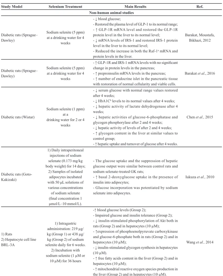
TABLE II - Selenium, glycemic parameters and insulin resistance: a summary of studies in cellular and non-human animal models
Study Model Selenium Treatment Main Results Ref.
Non-human animal studies
Diabetic rats (Sprague– Dawley)
Sodium selenite (5 ppm) at a drinking water for 4
weeks
- ↓ blood glucose;
- Restored the plasma level of GLP-1 to its normal range; - ↑ GLP-1R mRNA level and restored the GLP-1R protein level in the liver to its normal level;
- ↓ mRNA levels of IRS-1 and restored IRS-1 protein level in the liver to its normal level;
- Reduced the increase in both the Raf-1a mRNA and protein levels in the liver.
Barakat, Moustafa, Bikhazi, 2012
Diabetic rats (Sprague– Dawley)
Sodium selenite (5 ppm) at a drinking water for 4
weeks
- ↑ GLP-1R and IRS-1 mRNA levels with no significant change in protein levels in the pancreas;
- ↑ preproinsulin mRNA levels in the pancreas; - ↑ number of endocrine islet in the pancreatic tissue with restoration of normal cellularity and viable cells.
Barakat et al., 2016
Diabetic rats (Wistar)
Sodium selenite (1 ppm) at a
drinking water for 2 or 4 weeks
- ↓ serum glucose with normal range values restored after 4 weeks;
- ↓ HbA1Cb levels to its normal values after 4 weeks; - ↓ hepatic activity of lactate dehydrogenase after 4 weeks;
- ↓ hepatic activities of glucose-6-phosphatase and glycogen phosphorylase after 2 and 4 weeks;
- ↓ hepatic activity of levels of after 2 and 4 weeks; - ↑ glycogen content in the liver at similar values to control group;
- ↑ hepatic uptake and turnover of glucose after 4 weeks.
Chen et al., 2015
Diabetic rats (Goto-Kakizaki)
1) Daily intraperitoneal injections of sodium selenate (0.173 mg/kg body weight) for 14 days;
2) Samples of isolated adipocytes incubated with 50 μL solutions of various concentrations of sodium selenate (final concentration 1 μmol/L–10 mmol/L).
- The glucose uptake and the suppression of hepatic glucose output were similar between control rats and sodium selenate-treated GK rats;
- ↑ basal 2-deoxyglucose uptake in the presence of insulin into adipocytes;
- Glucose incorporation was potentiated by sodium selenate into adipocytes.
Iukuza et al., 2010
1) Rats
2) Hepatocyte cell line BRL-3A
1) Intragastric administration: 219 µg/ kg (Group 1) or 438 µg/ kg (Group 2) of sodium selenite daily for 6 weeks.
2) Incubation with sodium selenite (1 µM or
10 µM) for 36 hours
-↑ blood glucose levels (Group 2);
- Impaired glucose and insulin tolerance (Group 2); - ↓ insulin-stimulated phosphorylation of Akt both in rats (Group 2) and in hepatocytes (10 µM);
- ↑expression of phosphoenolpyruvate carboxykinase and glucose-6-phosphate both in rats (Group 2) and in hepatocytes (10 µM);
- ↓ insulin-stimulated glycogen synthesis in hepatocytes (10 µM).
- ↑ free fatty acids content in the liver (Group 2) and in hepatocytes (10 µM);
- ↑ mitochondrial reactive oxygen species production in the liver (Group 2) and in hepatocytes (10 µM).
Study Model Selenium Treatment Main Results Ref.
Female Wistar rats and its offspring*
Se-enriched yeast for 5 weeks Group 1 (deficient): basal diet (12 µg/kg of
selenium) Group 2 (adequate): basal diet + 0.3 mg/kg of
selenium Group 3 (high): basal diet
+ 3.0 mg/kg of selenium *The pups were fed the same diet as their
respective dam.
- ↑ insulin and glucose levels on day 19 of gestation and day 14 postpartum, respectively (Group 3);
- Glucose tolerance and insulin resistance impaired on day 19 of gestation (Grupo 3);
- Offspring were glucose intolerant and insulin resistant (Grupo 3);
- ↓ mRNA levels of Akt2, Insrc, Irs1 and FoxO1 in liver of the offspring (Grupo 3);
- ↓ mRNA levels of Irs2 and Pgc-1 in muscle of the offspring (Grupo 3).
Zeng et al., 2012
Diabetic rats (Wistar)
Sodium selenite (1 ppm) at a
drinking water for 2 or 4 weeks
- ↓ serum glucose after 2 and 4 weeks; - ↑ insulin levels after 2 and 4 weeks;
- Improved hepatic antioxidant status after 4 weeks; - Overall improvement in histoarchitecture of the liver, with reduction of lipid deposition and lymphocytic infiltration.
Zou et al., 2016
Cell studies (in vitro)
1) Mouse beta-cell line Min6
2) Isolated rat islets of Langerhans
1) Incubation with sodium selenite (30 nmol/L) for 3 hours.
2) Incubation with sodium selenite (30 nmol/L) for 72 hours.
- Significant 5-fold increase in GPx activity;
- 3.5-fold increase in the activity of the Ipf1d promoter and a 7-fold increase in the activity of the beta-cell specific section of this promoter;
- ↑ Ipf1 mRNA and protein levels; - ↑ insulin mRNA levels;
- ↑ insulin content and secretion from isolated rat islets of Langerhans;
Campbell et al., 2008
L6C11 rat skeletal muscle cell line
Incubation with 1 μM to 100 µM of various selenium compounds (L-selenomethionine,
sodium selenite, methylseleninic acid or sodium selenate) for 24
hours.
- ↓ intracellular reactive oxygen species generation with selenite (1 μM) exhibiting the most pronounced effect followed by selenomethionine (10 μM);
- ↓ insulin-induced phosphorylation of Akt by selenite and methylseleninic acid (1 μM);
- ↓ insulin-stimulated glucose uptake by selenite (5 μM) and methylseleninic acid (1 μM);
- ↑ i n s u l i n - s t i m u l a t e d g l u c o s e u p t a k e b y selenomethionine (100 μM).
Pinto et al., 2011
aRaf1: Raf-1 proto-oncogene, serine/threonine kinase; bHbA1C: Glycated hemoglobin A1C; cInsr: insulin receptor; dIpf1: insulin promoter factor 1.
TABLE II - Selenium, glycemic parameters and insulin resistance: a summary of studies in cellular and non-human animal models
(cont.)
Selenium’s antidiabetic action appears to result from its behaviour as an insulin-mimetic and as an antioxidant nutrient, since insulin signaling, and secretion is associated with the cellular redox state (Mao, Teng, 2013; Rayman, Stranges, 2013; Steinbrenner, 2013). In a study performed by Barakat, Moustafa and Bikhazi (2012), it was found that 4 weeks of treatment with sodium selenite (5 ppm in drinking water) increased mRNA and protein levels of glucagon-like peptide-1 receptor (GLP-1R) in the liver of diabetic rats, which may have favored the reduction of
blood glucose levels. It is worth mentioning that GLP-1 appears to reduce hepatic glucose production by promoting glycogen synthesis (D’Alessio, Vahl, Prigeon, 2004).
tissue (Barakat et al., 2016). The effects of GLP-1 on the biosynthesis and secretion of insulin, as well as the proliferation, differentiation and apoptosis of pancreatic β cells, are well known (Lee, Jun, 2014). In this regard, these results suggest a possible role of selenium in the physiological actions of incretin hormones.
Similarly, Campbell et al. (2008) found that selenium participates in insulin’s action on β-pancreatic cells, through its ability to regulate the gene expression of insulin promoter factor 1, and to increase the mRNA expression of insulin in the mouse beta cell line Min6. Selenium promotes the increased production and secretion of insulin in these cells.
In a study by Iukuza et al. (2010), 0.173 mg/ kg of sodium selenite (Na2SeO3) was administered
intraperitoneally to diabetic rats for 14 days. The result was increased glucose uptake by peripheral tissues and adipocytes. This was probably due to the action of selenium on the insulin receptor, thus increasing sensitivity to insulin and alleviating hyperglycemia, for regulation of hepatic glucose production.
It is important to mention selenium’s role in the regulation of the hepatic metabolism of carbohydrates. Chen et al. (2015) found that the administration of selenium to diabetic rats (through a 1 ppm dose in their drinking water) increased cellular glucose uptake and restored the activity of glucose-6-phosphatase and glycogen phosphorylase (enzymes involved in the hepatic glucose production) to normal levels.
Pancreatic β-cells are known for their reduced antioxidant protection, probably due to the important role played by physiological concentrations of reactive oxygen species in insulin signaling pathways. Research has revealed lower expression of GPx selenoproteins, and higher expression of Sepp1 in these cells. These findings are suggestive of a possible role of selenium in the regulation of the redox state of pancreatic β-cells (Lei, Vatamaniuk, 2011; Steinbrenner et al., 2013).
A study by Steinbrenner et al. (2013) showed that Sepp1 can provide additional antioxidant protection to the islets of Langerhans, serving as a selenium donor for the function of other antioxidant selenoproteins such as Gpx, and participating in the protection of cell membranes from oxidative damage. Sepp1 is also located in glucagon-secreting cells. Nevertheless, elevated glucose concentrations (11–22 mM) appear to dysregulate the expression of Sepp1 in β-cells, which can reduce antioxidant function and trigger endocrine dysfunction.
Labunskyy et al. (2011) found that a deficiency of Sepp1 and methionine sulfoxide reductase 1 (MsrB1) in transgenic mice induces glucose intolerance and
hyperinsulinemia. Research by Zou et al. (2016) showed that selenium treatment (a 1 ppm dose in drinking water) in diabetic rats reduces serum glucose concentration, lipid peroxidation, and the activity of the enzymes aspartate aminotransferase, alanine aminotransferase, and alkaline phosphatase. The authors also demonstrated that selenium supplementation increased the activity of major antioxidant enzymes, such as GPx, superoxide dismutase, and catalase. Figure 1 shows the participation of selenium in protection from insulin resistance.
On the other hand, selenium may also impair insulin’s action in target cells. Some studies have shown an association of high dietary selenium intake with increased risk of diabetes and insulin resistance (Table I and II) (Pounis et al., 2014; Stranges et al., 2007). The negative action of selenium on insulin signaling pathways appears to relate to dysregulation of the cellular redox state (Labunskyy et al., 2011; Wang et al., 2014).
In this context, the overexpression of antioxidant selenium-dependent enzymes promotes the removal of intracellular hydrogen peroxide, which is a reactive oxygen species that regulates key enzymes for insulin signaling. In turn, this promotes the dephosphorylation of the enzymes of insulin signaling, thereby inactivating them and changing the action of insulin (Steinbrenner, 2013; Wang et al., 2014; Zhou, Huang, Lei, 2013). In particular, some investigators have described the inhibition of phosphorylation of specific enzymes of the insulin signaling pathway, such as Akt and 5’-adenosine monophosphate-activated protein kinase (AMPK), caused by the elevated expression of GPx or Sepp1. Sepp1 also inhibits phosphorylation of the tyrosine residue on hepatocyte insulin receptors, and promotes the phosphorylation of a serine residue in IRS-1 (Misu et al., 2010; Steinbrenner, 2013; Wang et al., 2014).
Zeng et al. (2012) found that in rats, a diet rich in selenium (3.0 mg/kg per day) can induce hyperinsulinemia, insulin resistance, and glucose intolerance. High selenium intake also reduces the mRNA expression or protein levels of six factors that participate in insulin signaling pathways in the liver and muscle, and increases the activity of GPx1 in the pancreas, liver, and erythrocytes.
redox state, caused by high selenoprotein expression, can compromise the protein folding of insulin and its receptor.
Pinto et al. (2011) demonstrated the action of various compounds containing selenium (sodium selenite, sodium selenate, selenomethionine, and methylseleninic acid) on insulin sensitivity in vitro in myocytes. The authors found that > 1 μM doses of sodium selenite and methylseleninic acid were capable of inhibiting Akt phosphorylation, and attenuated the glucose uptake by decreasing the concentration of intracellular reactive oxygen species. This result can be attributed to the differences in bioavailability and metabolism of the selenium-containing compounds.
In addition, selenium may contribute to the induction of insulin resistance because of its role in the metabolism of carbohydrates and lipids (Steinbrenner, 2013). In a study on mice, Wang et al. (2014) found that high doses of selenium (200 mg/kg) can promote increased expression of carboxykinase phosphoenolpyruvate and glucose-6-phosphatase, a key enzyme for gluconeogenesis. The authors also found that selenium promotes the accumulation and metabolism of fatty acids in the liver; this effect is related to the increased production of reactive
oxygen species originating from mitochondrial oxidative phosphorylation, and hence the development of hepatic insulin resistance.
In this scenario, it is noteworthy that hyperglycemia favors hepatic Sepp1 transcription by activating the forkhead box class O 1α (FoxO1α) and hepatocyte nuclear factor 4α (HNF-4α) transcription factors, through the action of peroxisomal proliferator-activated receptor gamma coactivator 1α (PCG-1α). These transcription factors bind to a functional site in the Sepp1 promoter and upregulate gene expression. They are also involved in regulating the expression of hepatic glucose metabolism enzymes, and it is known that insulin promotes the inactivation of these enzymes. Therefore, under insulin resistance conditions, there may be an increase in Sepp1 concentrations concomitant with the increase in the activity of gluconeogenic enzymes. This situation may partially explain the associations between selenoprotein concentrations and the risk of type 2 diabetes mellitus (Jung et al., 2013; Mao, Teng, 2013; Steinbrenner et al., 2011; Steinbrenner, 2013).
A study by Jung et al. (2013) showed that induction of diabetes in rats by palmitate increases the expression of Sepp1 in HepG2 cells, thereby exacerbating insulin
FIGURE 1 - The role of selenium in protection from insulin resistance. Overall, selenium appears to improve insulin resistance
through three mechanisms. A) Firstly, in β-pancreatic cells, selenoproteins such as Sepp1 and Gpx exert antioxidant action, thereby
inhibiting the excessive production of reactive oxygen species, which may impair the insulin signaling pathway. Moreover, selenium
promotes the transcription and synthesis of proinsulin, insulin promoter factor 1, and GLP-1R. In the figure, we highlight the
participation of the GLP-1-activated protein kinase A pathway in the regulation of insulin secretion and synthesis. B) Secondly,
in the liver, selenium influences hepatic glucose production, by regulating glucose-6-phosphatase and glycogen phosphorylase
enzymatic activity, and increasing GLP-1R expression. C) Thirdly, in peripheral organs, selenium improves insulin sensitivity due to its antioxidant action. GLP-1: glucagon-like peptide 1; GLP-1R: GLP-1 receptor; GPx: glutathione peroxidase; IPF1: insulin
promoter factor 1; IRS-1: insulin receptor substrate 1; P: phosphorylation; PKA: protein kinase A; ROS: reactive oxygen species;
resistance. The activation of AMPK, induced by either salicylate or adiponectin, could reverse the increase in Sepp1 expression through phosphorylation of FoxO1α. Similar results were obtained by Takayama et al. (2014), wherein metformin inhibited the AMPK-mediated increased expression of Sepp1 in the H4IIEC3 hepatocytes of diabetic rats. On the other hand, coadministration of an AMPK antagonist reversed these results, indicating that this pathway can be a possible therapeutic target of insulin resistance and diabetes.
CONCLUSION
The action of selenium in the pathogenesis of insulin resistance is not fully elucidated, but our review has revealed that the main mechanisms involve the regulation of intracellular concentrations of reactive oxygen species, and carbohydrate and lipid metabolism. When ingested in sufficient amounts to maintain plasma concentrations in the range of 80-120 µg/L (Rayman, 2012), selenium acts as an antioxidant and insulin-mimetic nutrient, favoring the synthesis and action of insulin. However, when selenium intake results in plasma levels above 120 µg/L (Rayman, 2012), the cellular redox state may become dysregulated, which can compromise the chemical interactions involved in the insulin signaling cascade. Nevertheless, it is noteworthy that the effects of selenium on insulin resistance are dependent on the chemical form (organic or inorganic), dose, and exposure time; these factors are not yet fully established. Further studies are needed to clarify the relation between selenium and insulin resistance in humans.
REFERENCES
Alizadeh M, Safaeiyan A, Ostadrahimi A, Estakhri R,
Daneghian S, Ghaffari A, et al. Effect of L-arginine and selenium added to a hypocaloric diet enriched with legumes on cardiovascular disease risk factors in women with central obesity: a randomized, double-blind, placebo-controlled trial. Ann Nutr Metab. 2012;60(2):157-68.
Bahmani F, Kia M, Soleimani A, Asemi Z, Esmaillzadeh A.
Effect of selenium supplementation on glycemic control and lipid profiles in patients with diabetic nephropathy. Biol Trace
Elem. 2016;172(2):282-9.
Barakat GM, Moustafa ME, Bikhazi AB. Effects of selenium
and exendin-4 on glucagon-like peptide-1 receptor, IRS-1, and Raf-1 in the liver of diabetic rats. Biochem Genet. 2012;50(11-12):922-35.
Barakat GM, Moustafa ME, Khalifeh I, Hodroj MH, Bikhazi A,
Rizk S. Effects of exendin-4 and selenium on the expression of
GLP-1R, IRS-1, and preproinsulin in the pancreas of diabetic rats. J Physiol Biochem. 2016;73(3):387-94.
Bruce KD, Hanson MA. The developmental origins, mechanisms, and implications of metabolic syndrome. J Nutr. 2010;140(3):648-52.
Campbell SC, Aldibbiat A, Marriott CE, Landy C, Ali T, Ferris WF, et al. Selenium stimulates pancreatic beta-cell gene expression and enhances islet function. FEBS Lett. 2008;582(15):2333-7.
Chen H, Qiu Q, Zou C, Dou L, Liang J. Regulation of hepatic carbohydrate metabolism by selenium during diabetes. Chem Biol Interact. 2015;232:1-6.
Cominetti C, Bortoli MC, Abdalla DSP, Cozzolino SMF. Considerations about oxidative stress, selenium and nutrigenetics. Rev Nutr. 2011;36(3):131-53.
Cruz KJ, Oliveira AR, Pinto DP, Morais JB, Lima FS, Colli C, et al. Influence of magnesium on insulin resistance in obese
women. Biol Trace Elem Res. 2014;160(3):305-10.
Czernichow S, Couthouis A, Bertrais S, Vergnaud AC, Dauchet L, Galan P, et al. Antioxidant supplementation does not affect
fasting plasma glucose in the Supplementation with Antioxidant
Vitamins and Minerals (SU.VI.MAX) study in France:
association with dietary intake and plasma concentrations. Am J Clin Nutr. 2006;84(2):395-9.
D’Alessio D, Vahl T, Prigeon R. Effects of Glucagon-like
Peptide 1 on the hepatic glucose metabolism. Horm Metab Res. 2004;36(11-12):837-41.
Faghihi T, Radfar M, Barmal M, Amini P, Qorbani M, Abdollahi M, et al. A randomized, placebo-controlled trial of selenium supplementation in patients with type 2 diabetes: effects on
glucose homeostasis, oxidative stress, and lipid profile. Am J
Ther. 2014;21(6):491-495.
Farrokhian A, Bahmani F, Taghizadeh M, Mirhashemi SM, Aarabi MH, Raygan F, et al. Selenium supplementation
affects insulin resistance and serum hs-CRP in patients with
Feng W, Cui X, Liu B, Liu C, Xiao Y, Lu W, et al. Association
of urinary metal profiles with altered glucose levels and
diabetes risk: a population-based study in china. PLoS One.
2015;10(4):e0123742.
Gao H, Hägg S, Sjögren P, Lambert PC, Ingelsson E, Van
Dam RM. Serum selenium in relation to measures of glucose metabolism and incidence of Type 2 diabetes in an older Swedish population. Diabetic Med. 2014;31(7):787-793.
García OP, Ronquillo D, Caamaño MC, Martínez G, Camacho
M, López V, et al. Zinc, iron and vitamins A, C and E are
associated with obesity, inflammation, lipid profile and insulin resistance in mexican school-aged children. Nutrients. 2013;5(12):5012-30.
Henriksen EJ, Diamond-Stanic MK, Marchionne EM. Oxidative
stress and the etiology of insulin resistance and type 2 diabetes. Free Radic Biol Med. 2011;51(5):993-9.
Hirabara SM, Gorjão R, Vinolo MA, Rodrigues AC, Nachbar
RT, Curi, R. Molecular targets related to inflammation and insulin resistance and potential interventions. J Biomed Biotechnol. 2012;2012:379024.
Iizuka Y, Ueda Y, Yagi Y, Sakurai E. Significant improvement of
insulin resistance of GK rats by treatment with sodium selenate. Biol Trace Elem Res. 2010;138(1-3):265-71.
Jung TW, Choi HY, Lee SY, Hong HC, Yang SJ, Yoo HJ, et al.
Salsalate and adiponectin improve palmitate-induced insulin resistance via inhibition of selenoprotein P through the
AMPK-FOXO1α pathway. PLoS One. 2013;8(6):e66529.
Kim B, Feldman EL. Insulin resistance as a key link for the increased risk of cognitive impairment in the metabolic syndrome. Exp Mol Med. 2015;47(3):e149.
Kim HN, Song SW. Concentrations of chromium, selenium, and copper in the hair of viscerally obese adults are associated with insulin resistance. Biol Trace Elem Res. 2014;158(2):152-7.
Kwon H, Pessin JE. Adipokines mediate inflammation and insulin resistance. Front Endocrinol (Lausanne). 2013;4:71.
Labunskyy VM, Lee BC, Handy DE, Loscalzo J, Hatfield DL, Gladyshev VN. Both maximal expression of selenoproteins and selenoprotein deficiency can promote development of type
2 diabetes-like phenotype in mice. Antioxid Redox Signal. 2011;14(12):2327-36.
Lee YS, Jun HS. Anti-diabetic actions of glucagon-like peptide-1
on pancreatic beta-cells. Metabolism. 2014;63(1):9-19.
Lei XG, Vatamaniuk MZ. Two tales of antioxidant enzymes on β
cells and diabetes. Antioxid Redox Signal. 2011;14(3):489-503.
Litvinova LS, Kirienkova EV, Mazunin IO, Vasilenko MA,
Fattakhov NS. Pathogenesis of insulin resistance in metabolic obesity. Biochemistry (Moscow). 2014;8(3):191-202.
Mao J, Teng W. The relationship between selenoprotein P and glucose metabolism in experimental studies. Nutrients. 2013;5(6):1937-1948.
Misu H, Takamura T, Takayama H, Hayashi H, Matsuzawa-Nagata N, Kurita S, et al. A liver-derived secretory protein, selenoprotein P, causes insulin resistance. Cell Metab. 2010;12(5):483-95.
Muntoni S, Muntoni, S. Insulin resistance: pathophysiology and rationale for treatment. Ann Nutr Metab. 2011;58(1):25-36.
Mutakin, Meiliana A, Wijaya A, Kobayashi K, Yamazaki C,
Kameo S, et al. Association between selenium nutritional status and metabolic risk factors in men with visceral obesity. J Trace Elem Med Biol. 2013;27(2):112-116.
Pinto A, Speckmann B, Heisler M, Sies H, Steinbrenner, H. Delaying of insulin signal transduction in skeletal muscle cells by selenium compounds. J Inorg Biochem. 2011;105(6):812-20.
Pounis G, Costanzo S, Persichillo M, Curtis A, Sieri S, Vinceti
M et al. Mushroom and dietary selenium intakes in relation to fasting glucose levels in a free-living Italian adult population: The Moli-sani Project. Diabetes Metab. 2014;40(1):34-42.
Rains JL, Jain SK. Oxidative stress, insulin signaling, and
diabetes. Free Radic Biol Med. 2011;50(5):567-75.
R a y m a n M P. S e l e n i u m a n d h u m a n h e a l t h . L a n c e t . 2012;379(9822):1256-68.
Rayman MP, Stranges S. Epidemiology of selenium and type 2 diabetes: Can we make sense of it? Free Radic Biol Med. 2013;65:1557-64.
The role of selenium in insulin resistance
Steinbrenner H, Hotze AL, Speckmann B, Pinto A, Sies H, Schott M, et al. Localization and regulation of pancreatic selenoprotein P. J Mol Endocrinol. 2013;50(1):31-42.
Steinbrenner H, Speckmann B, Pinto A, Sies H. High selenium intake and increased diabetes risk: experimental evidence for interplay between selenium and carbohydrate metabolism. J Clin Biochem Nutr. 2011;48(1):40-5.
Steinbrenner, H. Interference of selenium and selenoproteins with the insulin-regulated carbohydrate and lipid metabolism. Free Radic Biol Med. 2013;65:1538-47.
Stranges S, Marshall JR, Natarajan R, Donahue RP, Trevisan M,
Combs GF, et al. Effects of long-term selenium supplementation
on the incidence of type 2 diabetes: a randomized trial. Ann Intern Med. 2007;147(4):217-23.
Takayama H, Misu H, Iwama H, Chikamoto K, Saito Y, Murao
K, et al. Metformin suppresses expression of the selenoprotein
P gene via an AMP-activated kinase (AMPK)/FoxO3a pathway
in H4IIEC3 hepatocytes. J Biol Chem. 2014;289(1):335-45.
Tencerova M, Aouadi M, Vangala P, Nicoloro SM, Yawe JC, Cohen JL, et al. Activated Kupffer cells inhibit insulin sensitivity
in obese mice. FASEB J. 2015;29(7):2959-69.
Wang X, Zhang W, Chen H, Liao N, Wang Z, Zhang X et al. High
selenium impairs hepatic insulin sensitivity through opposite
regulation of ROS. Toxicol Lett. 2014;224(1):16-23.
Wei Z, Yao M, Li Y, He X, Yang Z. Dietary selenium deficiency exacerbates lipopolysaccharide-induced inflammatory response in mouse mastitis models. Inflammation. 2014;37(6):1925-31.
Winnier DA, Fourcaudot M, Norton L, Abdul-Ghani MA, Hu
SL, Farook VS, et al. Transcriptomic identification of ADH1B
as a novel candidate gene for obesity and insulin resistance in
human adipose tissue in Mexican Americans from the Veterans Administration Genetic Epidemiology Study (VAGES). PLoS One. 2015;10(4):e0119941.
Zeng MS, Li X, Liu Y, Zhao H, Zhou JC, Li K, et al. A
high-selenium diet induces insulin resistance in gestating rats and
their offspring. Free Radic Biol Med. 2012;52(8):1335-42.
Zhou J, Huang K, Lei XG. Selenium and diabetes – evidence
from animal studies. Free Radic Biol Med. 2013;65:1548-56.
Zhou J, Xu G, Bai Z, Li K, Yan J, Li F, et al. Selenite exacerbates
hepatic insulin resistance in mouse model of type 2 diabetes through oxidative stress-mediated JNK pathway. Toxicol Appl Pharmacol. 2015;289(3):409-18.
Zou C, Qiu Q, Chen H, Dou L, Liang, J. Hepatoprotective
effects of selenium during diabetes in rats. Hum Exp Toxicol.
2016;35(2):114-23.