INTRODUCTION
Major Article
Address to: Dr. Raphael Sanzio Pimenta. Lab. Microbiologia Ambiental e Biotecnologia/UFT. Avenida NS 15, ALCNO 14, 109 Norte s/n, Bloco II/sala 05, 77001-090 Palmas, TO, Brasil. Phone: 55 63 3232-8007
e-mail: [email protected] Received in 27/03/2012
Accepted in 28/06/2012
The inluence of carbohydrates in the interacion of
Paracoccidioides brasiliensis
with CCL-6 cells
in vitro
Francisco Laurindo da Silva
[1], Raphael Sanzio Pimenta
[2], Juliana Fonseca Moreira da Silva
[2],
Déborah Aparecida Negrão Corrêa
[3]and Ary Corrêa Junior
[4][1]. Laboratório de Microbiologia, Centro de Estudos Superiores de Caxias, Universidade Estadual do Maranhão, Caxias, MA. [2]. Laboratório de Microbiologia Ambiental e Biotecnologia, Universidade Federal do Tocanins, Palmas, TO. [3]. Departamento de Parasitologia, Insituto de Ciências Biológicas, Universidade Federal de Minas Gerais, Belo Horizonte, MG. [4]. Departamento de Microbiologia, Insituto de Ciências Biológicas, Universidade Federal de Minas Gerais, Belo Horizonte, MG.
ABSTRACT
Introducion: Litle is known about the early events in the interacion between Paracoccidioides brasiliensis and its host. To understand the efect of carbohydrates in the interacion between the fungus and epithelial cell in culture, we analyzed the inluence of diferent carbohydrate soluions on the adhesion of P. brasiliensis yeast cells to CCL-6 cells in culture. Methods: Fungal cells were culivated with the epithelial cell line, and diferent concentraions of D-fucose, N-acetyl-glucosamine, D-mannose, D-glucosamine, D-galactosamine, sorbitol and fructose were added at the beginning of the experiment. Six hours ater the treatment, the cells were ixed and observed by light microscopy. The number of P. brasiliensis cells that were adhered to the CCL-6 monolayer was esimated. Results: The number of adhesion events was diminished following treatments with D-fucose, N-acetyl-glucosamine, D-mannose, D-glucosamine and D-galactosamine as compared to the untreated controls. Sorbitol and fructose-treated cells had the same adhesion behavior as the observed in the control.
P. brasiliensis propagules were treated with luorescent lecins. The FITC-labeled lecins WGA and Con-A bound to P. brasiliensis yeast cells, while SBA and PNA did not. Conclusions: The perceptual of adhesion between P. brasiliensis and CCL-6 cells decreased with the use of D-mannose, N-acetyl-glucosamine and D-glucosamine. The assay using FITC-labeled lecins suggests the presence of N-acetyl-glucosamine, α-mannose and α-glucose on the P. brasiliensis cell surface. An enhanced knowledge of the mediators of adhesion on P. brasiliensis could be useful in the future for the development of more eicient and less harmful methods for disease treatment and control.
Keywords: Paracoccidioidomycosis. Adhesion. CCL-6 cells. Lecin labeling.
Paracoccidioidomycosis (PCM) is a disease endemic in Latin America that is caused by a thermo-dimorphic fungus, Paracoccidioides brasiliensis1,2. The fungi initially infect the lungs, but can also
disseminate throughout the enire body3-5. Paracoccidioides brasiliensis
is a biotrophic pathogen, and an acute and sub-acute clinical form of PCM can be observed in paients3. Unfortunately, litle is known about
the ecological niche of the saprophyic phase of P. brasiliensis and its modes of infecion and disseminaion throughout the host. The cellular events that lead to infecion are also unclear, but undoubtedly the disease is established with the conversion of the ilamentous phase of the pathogen into its yeast phase, followed by the adhesion of the pathogenic yeast form to the hosts issue and fungal muliplicaion5.
The adhesion event is beter characterized in other pathogenic fungi, and there are only a few reports concerning the precise mechanism of binding between P. brasiliensis and its host. Most pathogenic fungi express surface factors that mediate the direct or indirect binding to host cells. For the later, binding to the host occurs through the interacion with adhesion components, such as extracellular matrix (ECM) proteins, which act as interlinking molecules6,7. For a number of pathogens, the ability to bind ECM
glycoproteins and the capability of internalizaion are considered to be
important characterisics, and molecules, such as laminin, ibronecin, integrin and carbohydrates, mediate these events6.
The diagnostic glycoprotein gp43, which is present on the
P. brasiliensis cell wall, has binds to laminin8. Other fungi, such as Aspergillus fumigatus, also bind to the laminin molecule9. Another
molecule that is associated with the interacion between the fungus and host is fibronectin. The participation of this molecule in the establishment of the host/pathogen interacion is evident in Candida albicans10. Laminin and ibronecin normally interact with integrins.
In fungi, the presence of integrin-like proteins was characterized in
C. albicans and Uromyces appendiculatus10,11. The regulaion of cell
adhesion by fungal surface carbohydrates has been characterized in
Fonsecaea pedrosoi and Trichophyton mentagrophytes conidia during the iniial stages of interacion with host cells12,13. However, litle is known
about the mechanism of carbohydrate-mediated adhesion in fungi.
In vitro studies are a helpful in the characterizaion of adhesion events and mediators. The adhesion P. brasiliensis to cultured cells has been previously reported6,14. Vero, LLC-MK
2 and Henle-407 epithelial cell lines
are able to support P. brasiliensis adhesion and infecion15. It has been
shown any evidence of the presence of mannose 43kDa glycoprotein (gp43) of P. brasiliensis and also their property of binding to Con A16.
To study the role of monosaccharides in the adhesion of P. brasiliensis
to its host, fungal propagules were culivated with CCL-6 cells with and without the presence of sorbitol, D-mannose, D-fucose, N-acetyl-glucosamine, D-N-acetyl-glucosamine, fructose and D-galactosamine soluions. The number of adhesion events was esimated. The lecins wheat germ aggluinin (WGA), Concanavalia eosiformes aggluinin (Con-A),
RESULTS METHODS
Microorganism
The P. brasiliensis (Pb18) strain was obtained from the Mycology Laboratory Collection in the Biological Sciences Institute at the Federal University of Minas Gerais, Minas Gerais, Brazil (ICB-UFMG) and maintained in its leveduriform state on parially solid Fava-Neto culture medium17 at 37oC. Prior to cellular interacions, the fungi were
adapted to Roswell Park Memorial Insitute (RPMI)-1640 (Sigma-Aldrich, St. Louis MO-USA) issue culture medium supplemented with 10% heat-inacivated fetal bovine serum (Sigma-Aldrich, St. Louis MO-USA) and anibioic soluion (Gentamicin 60mg/L - Ariston - Brazil) under agitaion (130rpm) at 37oC for 7 days. Ater incubaion, the cell viability
was esimated using a FDA/EB (luorescein diacetate 2.5mg/ml + ethidium bromide 20μg/ml) soluion for 30min; the green luorescent cells were considered viable (Olympus BX-41 microscope - WB ilter)18.
Host cell line
The host cell line (ATCC CCL-6) was kindly provided by Dr. Judith A. Apleton (Veterinary Medicine School, Cornell University - USA) and maintained in RPMI-1640 (Sigma-Aldrich, St. Louis MO-USA) in 25-ml lasks, supplemented with 10% heat-inacivated fetal bovine serum (Sigma-Aldrich, St. Louis MO-USA) and anibioic soluion (gentamicin 60mg/L - Ariston). The cells were incubated at 37oC in an atmosphere
containing 5% CO2. Cell viability was esimated using a 0.4% Trypan blue soluion and the non-stained cells were considered viable. Only cultures with more than 90% viability were uilized in the infecion assays. All experiments were performed with cells obtained from the second passage in RPMI culture medium19.
Host/pathogen interacion
The CCL-6 cell monolayer was grown on 24x24mm sterile cover slips on the botom of a 6-well culture plate and incubated in RPMI medium as previously reported for 24h. A fungal inoculum of 100µl of 1x10⁸ propagules/ml soluion was added to the culture plate and
incubated for 6h at 37oC in a 5 % CO
2 atmosphere. The test soluions
were added to the inoculums. Subsequent to treatment with
P. brasiliensis, the cover slips containing the CCL-6 cell monolayer were washed 3 imes in RPMI medium to remove the non-adhered fungal cells and ixed with 4% paraformaldehyde and 2.5% glutaraldehyde in 0.1M phosphate bufer for 2h. The adhesions events for each treatment were evaluated in triplicate by light microscopy using Nomarsky opics. Fity randomly chosen microscopic ields were assessed, and the adhesion events were counted. The adhesion inhibiion rates were esimated by the following equaion: IR =100 – [(AdCarb x 100)/AdCont], where: IR = inhibiion rate; AdCarb = number of fungal adhered cells on carbohydrates treated cells; and AdCont = number of fungal adhered cells on the control
Adhesion inhibiion assay
The ability of carbohydrates to interfere with the cellular interacion between P. brasiliensis and CCL-6 cell was assessed. The carbohydrates D-mannose, N-acetyl-glucosamine, D-fucose, sorbitol (Calbiochem Co. - San Jose CA, USA) at concentraions of 1, 25, 50 and 75mM, and D-glucosamine and D-galactosamine (5, 10 and 25mM) or fructose (75mM) (Sigma-Aldrich, St. Louis MO, USA) were added to the fungal inoculums prior to the infecions. No carbohydrates were added to the control group.
Immunolocalizaion
To beter characterize the adhesion of P. brasiliensis to host cells, 6h post-infecion, the cover slips containing the CCL-6 cell monolayer infected with the fungus were treated with a rabbit extract total ani-
P. brasiliensis anibody (produced at the Cell Immunology Laboratory at UFMG-Brazil and generously provided by Dr. Goes de Miranda) diluted 1:100 in PBS (0.1M) containing 5 % BSA for 2h at room temperature. Following incubaion, the cover slips were treated with rabbit ani-IgG monoclonal anibody conjugated to FITC (Sigma-Aldrich, St. Louis MO-USA) diluted 1:20 in PBS containing 5% BSA for 2h. The cover slips were examined for luorescence (Olympus IX70 - BW ilter). When necessary, the images obtained from the microscope were acquired using an Optomitrics camera (DMI - 470) and processed using the Image Pro-plus sotware (Media Cyberneics - London - England).
Labeling of the leveduriform cell with luorescein-labeled lecin
Leveduriform cells of P. brasiliensis were incubated in presence of FITC-labeled lecins [wheat germ aggluinin (WGA), concanavalin A (ConA) Arachis hypogea (PNA) and Glycine max (SBA) - (EY Laboratories, Inc - California (USA)] diluted in RPMI (sigma) supplemented with 10% FBS and containing 0.0005% Mg2+ and Ca2 at concentraion of 100μg/ml
for 1h at room temperature. The cells were washed three imes with RPMI and observed by luorescence microscopy (Olympus BX-41 - WB ilter). When necessary, the microphotographs were recorded using a DMI-470 video camera and the Image Pro-plus sotware.
Staisical analysis
The number of cell adhesions per ield of the treated and untreated cells were compared using the paired Student’s t test with a signiicance level of p<0.05 and converted into a percent of adhesion inhibiion. The staisic analysis was performed using BioEstat ver.5.0 (2007).
Host cell/parasite interacion
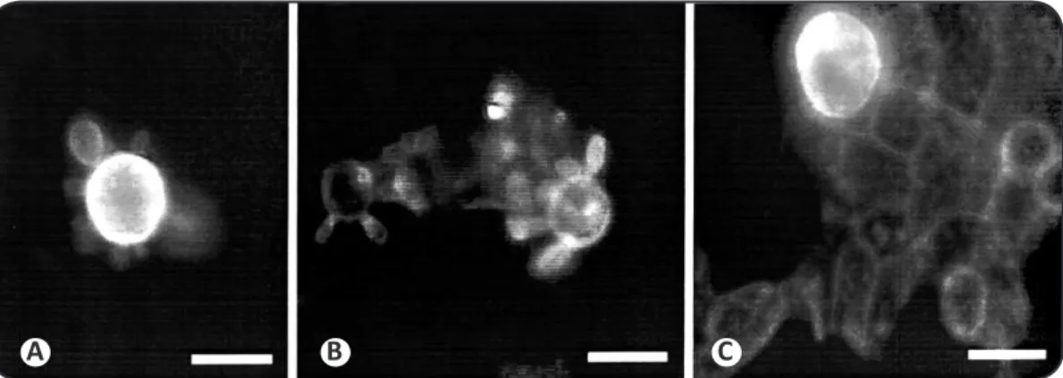
The P. brasiliensis cells that adhered to the CCL-6 monolayer were morphologically diferent from the non-adhered fungal cells
(Figure 1). The non-adherent cells had a tradiional shape of a pilot wheel(Figure 1A), and the propagules normally had muliple buds and pseudo-hyphae were rarely observed. However, the adhered cells had an abundant formaion of pseudo-hyphae (Figure 1B). These structures were longer than the usual pseudo-hyphae observed on
FIGURE 1 - Adhesion and invasion of Paracoccidioides brasiliensis cells on a CCL-6 cell layer. A: Nomarsky microphotograph of non-adhered Paracoccidioides brasiliensis cells in a tradiional pilot wheel shape. B: Nomarsky photomicrograph of a Paracoccidioides brasiliensis propagule with intense pseudo-hyphal formaion. Some of the pseudo-hyphae are growing underneath the CCL-6 layer (arrow). C: Cell layer treated with luorescent
ani-Paracoccidioides brasiliensis anibody. Arrowhead depicts fungal pseudo-hyphae growing underneath the cell layer; Arrows indicate luorescent fungal cells on the top of the monolayer. Bar=10µm.
A B C
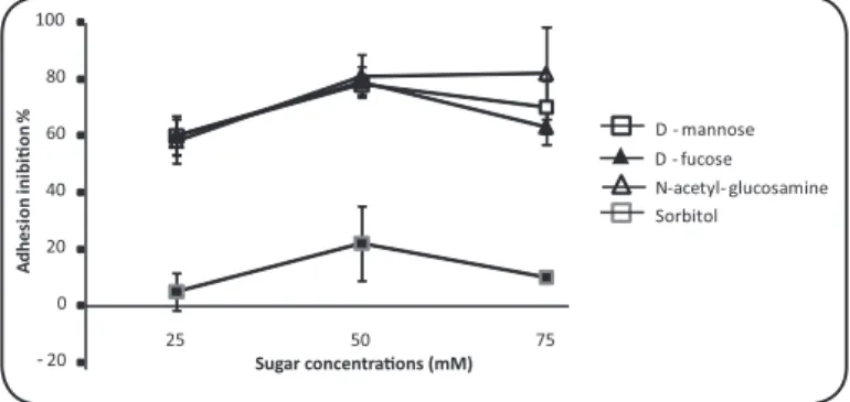
Adherence inhibiion
The addiion of D-mannose, D-fucose and N-acetyl-glucosamine at concentraions of 25, 50 and 75mM in the interacion medium signiicantly reduced the adhesion of P. brasiliensis to the CCL-6 cells. The rates of adhesion inhibiion in the treatments with D-mannose were reduced to 60, 78 and 70% at concentraions of 25, 50 and 75mM, respecively. The decrease in adhesion with the addiion of D-fucose and N-acetyl-glucosamine was 60, 79 and 63 and 58, 81 and 82% at the same concentraions, respecively. Pracically no efect was observed ater treatment with Sorbitol and Fructose Were observed signiicaive reducion in all concentraions and tested substances (p<0.05) in relaion with the control except the to sorbitol at 25mM (Figure 2). At concentraions of 1mM, no efect was observed, and at concentraions higher than 75mM, the monolayer cell death was higher than 80%. At 12h ater the addiion of carbohydrates, the adhesion rates were not signiicantly diferent from the untreated controls. To assess whether the decrease of adhesion inhibiion was due to the sequestraion or metabolization of soluble carbohydrates, mannose, N-acetyl-glucosamine and the non-metabolized carbohydrate fructose were freshly added at 8h ater the iniial carbohydrate treatment. Similar levels of adhesion inhibiion were observed ater the addiional carbohydrate treatment as compared with the 4h treated cells
(Figure 3).
The carbohydrates D-glucosamine and D-galactosamine were added at concentrations of 5, 10 and 25mM because higher
FIGURE 2 - Percentage adhesion inhibiion of Paracoccidioidesbrasiliensis cells to CCL-6 monolayer at 6 h ater treatment with Sorbitol ( ■ ), D-mannose (), D-fucose (▲), and N-acetyl-glucosamine () at concentraions ranging from 25 to 75 mM.
0 20 40 60 80
100
A B C
Mannose N-acetyl - glucosamine Fructose PBS
A
d
h
e
re
d
f
u
n
g
a
l
ce
ll
s/
fi
e
ld
B
B B
A A
A
A A
A
B B A
FIGURE 3 - Number of Paracoccidioides brasiliensis cells adhered to a CCL-6 monolayer ater treatment with 75 mM mannose (), N- acetyl-glucosamine (■), fructose (■) or PBS bufer (■) ater 4 (A), and 12 (B)h of sugar addiion or ater a second addiion of the sugar 8h ater incubaion (C).
concentrations were detrimental to the host cell. The adhesion reducions were esimated at 0, 30, 37% for D-glucosamine and 29, 40 and 37% for D-galactosamine for the corresponding concentraions, respecively. The results were signiicaive only for tests with 5 and 25 mM (Figure 4).
Lecin labeling
The ConA-FITC and WGA-FITC conjugates effectively labeled
- 20 0 20 40 60 80 100
5 10 25
D - glucosamine
D - galactosamine
A
dh
es
ion
in
hi
bi
�
on
%
FIGURE 4 - Percentage of adhesion inhibiion of Paracoccidioides brasiliensis
to the CCL-6 monolayer at 6h ater treatment with D-glucosamine (■) and D-galactosamine () at concentraions ranging from 5 to 25mM.
A B C
FIGURE 5 - Paracoccidioides brasiliensis cells ater treatment with luorescent lecins. A: Pilot wheel Paracoccidioides brasiliensis cells stained by WGA-FITC conjugate. Note the intense luorescence of the mother cell. B: Fungal cells labeled by ConA-FITC conjugate. The cells were evenly labeled. C: Paracoccidioides brasiliensis cell adhered to CCL-6 cells ater treatment with the WGA-FITC conjugate. Note the intense labeling of the fungal cell and the weak signal in the CCL-6 cell.
WGA: wheat germ aggluinin; Con-A: Concanavalia eosiformes aggluinin; FITC: luorescein isothiocyanate.
DISCUSSION
Most of our knowledge concerning the interaction between
Paracoccidioides brasiliensis and its host cell is derived from studies using in vitro cell culture models6,7,14. However, although insighful,
these reports did not assess the role of paricular components in the infecion. Litle is known about adhesion mediators produced by
P. brasiliensis during the course of the infecion. The gp43, is the only candidate molecule presumed to play a role in the regulaion of the fungal propagule to its host8. The efect of gp
43 in adhesion has been
inferred from indirect evidence due to the diiculty of providing direct proof. It has been previously shown that the puriied glycoprotein is able to bind to laminin in vitro, and P. brasiliensis cells pre-treated with laminin bind more efecively to Madin-Darby canine kidney cells. Furthermore, when P. brasiliensis laminin-treated cells were injected into guinea pigs, a higher number of granuloma was formed in the host in comparison with the guinea pig injected with the untreated
fungal cells8. Although the adhesion funcion of gp
43 adhesion funcion
was not directly tested. The gp43 is rich in mannose and mannose interfered in the adhesion of P. brasiliensis to CCL-6 cell. The addiion of monosaccharide D-mannose to the interacion medium prior to infecion signiicantly reduces the adhesion of P. brasiliensis to CCL-6 cells. The adhesion reducion was more signiicant at a concentraion of 50mM, reaching 79% inhibiion (Figure 2). The paricipaion of mannose residues in the interacion mechanism between the host and pathogen was also observed in other infecion models12,20.
Here, we report that some carbohydrates were able to diminish adhesion in vitro at rates as high as 90% and therefore, it’s the role of these carbohydrates in the natural adhesion process has to be invesigated. The role of carbohydrates in modulaing fungus/host cell adhesion has been reported previously9,21.
The addiion of monosaccharide D-mannose to the interacion medium prior to infection significantly reduces the adhesion of
The authors declare that there is no conlict of interest.
CONFLICT OF INTEREST
FINANCIAL SUPPORT
ABSTRACT IN PORTUgUESE
signiicant at a concentraion of 50mM, reaching 79% inhibiion
(Figure 2). The paricipaion of mannose residues in the interacion mechanism between the host and pathogen was also observed in other infecion models13,20. Most of the work, however, was conducted
using manopyranosides, lecins or glycosylated substrates as adhesion inhibitors. Normally, sugars linked to carrier protein are used to prevent the carbohydrate from being metabolized or sequestered by the cells during the assay. In our case the sugar itself has an efect on adhesion. Interesingly, if the co-culivaion was performed for a longer ime, the adhesion inhibiion would decrease but could be restored by the addiion of mannose to the culivaion media. The restoraion is probably due to the fact that the cell efecively processes mannose over ime and therefore, its concentraion decreases in the incubaion medium, resuling in the decrease of its efect on inhibiion of adhesion. An increase in mannose concentraion restores the inhibiion. Similar results were observed with N-acetyl-glucosamine
(Figure 3). Fructose is a sugar that is not metabolized by either the fungal or the host cells and did not have an efect on adhesion.
Another competitor of adhesion utilized in our studies was D-fucose. Fucose is a deoxyhexose that is present in a wide variety of organisms. In mammals, fucose-containing glycans have important roles in blood transfusion reacions, selecin-mediated leukocyte-endothelial adhesion, host-microbe interactions, and numerous ontogenic events, including signaling by the Notch receptor protein family21. The involvement of fucose in the interacion between fungi
and its host was established by the characterizaion of the fucose receptor in the germinaion tubes of C. albicans22 .
The addiion of N-acetyl-glucosamine to the interacion medium signiicantly decreases the adhesion percentage of the fungi to the cellular host type. The adhesion reducion was more signiicant at concentraions of 50 and 75mM (Figure 2). Results similar for
Fonsecaea pedrosoi pre-incubated with the N-acetyl-glucosamine-BSA conjugate13 .
D-glucosamine and D-galactosamine were also able to decrease the adhesion index of P. brasiliensis to CCL-6 cells (Figure 4). Those sugars have been implicated in the adhesion of Entamoeba histolyica
to epithelial cells and C. albicans to lymphocytes10,23.
Carbohydrates have been described as important adhesion mediators in many biological models, and this idea remains consistent with regard to the interacion of P. brasiliensis and epithelial CCL-6 cells. The cell treatment with those carbohydrates actually decreases the adhesion eiciency of the fungal cells. Unfortunately, even though and inhibitory efect was observed, we were not able to directly implicate the carbohydrates in the physiological event of adhesion. Several other molecules could also play a role in the process and would be not detected by our experimental approach.
Therefore, further studies are necessary to identify and characterize the molecules with ainity for carbohydrates that are present on the fungal surface and/or its host. Lecins are described as proteins that able to bind particular sugars. In several other biological systems, lecins are believed to be the cellular receptor for carbohydrates that modulate adhesion and cell recogniion, for example. To assess whether the lecins are capable of recognizing carbohydrates on the surface of P. brasiliensis, we labeled yeast cells with ConA, WGA, PNA and SBA lecins conjugated to FITC. Of the four lecins assessed, only ConA and WGA labeled P. brasiliensis cells.
The Con-A lecin speciically recognized α-mannose and α-glucose, while WGA bound to N-acetyl-glucosamine. These sugars were also able to inhibit P. brasiliensis adhesion to CCL-6 cells; therefore, one might suggest that those sugars are present on the P. brasiliensis surface and are the receptors responsible for cell-to-cell adhesion. However, PNA, which binds terminal galactose, and SBA, which recognizes N-acetyl-galactosamine and galactose, were not able to detect these sugars on the surface of P. brasiliensis cells. Inhibitory assays with N-acetyl-galactosamine showed a strong decrease of adhesion therefore, one would expect that the SBA lecin would recognize such a sugar on
P. brasiliensis cells. One possible argument for the apparent contradicion in the data is that the sugar would be present on the host cell surface.
However, SBA-FITC was also unable to show binding sites on the CCL-6 cells (data not shown). The role of galactosamine is therefore unclear. Adhesion is as early event in most of the pathogen/host interacions and is crucial for eicient disease development. Several mediators are constantly being implicated as having a role in the adhesion event, but a deiniive experiment is yet to be proposed. Our results, are circumstanial, and only emphasize the efect of P. brasiliensis surface carbohydrates in the adhesion/recognition process. An enhanced knowledge of the mediators of adhesion on the biotrophical fungus
P. brasiliensis will be useful in the future for the development of more eicient and less harmful methods for disease treatment and control.
This work was supported inancial by the Conselho Nacional de Desenvolvimento Cieníico e Tecnológico (CNPq).
A inluência de carboidratos na interação entre
Paracoccidioides brasiliensis
e células CCL-6 (
in vitro
)
Introdução: Pouco se conhece a respeito dos eventos iniciais que mediam as interações entre Paracoccidioides brasiliensis e seus hospedeiros. Com a intenção de compreender a importância de carboidratos junto a estas interações, foram analisados os efeitos de soluções de carboidratos sobre a adesão de células leveduriformes de P. brasiliensis sobre culturas de células CCL-6. Métodos: As células fúngicas foram cultivadas com as células epiteliais e diferentes concentrações de D-fucose, N-acetyl-glucosamina, D-manose, D-glicosamina, D-galactosamina, sorbitol e frutose foram adicionadas ao culivo no início da interação. Após 6h de tratamento, as células foram ixadas e observadas em microscópio ópico. Resultados: Os tratamentos uilizando D-fucose, N-aceil-glicosamina, D-manose, D-glicosamina e D-galactosamina reduziram os números de adesões quando comparados com o controle. Os tratamentos realizados com o uso de sorbitol e frutose apresentaram os mesmos resultados observados no controle. Para detectar a presença de carboidratos na superície do fungo, propágulos de P. brasiliensis foram tratados com lecinas luorescentes. WGA-FITC e Con-A-WGA-FITC se ligaram às células de P. brasiliensis ao contrário de SBA e PNA. Conclusões: O percentual de adesão entre P. brasiliensis e células CCL-6 foi reduzido com o uso de D-manose, N-aceil-glicosamina e D-glicosamina. O uso de lecinas marcadas sugeriu a presença de N-aceil-glicosamina, α-manose e α-glicose na superície de P. brasiliensis. Estes resultados contribuem para o aumento do conhecimento relacionado aos mediadores de adesão deP. brasiliensis, e poderão ser utilizados no futuro para o desenvolvimento de medidas mais eicientes para o controle e tratamento deste patógeno.
REFERENCES
1. Montenegro MR, Miyaji M, Franco M, Nishimura K, Coelho KI, Horie Y, et al. Isolaion of fungi from nature in the region of Botucatu state of São Paulo, Brazil, an endemic area of paraococcidioidomycosis. Mem Inst Osvaldo Cruz 1996; 91:665-670. 2. Loth EA, Castro SV, Silva JR, Gandra RF. Occurrence of 102 cases of
paracoccidioidomycosis in 18 months in the Itaipu Lake region, western Paraná. Rev Soc Bras Med Trop 2011; 44:636-637.
3. Borges-Walmsley MI, Chen D, Shu X, Walmsley AR. The pathobiology of
Paracoccidioides brasiliensis. Trends Microbiol 2002; 10:80-87.
4. Brummer E, Castaneda E, Restrepo A. Paracoccidioidomycosis: an update. Clin Microbiol 1993; 3:89-113.
5. Franco M. Host-parasite relaionships in paracoccidioidomycosis. J Med Vet Mycol 1986; 25: 5-18.
6. Mendes-Giannini MJS, Ricci LC, Vemura M, Toscano E, Arnus CW. Infecion and apparent invasion of Vero cells by Paracoccidioides brasiliensis. J Med Vet Mycol 1994; 32:189-195.
7. Sahai AS, Manocha MS. Chitinase of fungi and plants: their involvement in morphogenesis and host-parasite interacions. FEMS Microbiol 1993; 11:317-338. 8. Vicenini AP, Gesztesi JL, Franco MF, Sousa W, Moraes J, Travassos LR, et al. Binding of Paracoccidioides brasiliensis to laminin through surface glycoprotein gp-43 leads to enhancement of fungal pathogenesis. Infect Immun 1994; 62:1465-1469. 9. Trochin G, Esnault K, Renier G, Filmon R, Chabasse D, Bouchara JP. Expression and
ideniicaion of a laminin-binding protein in Aspergillus fumigatus. Infect Immun 1997; 65:9-15.
10. Forsyth CB, Mathews HL. Lymphocyte adhesion to Candida albicans. Infect Immun 2002; 70:517-527.
11. Corrêa AJ, Staples RC, Hoch HC. Inhibiion of thigmosimulated cell difereniaion with RGD-pepides in Uromyces germlings. Protoplasma 1996; 194:91-102.
12. Esquenazi D, Souza W, Alviano CS, Rozental S. The role of surface carbohydrates on the interacion of microconidia of Trichophyton mentagrophytes with epithelial cells. FEMS Immunol Med Microbiol 2003; 35:113-123.
13. Limongi CL, Rozental S, Alviano CS, Souza W. The inluence of surface carbohydrates on the interacion of Fonsecaea pedrosoi with Chinese hamster ovary glycosylaion mutant cell. Mycopathol 1997; 138:127-135.
14. Hanna SA, Juliana LMS, Mendes-Giannini MJ. Adherence and intracellular parasiism of Paracoccidioides brasiliensis in Vero cells. Microbes Infec 2000; 2:877-884.
15. Mendes-Giannini MJS, Taylor ML, Bouchara JB, Bugers E, Calich VLG, Escalante ED, et al. Pathogenesis II. Fungal responses to host responses: interacion of host cells with fungi. Med Mycol 2000; 1:113-123.
16. Franco M, Lacaz CS, Restrepo-Moreno A, Del Negro G. Paracoccidioidomycosis. Boca Raton: CRC Press; 1994.
17. Fava Neto C. Contribuição para o estudo imunológico da blastomicose de Lutz. Rev Inst Adolpho Lutz 1961; 21:99-194.
18. Calich VLG, Purchio A, Paula CR. A new luorescent viability test for fungi cells. Mycopathol1978; 66:175-177.
19. Silva FL. Estabelecimento in vitro de modelo para o estudo do processo infecivo de
Paracoccidioides brasiliensis em células hospedeiras de mamíferos. [Dissertaion]. [Belo Horizonte]: Universidade Federal de Minas Gerais; 2001; 104p.
20. Vardar-Üunlu G, McSharry C, Douglas LJ. Fucose-speciic adhesins on germ tubes of Candida albicans. FEMS Immunol Med Microbiol 1998; 20:55-67.
21. Agnani G, Tricot-Doleux S, Houalet S, Bonnaure-Mallet M. Epithelial cell surface sites involved in the polyvalent adherence of Porphyromonas gingivalis: a convincing role for neuraminic acid and glucuronic acid. Infecion Imm2003; 71:991-996. 22. Becker DJ, Lower JB. Fucose: biosynthesis and biological funcion in mammals.
Glycobiol 2003; 13:41-53.