Copyright © ABE&M todos os dir
eitos r
eser
vados.
Hypothalamic regulation of food intake
and clinical therapeutic applications
Regulação hipotalâmica da ingestão alimentar e suas aplicações terapêuticas clínicas
Katherine Anne Simpson1, Niamh M. Martin1, Stephen R. Bloom1
ABSTRACT
Current estimates suggest that over 1 billion people are overweight and over 300 million people are obese. Weight gain is due to an imbalance between energy expenditure and dietary intake. This review discusses the hypothalamic control of appetite and highlights key developments in research that have furthered our understanding of the complex pathways involved. Nuclei within the hypothalamus integrate peripheral signals such as adiposity and caloric intake to regulate important pathways within the central nervous system controlling food intake and energy expenditure. Firmly established pathways involve the orexigenic NPY/AgRP and the anorexigenic POMC/CART neurons in the arcuate nucleus (ARC) of the hypothalamus. These project from the ARC to other important hypothalamic nuclei, including the paraventricular, dorsomedial, ventromedial and lateral hypothalamic nuclei. In addition there are many pro-jections to and from the brainstem, cortical areas and reward pathways, which modulate food intake. Arq Bras Endocrinol Metab. 2009;53(2):120-128.
Keywords
Hypothalamus; obesity; appetite; arcuate nucleus; orlistat; sibutramine
RESUMO
As estimativas atuais sugerem que mais de 1 bilhão de pessoas apresentam sobrepeso e 300 milhões são obesas. O ganho de peso representa um desequilíbrio entre o gasto energéti-co e o energéti-consumo alimentar. Esta revisão discute o energéti-controle hipotalâmienergéti-co do apetite e destaca os pontos-chave no desenvolvimento de pesquisas para ampliar o nosso entendimento dos complexos mecanismos envolvidos nesta regulação. Núcleos situados no hipotálamo integram uma série de sinais com o sistema nervoso central controlando a ingestão alimentar e o gasto energético. As vias mais estabelecidas envolvem os neurônios orexigênicos NPY/AgRP e os neurônios anorexigênicos POMC/CART no núcleo arqueado (ARC) do hipotálamo. Esses neurô-nios se projetam do ARC para outros importantes núcleos hipotalâmicos, tais quais: paraventri-cular, dorsomedial, ventromedial e lateral. Além disso, existem várias projeções que vão e vem do tronco cerebral, das áreas corticais e das vias de retroalimentação que modulam o consumo alimentar. Arq Bras Endocrinol Metab. 2009;53(2):120-128.
Descritores
Hipolátamo; obesidade; apetite; núcleo arqueado; orlistate, sibutramina
INTRODUCTION
C
urrent estimates suggest that over 1 billion people are overweight and over 300 million people are obese (1). Furthermore, 80% of obese adults have at least one or more co-morbidities including diabetes mellitus, hyperlipidaemia, hypertension, cardiovascular disease and have a significant increase in many forms of cancer (2). The increasing prevalence of obesity is partlyattrib-utable to a lack of exercise and partly to the availability of high calorie palatable food. In addition family, twin and adoption studies indicate that obesity is highly heritable, with the estimated genetic contribution ranging from 60-84% (3). The concept of a “thrifty phenotype” was contemplated in the 1950s and suggested that carriers of genes that enabled storage of energy more efficiently during periods of abundant food supply increased their
1 Department of Investigative
Medicine, Imperial College, London, United Kingdom
Correspondence to: Stephen R. Bloom Department of Investigative Medicine, Imperial College London W12 ONN, UK [email protected]
Copyright © ABE&M todos os dir
eitos r
eser
vados.
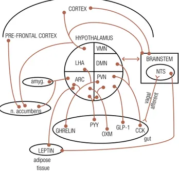
odds of survival during famine. However this “thrifty genotype” becomes a disadvantage at times of abundant energy supply, resulting in obesity. A series of complex systems maintain energy homeostasis in order that suffi-cient energy is available and body weight remains stable. Central circuits in the brain rely on peripheral signals in-dicating satiety levels and energy stores, as well as higher cortical factors such as emotional and reward pathways. As illustrated in Figure 1, the hypothalamus is critical in the relaying of afferent signals from the gut and brain-stem as well as processing efferent signals that modulate food intake and energy expenditure. The hypothalamus is subdivided into interconnecting nuclei, including the arcuate nucleus (ARC), paraventricular nucleus (PVN), ventromedial nucleus (VMN), dorsomedial nucleus (DMN) and lateral hypothalamic area (LHA). Neuronal pathways between these nuclei are organised into a complex network in which orexigenic and anorexigenic circuits influence food intake and energy expenditure. The purpose of this review is to provide some clarity of this complex network and its role in appetite, highlighting areas of potential therapeutic targets for obesity.
HYPOTHALAMIC NUCLEI INVOLVED IN APPETITE
CONTROL
Arcuate nucleus (ARC)
The ARC is a key hypothalamic nucleus in the regula-tion of appetite. In mice, lesions of the ARC result in obesity and hyperphagia (4). Its proximity to the me-dian eminence and the fact that the ARC is not fully in-sulated from the circulation by the blood brain barrier means it is strategically positioned to integrate a num-ber of peripheral signals controlling food intake (Fig-ure 2). There are two major neuronal populations in the ARC implicated in the regulation of feeding. One population increases food intake and co-expresses neu-ropeptide Y (NPY) and agouti-related protein (AgRP). The second population of neurons expresses co-caine- and amphetamine-related transcript (CART) and pro-opiomelanocortin (POMC) and inhibits food intake. Neuronal projections from these two popula-tions then communicate with other hypothalamic areas involved in appetite regulation such as the PVN, DMN and LHA (5).
CART/POMC neurons
Cleavage of POMC by prohormone convertases PC1 and PC2 produces melanocortins which exert their ef-fects through binding to G-protein coupled melano-cortin receptors (MC-Rs). Five melanomelano-cortin receptors have been cloned however only the MC3-R and MC4-R are expressed in the brain (6). The MC4-R is highly expressed in the hypothalamus, most notably the PVN (6). Targeted deletion of the MC4-R in mice results in hyperphagia, reduced energy expenditure and obesity, underlying the importance of this receptor in appetite regulation (7). The role of the MC3-R remains less clear however MC3 receptor (Mc3r) knockout mice have a higher fat content and reduced lean body mass (8).
Melanocortin peptides, including α-MSH, released from ARC POMC neurons bind to downstream MC4-Rs to inhibit food intake (9). Consistent with this, knockout mice lacking all POMC derived peptides display increased food intake and weight gain (10). AgRP is the endogenous antagonist at the MC3-R and MC4-R (11) suggesting that melanocortinergic neu-rons may exert a “tonic” inhibition on feeding and permit increased energy expenditure, which is relaxed following AgRP antagonism of the MC3 and MC4-Rs, ultimately resulting in stimulation of feeding and a lower metabolic rate.
Figure 1.Pathways are shown between the brainstem, hypothalamus, cortical areas and reward circuitry known to regulate appetite control. There are also projections from hypothalamic nuclei to the pre-frontal cortex, involved in conditioned taste aversion, as well as reward centres such as the amygdala and nucleus accumbens. Gut hormones acting via vagal afferents act on nuclei within the brainstem which in turn signal to the hypothalamus. Some gut hormones may also act directly on hypothalamic nuclei via the circulation and across an incomplete blood brain barrier. Leptin is also thought to act directly on the brainstem nuclei as well as hypothalamic nuclei suggesting that it can modulate appetite through different pathways.
NTS = nucleus tractus solitarius; amyg = amygdala; n. accumbens = nucleus accumbens.
ARC
LHA DMN
PVN
CCK GLP-1 OXM PYY GHRELIN n. accumbens
amyg. PRE-FRONTAL CORTEX
CORTEX
LEPTIN adipose
tissue
gut NTS BRAINSTEM HYPOTHALAMUS
VMN
Copyright © ABE&M todos os dir
eitos r
eser
vados.
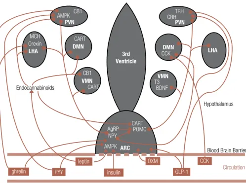
Figure 2. The main hypothalamic nuclei, neuropeptides and pathways involved in the regulation of appetite. Circulating hormones act directly on the ARC affecting downstream pathways which modulate appetite control. In the ARC, orexigenic neurons co-express NPY and AgRP, whereas neurons co-expressing POMC and CART are anorexigenic.
ARC = arcuate nucleus; PVN = paraventricular nucleus; VMN = ventromedial nucleus; DMN = dorsomedial nucleus; LHA = lateral hypothalamic area; BDNF = brain-derived neurotrophic factor; CB1= endocannabinoid receptor 1; MCH = melanin concentrating hormone; CCK = cholecystokinin; GLP-1 = glucagon-like peptide 1; OXM = oxyntomodulin; PYY = peptide YY; AgRP = agouti related protein; NPY = neuropeptide Y; POMC = pro-opiomelanocortin; CART = cocaine- and amphetamine-related transcript; AMPK = adenosine mono-phosphate protein kinase.
SON
PVN
LHA
DMN
VMN
3rd Ventricle
ARC
OXM
insulin
CCK
Circulation Endocannabinoids
leptin
PVN
LHA DMN
VMN
AgRP CARTPOMC NPY
Blood Brain Barrier Hypothalamus
AMPK
CB1
BDNF MCH
Orexin CART
AMPK
ghrelin PYY GLP-1
CB1
CART T3
CCK CRHTRH
Several recent studies have demonstrated the impor-tance of the melanocortin system in regulating energy homeostasis in humans. MC4R mutations account for approximately 6% of severe early onset human obesity and as many as 90 different mutations have been associ-ated with obesity (12). Homozygous mutations in the
POMC gene in humans results in early onset obesity, adrenal insufficiency and red hair pigmentation (13).
The majority of POMC neurons in the ARC also co-express CART mRNA. Animal studies have shown that ICV administration of CART inhibits food intake, whereas ICV injection of CART antiserum increases food intake (14). However, preventing CSF flow be-tween the 3rd and 4th ventricles by plugging the
cere-bral aqueduct abolishes the anorectic effect of CART following its administration into the 3rd ventricle (15).
This suggests that the anorectic effects of CART occur through the hindbrain rather than the hypothalamus.
Interestingly, transgenic mice which are CART-deficient do not demonstrate significant alterations in feeding behaviour or body weight when fed a normal diet (16). In addition, recent studies have proposed that CART may also have an orexigenic role since CART
injected directly into the VMN or ARC of fasted rats causes a significant increase in food intake at 1-2 hours (17). Similarly, twice daily intra-ARC injection of CART for one week in rats results in a 60% increase in food in-take and CART overexpression using a CART transgene construct increases cumulative food intake and weight gain (18). This suggests that there may be distinct neu-ronal circuits within the hypothalamus in which CART can act as an orexigenic or anorexigenic signal.
NPY/AgRP neurons
Copyright © ABE&M todos os dir
eitos r
eser
vados.
neurons have extensive projections within the hypothal-amus including the PVN, DMN and LHA which appear to be the main targets for the orexigenic effects of NPY (11,19,22). Approximately 20% of ARC NPY neurons innervate the PVN and DMN (22). Stimulation of this pathway leads to increased food intake through direct stimulation of Y1 and Y5 receptors in addition to AgRP antagonism of MC3 and MC4-Rs in the PVN.
Paraventricular nucleus (PVN)
Microinjection of almost all known orexigenic peptides into the PVN, including NPY and AgRP stimulate feed-ing (23,24). NPY/AgRP neurons from the ARC com-municate with PVN neurons containing thyrotrophin releasing hormone (TRH) (25) which has been impli-cated in the control of energy balance, by contributions to both food intake and energy expenditure (26).
Lateral hypothalamic area (LHA)
The LHA receives neuronal projections from the ARC and contains the orexigenic neuropeptides melanin con-centrating hormone (MCH) and orexins. NPY, AgRP and α-MSH immunoreactive terminals are extensive in the LHA and are in contact with MCH and orexin-expressing cells (27). MCH immunoreactive fibres also project to the cortex, brainstem and spinal cord (28).
In humans, two MCH receptors have been cloned in humans, Mchr1 and Mchr2 whereas in rodents only
Mchr1 has been identified. Mchr1 knockout mice have increased energy expenditure, locomotor activity and are resistant to diet-induced obesity (29). In contrast, injection of MCH into the lateral ventricle of rats in-creases food intake and fasting inin-creases the expression of Mch mRNA (30). Orexin A and B act via two recep-tors, OX1R and OX2R and ICV administration of these peptides increases food intake (31). However, subse-quent studies have proposed that this may reflect associ-ated heightened arousal and reduced sleep (32).
Dorsomedial nucleus (DMN)
Destruction of the DMN results in hyperphagia and obesity (33). The DMN contains a high level of NPY terminals (19) and α-MSH terminals originating in the ARC (34). α-MSH fibres also project from the DMN to the PVN terminating on TRH-containing neurons (35). In diet-induced obesity, obese agouti mice and Mc4r
knockout mice, NPY mRNA expression is increased in the DMN (36,37).
Ventromedial nucleus (VMN)
Neuroimaging studies in humans have shown increased signal in the area of the VMN following an oral glu-cose load (38). The VMN contains a large population of glucoresponsive neurons and receives NPY, AgRP and POMC neuronal projections from the ARC. Brain-de-rived neurotrophic factor (BDNF) is highly expressed in the VMN and lateral ventricle administration of BDNF reduces food intake and body weight (39). It is thought that ARC POMC neurons have a role in activating VMN BDNF neurons to decrease food intake (40).
ADIPOSITY SIGNALS ACTING ON THE
HYPOTHALAMUS
Adipokines are secreted by adipose tissue and include leptin, adiponectin and resistin. They have been shown to act via the hypothalamus to affect food intake and energy expenditure (41). Leptin is secreted by adipo-cytes and circulates at concentrations proportional to fat mass. Rodents lacking leptin (ob/ob mice) or the leptin receptor (db/db mice and Zucker fa/fa rats) are obese and hyperphagic. In humans, the rare condition of leptin deficiency causes severe obesity which can be ameliorated by peripheral leptin administration (42).
Circulating leptin crosses the blood brain barrier and binds to the long form of the leptin receptor, Ob-Rb, in the hypothalamus (43). The Obr receptor is ex-pressed widely within the hypothalamus but particularly in the ARC, VMN, DMN and LHA. Using viral me-diated gene expression, chronic leptin over-expression in the ARC, PVN and VMN results in reduced food intake (44). In the ARC, Ob-Rb mRNA is expressed by both NPY/AgRP and CART/POMC neurons. Leptin directly activates anorectic POMC neurons and inhibits orexigenic AgRP/NPY neurons resulting in an overall reduction in food intake (45).
Copyright © ABE&M todos os dir
eitos r
eser
vados.
precise mechanism by which insulin inhibits food intake is still unclear although administration of insulin into the 3rd ventricle of fasted rats increases ARC POMC
mRNA expression and reduces food intake (47). This anorexigenic effect of insulin is blocked by melanocor-tin antagonists (47).
INTERACTIONS BETWEEN THE BRAINSTEM AND
HYPOTHALAMUS
The hypothalamus is often regarded as the “gate keep-er” of appetite signalling as it also receives input from the cortex, brain stem and the periphery (Figures 1 and 2). Similarly to the ARC, the area postrema of the brain stem also possesses an incomplete blood brain barrier. As such, peripheral satiety signals can also act directly on brainstem structures. Extensive reciprocal neuronal pathways exist between brainstem and hypothalamic ap-petite circuits to provide an alternative pathway through which circulating satiety factors can communicate with the hypothalamus (48,49). An additional major link between the gastrointestinal tract and the brain exists via the vagus nerve. Cell bodies of afferent fibers of the abdominal vagus nerve are located in the nodose gan-glia, which project onto the brainstem. Here, the dorsal vagal complex (DVC) (consisting of the dorsal motor nucleus, the area postrema, and the sensory nucleus of the tractus solitarius (NTS)) contains projections to hy-pothalamic and higher centers (48,49).
GUT HORMONES
The gastrointestinal tract releases an array of peptide hormones that are sensitive to gut nutrient content. Furthermore, short-term feelings of hunger and sati-ety are believed to be partly mediated by co-ordinated changes in circulating gut hormone concentrations.
Cholecystokinin (CCK)
CCK was the first gut hormone demonstrated to have an effect on food intake. CCK is released post-pran-dially and in addition to local effects within the gut, inhibits food intake in rodents and humans (50,51). CCK1 receptor knockout rats and intraperitoneal de-livery of CCK1 antagonists results in obesity, partly due to hyperphagia (52). The anorectic effects of peripher-ally administered CCK are thought to be mediated via CCK 1 receptors on vagal afferent fibres that relay to
the brainstem. Interestingly, intraperitoneal CCK ad-ministration also increases c-fos expression in the DMN and PVN of the hypothalamus (53). Direct administra-tion of CCK into the DMN decreases food intake and down-regulates NPY gene expression (53).
Glucagon like peptide-1 (GLP-1)
The pre-pro-glucagon gene is widely expressed in the enteroendocrine L cells of the intestine, pancreas and brainstem. It is cleaved by pro-hormone convertases 1 and 2 to produce mainly glucagon in the pancreas, and GLP-1, GLP-2 and oxyntomodulin in the CNS and in-testine. GLP-1 is released into the circulation following a meal in proportion to the calories consumed and acts via the vagus nerve to inhibit food intake (54). Central administration of GLP-1 to rats inhibits food intake and activates c-fos expression in the ARC, amygdala and PVN (54,55). GLP-1 receptor mRNA is densely ex-pressed in the ARC and over 60% appears to be co-lo-calized with POMC neurons (56). Peripherally injected GLP-1 also induces expression of c-fos in the ARC and has an anorectic effect (57). However, this is thought to be mediated, in part, via the vagus nerve since vago-tomy or ablation of the brainstem-hypothalamus path-ways attenuates the anorectic effect of GLP-1 (57).
Oxyntomodulin
Like GLP-1, oxyntomodulin is secreted from intestinal L cells post-prandially and reduces food intake when administered peripherally or ICV to rodents (58). Pe-ripheral administration of oxyntomodulin activates c-fos
expression in the ARC and its anorectic effects can be blocked through the use of a GLP-1 antagonist (58). As a member of the secretin glucagon family of pep-tides, oxyntomodulin differs in producing a stronger inhibition of food intake than other members and has an anorectic action disproportionate to its binding to the GLP-1 receptor suggesting the possibility of an ad-ditional mode of action.
Ghrelin
Copyright © ABE&M todos os dir
eitos r
eser
vados.
neurons (59), as well as orexin fibres in the LHA (60). Ghrelin initiates hunger prior to a meal and stimulates food intake when injected directly into the PVN (61). Peripheral and central administration of ghrelin increas-es c-fos expression in ARC NPY/AgRP neurons and in-creases hypothalamic NPY mRNA expression (62). Al-though, ghrelin has potent actions on appetite, ghrelin null mice have normal appetite and body weight when fed a standard diet however do resist diet-induced obe-sity (63). This may be due to up-regulation of alterna-tive systems controlling appetite or perhaps ghrelin has only short term effects on food intake, playing a smaller role in the overall regulation of appetite.
Peptide YY (PYY)
PYY3-36 is a member of the PP-fold family of peptides released by L-cells in the gut, into the circulation fol-lowing a meal. The PP-fold family comprises NPY, PYY and pancreatic polypeptide (PP) although PYY3-36 is rel-atively selective for the Y2 receptor. Peripheral adminis-tration of PYY3-36 reduces food intake in rodents and hu-mans and PYY knockout mice develop obesity (64,65). Although the exact mechanisms are unclear, the ano-rectic effects of PYY3-36 are thought to occur via the Y2 receptor since this is abolished in Y2 receptor knockout mice (64). The Y2 receptor is highly expressed on ARC NPY neurons and PYY3-36 may reduce food intake by inhibiting NPY release via autoinhibitory Y2 receptors. Interestingly vagotomy or lesioning of the brainstem-hypothalamic neuronal pathways abolishes the anorec-tic effects of peripheral PYY3-36 (57). This observation, combined with evidence for Y2 receptor expression in the NTS and nodose ganglion of the vagus nerve, has led to the proposal that PYY3-36 may regulateARC neu-ronal activity indirectly via vagal-brainstem pathways.
Pancreatic polypeptide (PP)
The anorectic gut hormone PP is released from the pancreas into the circulation after a meal and like PYY, is released in proportion to calories ingested. Peripheral injection of PP to rodents and humans reduces food intake (66,67). Peripheral PP administration activates neurons in the area postrema of the brainstem, an area with a high density of Y4 receptors and reduces hypo-thalamic NPY and orexin mRNA expression (66). Like PYY, the reduction of food intake by intraperitoneal PP is abolished by vagotomy in rodents (66).
NUTRIENT SENSING
There is evidence that the hypothalamus can also sense nutrients and adjust food intake accordingly. When cel-lular energy stores are deplete, the enzyme adenosine monophosphate-activated protein kinase (AMPK) is activated in order to increase substrate uptake (68). In the ARC, activation of AMPK leads to increased food intake and body weight; an effect which is inhibited by both insulin and leptin (69). AMPK in the VMN also appears to play a key role in the detection of acute hy-poglycaemia and initiation of the glucose counter-regu-latory response (70). Acute hypoglycaemia also increas-es hypothalamic NPY and AgRP and reducincreas-es POMC expression (71). Other nutrients such as plasma long chain fatty acids and the amino acid leucine can regu-late food intake via the hypothalamus. ICV administra-tion of the long chain fatty acid, oleic acid inhibits food intake by reduction of ARC AgRP and NPY expression (72) and ICV administration of leucine reduces food intake in rats (73).
REWARD MECHANISMS AND HYPOTHALAMIC
APPETITE REGULATION
Reward mechanisms are thought to predominantly in-volve the mesolimbic system in the brain. Conditioned Taste Aversion (CTA) and lesioning experiments suggest the orbitofrontal cortex and amygdala are important in learning and experiencing food and its subsequent ef-fect on food intake. MCH and orexin fibres in the LHA transmit and receive information from the cerebral cor-tex (28). In addition, the LHA receives an inhibitory input from the shell of the nucleus accumbens which in turn receives inputs from the prefrontal cortex (74).
Reward pathways utilise dopamine, opioids, sero-tonin and noradrenaline neuronal fibres which con-nect the hindbrain and midbrain to the hypothalamus and all are known to affect appetite when injected into hypothalamic nuclei. In addition, orexigenic NPY and anorexigenic POMC neurons in the ARC have projec-tions throughout the brain including the serotonergic system in the raphe nuclei and areas involved in reward such as the amygdala.
Copyright © ABE&M todos os dir
eitos r
eser
vados.
CB1 receptors inhibits food intake and causes weight loss in rodents (76). The weight-reducing effect of CB1 antagonists (e.g. rimonabant) has been used in the treatment of human obesity until recently.
CLINICAL THERAPEUTIC APPLICATIONS
The hypothalamic control of appetite is complex and relies not just on signalling pathways within the brain but also peripheral signals acting via the brainstem and reward circuitries. As such, there are multiple poten-tial targets for developing anti-obesity agents. At pres-ent only two drugs are licensed by the Food and Drug Administration for long term therapy against obesity: orlistat and sibutramine. Orlistat is an inhibitor of pan-creatic and gastrointestinal lipases preventing the ab-sorption of dietary fat. The gastrointestinal side effects of diarrhoea and oily stools reduces compliance. Inter-estingly, a recent study has shown reduced plasma levels of CCK, PYY and GLP-1 following orlistat treatment in humans (77). Sibutramine is a serotonin and nora-drenaline reuptake inhibitor and is contra-indicated in patients with hypertension. Both drugs result in very modest weight loss in clinical trials, perhaps between 4%-8%. Work is currently underway to identify novel treatments that act within the CNS to control appetite. MC4 receptor agonists (78) and drugs that modulate NPY (79) and serotonergic (80) signalling are currently being investigated however they have the disadvantage of affecting more functions than just appetite. Further, due to the complexity of neuronal circuits involved in appetite control, it is unlikely that targeting one specific pathway will result in prolonged and clinically relevant weight loss. The ability to modulate central pathways using peripherally administered physiological appetite regulating agents is more likely to be a successful, low side effect, approach. If we can mimic the success of by-pass surgery by administering the responsible gut hor-mones we may be able to provide real hope for effective treatments for obese patients.
Disclosure: No potential conflict of interest relevant to this arti-cle was reported.
REFERENCES
World Health Organisation. Genomics and World Health, Report 1.
of the Advisory Committee on Health Research – Summary. 2002. Must A, Spadano J, Coakley EH, Field AE, Colditz G, Dietz WH. 2.
The disease burden associated with overweight and obesity. JAMA. 1999;282(16):1523-9.
Miller J, Rosenbloom A, Silverstein J. Childhood obesity. J Clin 3.
Endocrinol Metab. 2004;89(9):4211-8. Olney JW. Brain
4. lesions, obesity, and other disturbances in mice tre-ated with monosodium glutamate. Science. 1969;164(880):719-21. Bouret SG, Draper SJ, Simerly RB. Formation of projection pa-5.
thways from the arcuate nucleus of the hypothalamus to hypo-thalamic regions implicated in the neural control of feeding beha-vior in mice. J Neurosci. 2004;24(11):2797-805.
Kishi T, Aschkenasi CJ, Lee CE, Mountjoy KG, Saper CB, Elmquist 6.
JK. Expression of melanocortin 4 receptor mRNA in the central nervous system of the rat. J Comp Neurol. 2003;457(3):213-35. Huszar D, Lynch CA, Fairchild-Huntress V, Dunmore JH, Fang Q, 7.
Berkemeier LR, et al. Targeted disruption of the melanocortin-4 receptor results in obesity in mice. Cell. 1997;88(1):131-41. Chen AS, Marsh DJ, Trumbauer ME, Frazier EG, Guan XM, Yu H, 8.
et al. Inactivation of the mouse melanocortin-3 receptor results in increased fat mass and reduced lean body mass. Nat Genet. 2000;26(1):97-102.
Cone RD. Studies on the physiological fun
9. ctions of the melano-cortin system. Endocr Rev. 2006;27(7):736-49.
Yas
10. wen L, Diehl N, Brennan MB, Hochgeschwender U. Obesity in the mouse model of pro-opiomelanocortin deficiency responds to peripheral melanocortin. Nat Med. 1999;5(9):1066-70. Bagnol D, Lu XY, Kaelin CB, Day HE, Ollmann M, Gantz I, et al. 11.
Anatomy of an endogenous antagonist: relationship between Agouti-related protein and proopiomelanocortin in brain. J Neu-rosci. 1999;19(18):RC26.
Farooqi IS, Keogh JM, Yeo GS, Lank EJ, Cheetham T, O’Rahilly S. 12.
Clinical spectrum of obesity and mutations in the melanocortin 4 receptor gene. N Engl J Med. 2003;348(12):1085-95.
Krude H, Biebermann H, Luck W, Horn R, Brabant G, Gruters A. 13.
Severe early-onset obesity, adrenal insufficiency and red hair pigmentation caused by POMC mutations in humans. Nat Genet. 1998;19(2):155-7.
Kristensen P, Judge ME, Thim L, Ribel U, Christjansen KN, Wulff 14.
BS, et al. Hypothalamic CART is a new anorectic peptide regula-ted by leptin. Nature. 1998;393(6680):72-6.
Aja S, Sahandy S, Ladenheim EE, Schwartz GJ, Moran TH. Intra-15.
cerebroventricular CART peptide reduces food intake and alters motor behavior at a hindbrain site. Am J Physiol Regul Integr Comp Physiol. 2001;281(6):R1862-R1867.
Asnicar MA, Smith DP, Yang DD, Heiman ML, Fox N, Chen YF, et 16.
al. Absence of cocaine- and amphetamine-regulated transcript results in obesity in mice fed a high caloric diet. Endocrinology. 2001;142(10):4394-400.
Abbott CR, Rossi M, Wren AM, Murphy KG, Kennedy AR, Stanley 17.
SA, et al. Evidence of an orexigenic role for cocaine- and am-phetamine-regulated transcript after administration into discrete hypothalamic nuclei. Endocrinology. 2001;142(8):3457-63. Kong WM, Stanley S, Gardiner J, Abbott C, Murphy K, Seth A, et 18.
al. A role for arcuate cocaine and amphetamine-regulated trans-cript in hyperphagia, thermogenesis, and cold adaptation. FASEB J. 2003;17(12):1688-90.
Broberger C, Johansen J, Johansson C, Schalling M, Hokfelt T. 19.
The neuropeptide Y/agouti gene-related protein (AGRP) brain cir-cuitry in normal, anorectic, and monosodium glutamate-treated mice. Proc Natl Acad Sci USA. 1998;95(25):15043-8.
Bewick GA, Gardiner JV, Dhillo WS, Kent AS, White NE, Webster 20.
Z, et al. Post-embryonic ablation of AgRP neurons in mice leads to a lean, hypophagic phenotype. FASEB J. 2005;19(12):1680-2. Clark JT, Kalra PS, Crowley WR, Kalra SP. Neuropeptide Y and 21.
Copyright © ABE&M todos os dir
eitos r
eser
vados.
Baker RA, Herkenham M. Arcuate nucleus neurons that project 22.
to the hypothalamic paraventricular nucleus: neuropeptidergic identity and consequences of adrenalectomy on mRNA levels in the rat. J Comp Neurol. 1995;358(4):518-30.
Kim MS, Rossi M, Abusnana S, Sunter D, Morgan DG, Small CJ, 23.
et al. Hypothalamic localization of the feeding effect of agouti-related peptide and alpha-melanocyte-stimulating hormone. Dia-betes. 2000;49(2):177-82.
Stanley BG, Chin AS, Leibowitz SF. Feeding and drinking elicited 24.
by central injection of neuropeptide Y: evidence for a hypothala-mic site(s) of action. Brain Res Bull. 1985;14(6):521-4.
Legradi G, Lechan RM. Agouti-related protein containing ner-25.
ve terminals innervate thyrotropin-releasing hormone neurons in the hypothalamic paraventricular nucleus. Endocrinology. 1999;140(8):3643-52.
Martin NM, Smith KL, Bloom SR, Small CJ. Interactions between 26.
the melanocortin system and the hypothalamo-pituitary-thyroid axis. Peptides. 2006;27(2):333-9.
Broberger C, De LL, Sutcliffe JG, Hokfelt T. Hypocretin/orexin- 27.
and melanin-concentrating hormone-expressing cells form dis-tinct populations in the rodent lateral hypothalamus: relationship to the neuropeptide Y and agouti gene-related protein systems. J Comp Neurol. 1998;402(4):460-74.
Bittenco
28. urt JC, Presse F, Arias C, Peto C, Vaughan J, Nahon JL, et al. The melanin-concentrating hormone system of the rat brain: an immuno- and hybridization histochemical characterization. J Comp Neurol. 1992;319(2):218-45.
Chen Y, Hu C, Hsu CK, Zhang Q, Bi C, Asnicar M, et al. Targeted 29.
disruption of the melanin-concentrating hormone receptor-1 re-sults in hyperphagia and resistance to diet-induced obesity. En-docrinology. 2003;143(7):2469-77.
Qu D, Ludwig DS, Gammeltoft S, Piper M, Pelleymounter 30.
MA, Cullen MJ, et al. A role for melanin-concentrating hor-mone in the central regulation of feeding behaviour. Nature. 1996;380(6571):243-7.
Sakurai T, Amemiya A, Ishii M, Matsuzaki I, Chemelli RM, Tanaka 31.
H, et al. Orexins and orexin receptors: a family of hypothalamic neuropeptides and G protein-coupled receptors that regulate fee-ding behavior. Cell. 1998;92(4):573-85.
Hagan JJ, Leslie RA, Patel S, Evans ML, Wattam TA, Holmes S, 32.
et al. Orexin A activates locus coeruleus cell firing and increases arousal in the rat. Proc Natl Acad Sci USA. 1999;96(19):10911-6. Bernardis LL, Bellinger LL. The dorsomedial hypothalamic nu-33.
cleus revisited: 1986 update. Brain Res. 1987;434(3):321-81. Jacobowitz DM, O’Donohue TL. Alpha-melanocyte stimulating hor-34.
mone: immunohistochemical identification and mapping in neu-rons of rat brain. Proc Natl Acad. Sci USA. 1978;75(12):6300-4. Mihaly E, Fekete C, Legradi G, Lechan RM. Hypothalamic dorso-35.
medial nucleus neurons innervate thyrotropin-releasing hormo-ne-synthesizing neurons in the paraventricular nucleus. Brain Res. 2001;891(1-):20-31.
Guan XM, Yu H, Trumbauer M, Frazier E, Van der Ploeg LH, 36.
Chen H. Induction of neuropeptide Y expression in dorsome-dial hypothalamus of diet-induced obese mice. Neuroreport. 1998;9(15):3415-9.
Kesterson RA, Huszar D, Lynch CA, Simerly RB, Cone RD. Induc-37.
tion of neuropeptide Y gene expression in the dorsal medial hy-pothalamic nucleus in two models of the agouti obesity syndro-me. Mol Endocrinol. 1997;11(5):630-7.
Matsuda M, Liu Y, Mahankali S, Pu Y, Mahankali A, Wang J, et al. 38.
Altered hypothalamic function in response to glucose ingestion in obese humans. Diabetes. 1999;48(9):1801-6.
Pelleymounter MA, Cullen MJ, Wellman C
39. L. Characteristics of BDNF-induced weight loss. Exp Neurol. 1995;131(2):229-38.
Xu B, Goulding EH, Zang K, Cepoi D, Cone RD, Jones KR, et al
40. .
Brain-derived neurotrophic factor regulates energy balance downs-tream of melanocortin-4 receptor. Nat Neurosci. 2003;6(7):736-42. Ahima RS, Lazar MA. Adipokines and the peripheral and neural 41.
control of energy balance. Mol Endocrinol. 2008;22(5):1023-31. Farooqi IS, Jebb SA, Langmack G, Lawrence E, Cheetham CH, Pren-42.
tice AM, et al. Effects of recombinant leptin therapy in a child with congenital leptin deficiency. N Engl J Med. 1999;341(12):879-84. Faouzi M, Leshan R, Bjornholm M, Hennessey T, Jones J, Munz-43.
berg H. Differential accessibility of circulating leptin to individual hypothalamic sites. Endocrinology. 2007;148(11):5414-23. Bagnasco M, Dube MG, Kalra PS, Kalra SP. Evidence for the exis-44.
tence of distinct central appetite, energy expenditure, and ghrelin stimulation pathways as revealed by hypothalamic site-specific leptin gene therapy. Endocrinology. 2002;143(11):4409-21. Cowley MA, Smart JL, Rubinstein M, Cerdan MG, Diano S, Horvath 45.
TL, et al. Leptin activates anorexigenic POMC neurons through a neu-ral network in the arcuate nucleus. Nature. 2001;411(6836):480-4. McGowan MK, Andrews KM, Grossman SP. Chronic intrahypo-46.
thalamic infusions of insulin or insulin antibodies alter body wei-ght and food intake in the rat. Physiol Behav. 1992;51(4):753-66. Benoit SC, Air EL, Coolen LM, Strauss R, Jackman A, Clegg DJ, 47.
et al. The catabolic action of insulin in the brain is mediated by melanocortins. J Neurosci. 2002;22(20):9048-52.
ter Horst GJ, Luiten PG, Kuipers F. Descending pathways from 48.
hypothalamus to dorsal motor vagus and ambiguus nuclei in the rat. J Auton Nerv Syst. 1984;11(1):59-75.
ter Horst GJ, de BP, Luiten PG, van Willigen JD. Ascending pro-49.
jections from the solitary tract nucleus to the hypothalamus. A Phaseolus vulgaris lectin tracing study in the rat. Neuroscience. 1989;31(3):785-97.
Gibbs J, Young RC, Smith GP. Cholecystokinin decreases food in-50.
take in rats. J Comp Physiol Psychol. 1973 84(3):488-95. Kissileff HR, Pi-Sunyer FX, Thornton J, Smith GP. C-terminal oc-51.
tapeptide of cholecystokinin decreases food intake in man. Am J Clin Nutr. 1981;34(2):154-60.
Silver AJ, Flood JF, Song AM, Morley JE. Evidence for a physio-52.
logical role for CCK in the regulation of food intake in mice. Am J Physiol. 1989;256(3 Pt 2):R646-R652.
Chen J, Scott KA, Zhao Z, Moran TH, Bi S. Characterization of the 53.
feeding inhibition and neural activation produced by dorsome-dial hypothalamic cholecystokinin administration. Neuroscience. 2008;152(1):178-88.
Turton MD, O’Shea D, Gunn I, Beak SA, Edwards CM, Meeran K, 54.
et al. A role for glucagon-like peptide-1 in the central regulation of feeding. Nature. 1996;379(6560):69-72.
Larsen PJ, Tang-Christensen M, Jessop DS. Central administra-55.
tion of glucagon-like peptide-1 activates hypothalamic neuroen-docrine neurons in the rat. Endocrinology. 1997;138(10):4445-55. Sandoval DA, Bagnol D, Woods SC, D’Alessio DA, Seeley RJ. Ar-56.
cuate GLP-1 receptors regulate glucose homeostasis but not food intake. Diabetes. 2008;57(8):2046-54.
Abbott CR, Monteiro M, Small CJ, Sajedi A, Smith KL, Parkinson 57.
JR, et al. The inhibitory effects of peripheral administration of peptide YY(3-36) and glucagon-like peptide-1 on food intake are attenuated by ablation of the vagal-brainstem-hypothalamic pa-thway. Brain Res. 2005;1044(1):127-31.
Dakin CL, Small CJ, Batterham RL, Neary NM, Cohen MA, Patter-58.
son M, et al. Peripheral oxyntomodulin reduces food intake and body weight gain in rats. Endocrinology. 2004;145(6):2687-95. Cowley MA, Smith RG, Diano S, Tschop M, Pronchuk N, Grove 59.
Copyright © ABE&M todos os dir
eitos r
eser
vados.
Toshinai K, Date Y, Murakami N, Shimada M, Mondal MS, Shim-60.
bara T, et al. Ghrelin-induced food intake is mediated via the ore-xin pathway. Endocrinology. 2003;144(4):1506-12.
Shrestha YB, Wickwire K, Giraudo SQ. Action of MT-II on ghrelin-61.
induced feeding in the paraventricular nucleus of the hypothala-mus. Neuroreport. 2004;15(8):1365-7.
Nakazato M, Murakami N, Date Y, Kojima M, Matsuo H, Kangawa 62.
K, et al. A role for ghrelin in the central regulation of feeding. Nature. 2004;409(6817):194-8.
Sun Y, Ahmed S, Smith RG. Deletion of ghrelin impairs neither 63.
growth nor appetite. Mol Cell Biol. 2003;23(22):7973-81. Batterham RL, Cowley MA, Small CJ, Herzog H, Cohen MA, Dakin 64.
CL, et al. Gut hormone PYY(3-36) physiologically inhibits food in-take. Nature. 2002;418(6898):650-4.
Boey D, Lin S, Karl T, Baldock P, Lee N, Enriquez R, et al. Peptide 65.
YY ablation in mice leads to the development of hyperinsulinae-mia and obesity. Diabetologia. 2006;49(6):1360-70.
Asakawa A, Inui A, Yuzuriha H, Ueno N, Katsuura G, Fujimiya M, et 66.
al. Characterization of the effects of pancreatic polypeptide in the re-gulation of energy balance. Gastroenterology. 2003;124(5):1325-36. Batterham RL, Le Roux CW, Cohen MA, Park AJ, Ellis SM, Patter-67.
son M, et al. Pancreatic polypeptide reduces appetite and food intake in humans. J Clin Endocrinol Metab. 2003;88(8):3989-92. Hardie DG. The AMP-activated protein kinase pathway–new players 68.
upstream and downstream. J Cell Sci. 2004;117(Pt 23):5479-87. Minokoshi Y, Alquier T, Furukawa N, Kim YB, Lee A, Xue B, et al. AMP-69.
kinase regulates food intake by responding to hormonal and nutrient signals in the hypothalamus. Nature. 2004;428(6982):569-74. McCrimmon RJ, Shaw M, Fan X, Cheng H, Ding Y, Vella MC, et al. 70.
Key role for AMP-activated protein kinase in the ventromedial hy-pothalamus in regulating counterregulatory hormone responses to acute hypoglycemia. Diabetes. 2008;57(2):444-50.
Ritter S, Dinh TT, Li AJ. Hindbrain catecholamine neurons 71.
control multiple glucoregulatory responses. Physiol Behav. 2006;89(4):490-500.
Morgan K, Obici S, Rossetti L. Hypothalamic responses to 72.
long-chain fatty acids are nutritionally regulated. J Biol Chem. 2004;279(30):31139-48.
Cota D, Proulx K, Smith KA, Kozma SC, Thomas G, Woods SC, et 73.
al. Hypothalamic mTOR signaling regulates food intake. Science. 2006;312(5775):927-30.
Kelley AE. Ventral striatal control of appetitive motivation: role 74.
in ingestive behavior and reward-related learning. Neurosci Bio-behav Rev. 2004;27(8):765-76.
Pagotto U, Marsicano G, Cota D, Lutz B, Pasquali R. The emerging 75.
role of the endocannabinoid system in endocrine regulation and energy balance. Endocr Rev. 2006;27(1):73-100.
Osei-Hyiaman D, Depetrillo M, Harvey-White J, Bannon AW, 76.
Cravatt BF, Kuhar MJ, et al. Cocaine- and amphetamine-related transcript is involved in the orexigenic effect of endogenous anandamide. Neuroendocrinology. 2005;81(4):273-82.
Ellrichman M, Kapelle M, Ritter PR, Holst JJ, Herzig KH, Schmidt 77.
WE, et al. Orlistat inhibition of intestinal lipase acutely increases appetite and attenuates postprandial glucagon-like peptide-1-(7-36)-amide-1, cholecystokinin, and peptide YY concentrations. J Clin Endocrinol Metab. 2008;93(10):3995-8.
Wikberg JE, Mutulis F. Targeting melanocortin receptors: an ap-78.
proach to treat weight disorders and sexual dysfunction. Nat Rev Drug Discov. 2008;7(4):307-23.
Kamiji MM, Inui A. Neuropep
79. tide Y receptor selective ligands in the treatment of obesity. Endocr Rev. 2007;28(6):664-84. Halford JC, Harrold JA, Lawton CL, Blundell JE. Serotonin (5-HT) 80.