THE DORSOMEDIAL HYPOTHALAMUS AND THE CENTRAL
PATHWAYS INVOLVED IN THE CARDIOVASCULAR RESPONSE
TO EMOTIONAL STRESS
M. A. P. FONTES,a* C. H. XAVIER,a
R. C. A. DE MENEZESbAND J. A. DIMICCOc
aLaboratório de Hipertensão, Departamento de Fisiologia e Biofísica, Instituto de Ciências Biológicas (ICB), Universidade Federal de Minas Gerais (UFMG), Minas Gerais, Brazil
bDepartamento de Ciências Biológicas, Instituto de Ciências Exatas e Biológicas, Universidade Federal de Ouro Preto (UFOP), Ouro Preto, Brazil
cDepartment of Pharmacology and Toxicology, Indiana University School of Medicine, Indianapolis, IN, USA
Abstract—Psychological stress elicits increases in sympa-thetic activity accompanied by a marked cardiovascular re-sponse. Revealing the relevant central mechanisms involved in this phenomenon could contribute significantly to our un-derstanding of the pathogenesis of stress-related cardiovas-cular diseases, and the key to this understanding is the identification of the nuclei, pathways and neurotransmitters involved in the organization of the cardiovascular response to stress. The present review will focus specifically on the dorsomedial hypothalamus, a brain region now known to play a primary role in the synaptic integration underlying the cardiovascular response to emotional stress. © 2011 IBRO. Published by Elsevier Ltd. All rights reserved.
Key words: dorsomedial hypothalamus, stress, cardiovascu-lar system, central pathways.
Contents
Stress: impact on the cardiovascular system 64
DMH: Anatomical organization 65
DMH: A Key region in the cardiovascular response to stress 65 Rostral ventrolateral medulla and the vasomotor component of
the response to activation of the DMH 66 Raphe pallidus and the cardiac component of the response to
activation of the DMH 66
Periaqueductal gray: a source of excitatory input to neurons
in the DMH? 69
Nucleus tractus solitarius: stress, DMH and baroreflex
modulation 70
Brain functional asymmetry and DMH 70
Conclusion and perspectives 71
Acknowledgments 71
References 71
STRESS: IMPACT ON THE
CARDIOVASCULAR SYSTEM
Psychological stress elicits increases in sympathetic activity that result in changes in the level of cardiac function and vascular resistance with consequent redis-tribution of blood flow. This physiological strategy en-hanced the probability of survival for mammals faced with a physical threat in nature. However, with one-half of the world’s population living in the cities (Ginkel, 2008), the impact of psychosocial stress has undoubt-edly been a challenge for the cardiovascular system and body homeostasis. Indeed, psychological stress is con-sidered a component of the so called cardiovascular risk (Lloyd-Jones et al., 2009), and examples of such stres-sors in modern society are numerous. Mittleman and colleagues reported that the relative risk of acute myo-cardial infarction in the 2 h after an episode of anger was more than double compared with no anger (Mittleman et al., 1995). The number of sudden deaths resulting from cardiac causes sharply increased on the day of the Northridge earthquake that struck the Los Angeles area in 1994 (Leor et al., 1996). Signs of elevated sympa-thetic activity are commonly observed in patients with white coat hypertension (Smith et al., 2004), a phenom-enon in which patients exhibit elevated blood pressure (BP) that is likely a consequence of increased anxiety in a clinical setting. These examples illustrate the potential contribution of emotional stress in precipitating adverse cardiovascular events.
According to the reactivity hypothesis, persistently ex-aggerated psychological stress responses might be a marker of individuals or subgroups with increased risk of cardiovascular disease (Lovallo and Gerin, 2003). Al-though the potential causes of the individual differences in reactivity remain poorly understood, the possibility that prolonged stress might cause perpetuated changes in crit-ical groups of neurons in the CNS, resulting in sympathetic overreactivity, overactivity or autonomic imbalance is plau-sible. Thus, to understand how psychological stress af-fects the cardiovascular system, it is necessary first to identify the nuclei involved and the central pathways that control the cardiac and vascular sympathetic outflows. The present brief review summarizes our current under-standing of a central circuit that integrates the cardio-vascular response to acute stress. The focus is the region of dorsomedial hypothalamus (DMH), which plays a key role within this circuit.
*Corresponding author. Tel:⫹55-31-3409-2953; fax:⫹55-31-3409-2924. E-mail address:peliky@icb.ufmg.br(M. A. Peliky Fontes).
Abbreviations:BMI, bicuculline methiodide; BP, blood pressure; DMH, dorsomedial hypothalamus; DMN, dorsomedial hypothalamic nucleus; HR, heart rate; l/dlPAG, lateral/dorsolateral region of PAG; NTS, nu-cleus tractus solitarius; PAG, periaqueductal gray region; PVN, para-ventricular nucleus; RPa, raphe pallidus; RVLM, rostral ventrolateral medulla.
0306-4522/11 $ - see front matter © 2011 IBRO. Published by Elsevier Ltd. All rights reserved. doi:10.1016/j.neuroscience.2011.03.018
DMH: ANATOMICAL ORGANIZATION
As functional studies involving the human hypothalamus are rare, comparison of the structural organization of the human hypothalamus with the hypothalamus of other spe-cies could provide a meaningful reference for extrapolating physiological findings obtained in studies involving hypo-thalamus of experimental animals to humans. In this re-gard, the human hypothalamus is now known to be signif-icantly more homologous to the hypothalamus of the rat than was previously thought, and this seems to be partic-ularly true regarding the DMH (Koutcherov et al., 2003, 2004). In this review, we refer to DMH to indicate a region of the hypothalamus that includes the dorsomedial hypo-thalamic nucleus (DMN) but also adjoining areas, particu-larly dorsal and posterior to the nucleus itself as well as laterally including the medial part of the perifornical area. In the rat, the DMH lies adjacent to the third ventricle, caudal and ventral to the hypothalamic paraventricular nucleus (PVN), dorsal to the ventromedial hypothalamic nucleus (VMH) and ventral to the mamillothalamic tract. Laterally, the DMH is bounded by the fornix and the lateral hypotha-lamic area (Fig. 1). Its caudal border is far less distinct and
is loosely delimited with the posterior hypothalamic area. The DMN itself is subdivided in two distinct portions, a poorly defined diffuse portion and a cell dense compact portion or zona compacta (Paxinos and Watson, 1986), the latter being clearly delimited in the posterior part of the DMH. Since this subcompartmental organization is homol-ogous to that found in monkeys and humans (Koutcherov et al., 2004), the DMH seems to be highly conserved during the course of the mammalian evolution. This obser-vation fuels speculation that the same may be true for its functional role in the cardiovascular response to emotional stress.
DMH: A KEY REGION IN THE CARDIOVASCULAR
RESPONSE TO STRESS
The DMH plays a key role in coordinating the neuroendo-crine, autonomic and behavioral responses to emotional stress (DiMicco et al., 2002). Similarly, the DMH has also been implicated as a key component of the “panic circuit”. Chronic disruption of GABAergic inhibition in the DMH leads to panic-like responses in rats (Johnson and Shek-har, 2006; Shekhar et al., 2006). In the pioneering
iments demonstrating a crucial role of DMH neurons in the cardiovascular response to acute stress, Lisa and col-leagues (Lisa et al., 1989a)demonstrated that inhibition of DMH neurons with the GABAAagonist, muscimol, failed to
influence baroreflex-induced tachycardia but abolished the increases in heart rate (HR) normally seen in an air stress paradigm (Lisa et al., 1989a). The site of action for mus-cimol was demonstrated to be specifically the DMH and not the paraventricular nucleus, another region potentially involved in the physiological responses to stress ( Stotz-Potter et al., 1996b).
Conversely, DiMicco and colleagues demonstrated that microinjection of bicuculline methiodide (BMI), a GABAA
re-ceptor antagonist, into the DMH of conscious rats evokes marked increases in heart rate and pressor responses in conscious rats in a pattern that mimics the cardiovascular response to emotional stress (DiMicco et al., 2002). Thus, DMH neurons are under powerful GABAergic inhibition. Injections of BMI at doses ranging from 0.1 to 40 pmol that targeted the region of DMH evoke dose-related increases in mean arterial pressure (MAP), HR and renal sympa-thetic nerve activity (Horiuchi et al., 2004b). Moreover, injections of excitatory aminoacids (EEA) into the DMH also produce increases in HR and blood pressure (Soltis and DiMicco, 1991a, 1992; De Novellis et al., 1995). In addition, blockade of EEA receptors in the DMH sup-presses the cardiovascular response evoked by BMI injec-tions into the same region (Soltis and DiMicco, 1991b). These findings suggest that the response caused by block-ade of GABAergic inhibition in the DMH of the rat is de-pendent on activation of local EAA receptors.
Although several studies have reported that disinhibi-tion or excitadisinhibi-tion of DMH neurons can produce changes in blood pressure or heart rate similar to the ones seen during emotional stress, the precise location of these sites were not well characterized. Nonetheless, Samuels and col-leagues (2004)have shown that injections (2 pmol/5 nl) of BMI into a specific area, dorsal to the DMN, called dorsal hypothalamic area (DA,Fig. 1) evoke increases in HR that are significantly greater than the increases observed after injection into the DMN itself (Samuels et al., 2004). They have also observed that the site of these injections corre-sponded to the location of neurons which project directly to the RP, which has been shown to be fundamental to the tachycardia evoked during stress (Zaretsky et al., 2003b). These data were later confirmed byTanaka and McAllen (2008), who demonstrated that injections ofD,L
-homocys-teic acid (50 mM in 15 nl) in the DA and dorsal parts of the DMN produced increases in heart rate, however, injections in the ventral parts of the DMN did not change it (Tanaka and McAllen, 2008). On the other hand, increases in blood pressure could be achieved by activating different areas of the DMH. Hence, even with high spatial resolution data, it is still not possible to determine the exact sites responsible for the changes in specific physiological variables evoked by DMH (or DA) activation.
It could be speculated that during acute psychological stress, the sensory input from the environment overcomes or reduces the tonic inhibition of neurons in the DMH,
resulting in a characteristic cardiovascular response. Al-though the DMH receives inputs from several forebrain regions including amygdala (Soltis et al., 1998), that play roles in the physiological responses to stress (Fig. 2), the origin of the GABAergic input to neurons in the DMH remains unknown. The medial preoptic area (mPOA) may represent a significant source of inhibitory tone to key neurons in the DMH (Hunt et al., 2010). Another possibility is the medial prefrontal cortex (mPFC) which is a limbic structure involved in the regulation of cognitive and emo-tional information (Bush et al., 2000) and in the regulation of stress-induced neural activity (Amat et al., 2005). The DMH also receives projections from neurons in the mPFC, specifically in the infralimbic division (Hurley et al., 1991; Vertes, 2004). However, the only two functional studies offering a clue about a possible mPFC-DMH inhibitory projection contain divergent findings (McDougall et al., 2004; Radley et al., 2009).
ROSTRAL VENTROLATERAL MEDULLA AND
THE VASOMOTOR COMPONENT OF THE
RESPONSE TO ACTIVATION OF THE DMH
In the past decade, much has been learned about the descending pathways that mediate the sympathoexcitatory response evoked from the DMH. Previous anatomic stud-ies indicated that the DMH contains no or very few neurons that project directly to the spinal cord (ter Horst and Luiten, 1986; Hosoya et al., 1987; Thompson et al., 1996). There-fore, the descending sympathoexcitatory pathway from the DMH should include one or more synaptic connections in supraspinal regions, such as the rostral ventrolateral me-dulla (Fontes et al., 2001), since this brainstem region contains sympathetic premotor neurons involved in the maintenance of vasomotor sympathetic activity and blood pressure (Dampney et al., 2000). The pressor and sympa-thoexcitatory responses, but not the tachycardic response evoked by activation of neurons in the DMH with BMI were greatly reduced after bilateral microinjection of muscimol into the rostral ventrolateral medulla (Fig. 1B). Thus, as shown inFig. 2(pathway 7), the vasomotor component of the response evoked from DMH is dependent on neuronal activity in the rostral ventrolateral medulla (RVLM) (Fontes et al., 2001). Indeed, in response to activation of the DMH, the firing rate of RVLM neurons could be increased by as much as 400% (Horiuchi et al., 2004b).
RAPHE PALLIDUS AND THE CARDIAC
COMPONENT OF THE RESPONSE TO
ACTIVATION OF THE DMH
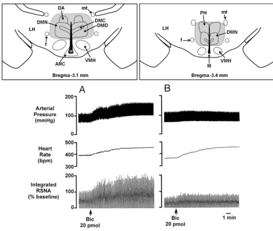
car-Fig. 2. Schematic diagram based on functional and anatomic studies showing descending pathways involved in the organization of the cardiovascular response to emotional stress at different levels of the neuraxis. The DMH is showed as a key integrative region in this response (Lumb, 1990; Thompson and Swanson, 1998; DiMicco et al.,2002), which also involves higher and lower brain regions (Kober et al., 2008; Cechetto and Shoemaker, 2009). From the DMH, descending pathways are represented bilaterally to better illustrate our recent functional findings and hypothesis (see text for details). 1—Amygdala/DMH (Nalivaiko and Blessing, 2001; Quirk and Gehlert, 2003; Quirk et al., 2003); 2—Insular cortex/DMH (Cechetto and Chen,1990;Oppenheimer and Cechetto, 1990; Yasui et al., 1991; Butcher and Cechetto, 1998); 3—Medial prefrontal cortex/DMH (Vertes,2004,
diac sympathetic preganglionic neurons (Amendt et al., 1979; Loewy, 1981; Ter Horst et al., 1996). As demon-strated by Samuels and colleagues (2002), the tachy-cardia evoked by activation of neurons in the DMH with BMI was markedly suppressed after inhibition of neu-rons in the RPa with muscimol (Fig. 3A). Subsequent experiments in conscious rats demonstrated that inhibi-tion of the RPa virtually abolished the tachycardia evoked by acute stress (Fig. 3B) but failed to influence the tachycardia produced by baroreceptor unloading (Zaretsky et al., 2003b). Further support for the involve-ment of neurons in the RPa in the tachycardia evoked by stress comes from experiments showing that direct in-jection of BMI into RPa neurons evokes tachycardia of similar magnitude as that evoked by activation of neu-rons in the DMH (Samuels et al., 2002; Cao and
Morri-son, 2003). Interestingly, inhibition of the RPa in con-scious rats has no effect on baseline HR (Zaretsky et al., 2003b), but blockade of GABAA receptors in the RPa produces sustained increases in cardiac sympathetic activity and in HR even after complete suppression of activity in sympathoexcitatory neurons in the RVLM with muscimol (Cao and Morrison, 2003). Therefore, the car-diac sympathoexcitation and tachycardia evoked by ac-tivation of neurons in the RPa can occur independently of excitation of sympathetic premotor neurons in the RVLM that normally provide the excitatory drive to sup-port basal cardiac sympathetic activity and HR. As pro-posed byCao and Morrison (2003), dorsomedial hypo-thalamic neurons apparently act to reduce or overcome the tonic inhibition of these RPa neurons, which in turn provide an excitatory drive to spinal cardiac sympathetic
Fig. 3.Raphe neurons are a crucial relay in the pathway responsible for stress-induced tachycardia. (A) Inhibition of raphe neurons, through microinjection of the GABAAreceptor agonist muscimol, reduces the increase in HR, but not in BP, evoked by disinhibition of the DMH (by
microinjection of the GABAAreceptor antagonist bicuculline). Left: Example of an original tracing from a representative experiment. Right: Grouped
preganglionic neurons to augment cardiac sympathetic activity and HR (Fig. 2, pathway 9).
PERIAQUEDUCTAL GRAY: A SOURCE OF
EXCITATORY INPUT TO NEURONS
IN THE DMH?
Ultimately, a model that relies on the regulation of neuronal activity through disinhibition must include a mechanism responsible for excitation of the neuronal population being studied (Morrison, 2004). As is seen after acute stress or disinhibition of neurons in the DMH with BMI, a tachycardic response can also be induced by stimulating neurons in the DMH with agonists of EAA receptors (Soltis and DiMicco, 1991a, 1992; Tanaka and McAllen, 2008). The first structure evaluated as a source of excitatory input to DMH neurons was the amygdala, a structure well-known to be involved in stress and anxiety (LeDoux, 2007). Chem-ical stimulation of the amygdala results in cardiovascular changes that are abolished after blockade of glutamatergic receptors in the DMH (Soltis et al., 1998). However, recent attempts to reveal the descending cardiovascular connec-tions from DMH led us also to consider the periaqueductal gray region (PAG) (da Silva et al., 2003, 2006) (Fig. 2
pathway 6).
Our findings that increases in HR and MAP seen in air jet stress were reduced by microinjection of muscimol into the lateral/dorsolateral region of PAG (l/dlPAG) (de Me-nezes et al., 2008), in the same manner that the inhibition of DMH neurons alters the cardiovascular response to air jet stress (Stotz-Potter et al., 1996b), suggested that neu-rons in the l/dlPAG constitute downstream effectors for cardiovascular changes evoked from the DMH. Surpris-ingly, however, we also observed that microinjection of muscimol into the l/dlPAG reduced the increases in plasma adrenocorticotropic hormone (ACTH) evoked by air jet stress. Increases in plasma ACTH seen in this paradigm represent activation of the hypothalamic-pituitary-adrenal axis, a hallmark of the response to stress, and have been proposed to be mediated in large part through a direct projection from neurons in the DMH to the hypothalamic paraventricular nucleus [PVN; for review, see (DiMicco et al., 2002)]. On the other hand, neurons in the l/dlPAG do not project to the PVN (Cameron et al., 1995).
This hypothesis that the PAG is a source of excitatory input to neurons in the DMH during stress was validated by demonstrating that the increases in HR, BP and core body temperature produced by microinjection of the excitatory amino acid (NMDA) into l/dlPAG in conscious rats were markedly attenuated either by neuronal inhibition (micro-injection of muscimol) or by blockade of glutamate trans-mission (microinjection of NBQX⫹Ap5) within the DMH, but not within the PVN (de Menezes et al., 2009). Likewise, microinjection of muscimol into the DMH of anaesthetized rats reduced the increases in BP as well as the increases in phrenic and renal sympathetic nerve activity produced by the activation of the dlPAG (Horiuchi et al., 2009).
Taken together, these data demonstrated that the physiological responses produced by activation of the
l/dl-PAG depend on neuronal activity in the DMH. Thus, the l/dlPAG may represent one of several regions that provide glutamatergic excitation to neurons in the DMH (Fig. 2, pathway 5) whose activation is ultimately responsible for physiological changes seen in experimental stress. Previ-ous data from anatomical studies are consistent with this notion. For instance, it is known that neurons in the l/dlPAG send axonal projections to neurons located in the region of the DMH (Shaikh et al., 1987; Cameron et al., 1995; Siegel et al., 1997). Also, chemical or electrical stimulation of the l/dlPAG increases the expression of c-fos, a marker for neuronal activation (Dragunow and Faull, 1989), in the DMH, where the terminals of projections from the l/dlPAG can also be found (de Oliveira et al., 2000; Borelli et al., 2006). It important to observe that, in the study of de Oliveira, the increase in c-fos expression was restricted to the dorsomedial nucleus and occurred mainly on the side ipsilateral to the stimulation site in the dlPAG. This fact suggests that the increase in c-fos expression within the DMH was due to the specific activation of the neurons in the PAG and not to the generalized behavioral arousal that was also produced. Thus, during stress, afferents from neurons in the l/dlPAG, perhaps along with those from other regions, might act to excite neurons in the DMH. On the other hand, the tonic inhibitory drive that is present under resting conditions might at the same time be with-drawn, thus changing the balance between GABAergic and glutamatergic transmission that occurs in the DMH [see (DiMicco et al., 2002)]. These shifts would then lead to activation of (1) CRH-containing neurons in the PVN to stimulate the secretion of ACTH, and (2) autonomic cen-ters in the brainstem to increase HR, MAP, temperature and respiratory rate. It is important to consider that the projections through which the l/dlPAG influences the DMH do not necessarily have to be direct. For example, the dlPAG has major projections to the cuneiform nucleus and to the superior lateral parabrachial nucleus in the pons (Lisa et al., 1989b; Carrive, 1993; Krout et al., 1998), and these in turn have projections to the hypothalamus, includ-ing the DMH (Bester et al., 1997; Lam et al., 1997).
The results of the studies showing that the physiolog-ical changes produced by the activation of the l/dlPAG neurons depend on neuronal activity in the DMH (de Me-nezes et al., 2009; Horiuchi et al., 2009), combined with data from the earlier studies showing that the changes evoked by disinhibition of the DMH are, also, dependent on the activation of l/dlPAG neurons (da Silva et al., 2003, 2006; de Menezes et al., 2006), requires alternative expla-nations to the hypothesis presented above. In this regard,
evoked from either region. Once again, loss of either source of background facilitation may effectively weaken responses evoked from the other.
NUCLEUS TRACTUS SOLITARIUS: STRESS,
DMH AND BAROREFLEX MODULATION
Acute psychological stress and stimulation of the DMH can both generate physiological and behavioral responses, as described above, with the main cardiovascular effect being increases in HR and BP (Stotz-Potter et al., 1996a,b;
Fontes et al., 2001; DiMicco et al., 2002; da Silva et al., 2003; de Menezes et al., 2006). In addition to these changes, it is known that both stress and stimulation of the DMH can modulate the baroreceptor reflex (Kunos and Varga, 1995; Hatton et al., 1997; Schadt and Hasser, 1998; Sevoz-Couche et al., 2003; McDowall et al., 2006). This modulation during the defense reaction is necessary to ensure that the changes in HR and BP can occur simul-taneously, without compromise of either response (i.e. increases in HR or BP). Studies in conscious animals indicate that during stress the increases in HR and BP are accompanied by resetting of the baroreceptor reflex ( Hat-ton et al., 1997; Schadt and Hasser, 1998). In these stud-ies, the reflex control of HR was reset to higher levels of arterial pressure without any changes in the gain. In addi-tion, recent evidence by Kanbar and colleagues (2007)
demonstrated that the baroreflex control of sympathetic activity is reset and sensitized during emotional stress (Kanbar et al., 2007).
The exact nature of DMH influence in the baroreceptor reflex remains to be determined. An early report by Kunos and Varga showed that ipsilateral intra-nucleus tractus solitarius (NTS) injection of BMI or 2-OH-saclofen (GABAB
antagonist), attenuated the tachycardia elicited by BMI injection into the DMH. The tachycardia was also inhibited by intra-NTS administration of EEA receptor channel blockers. Authors concluded that the descending input from the DMH to the NTS releases GABA via glutamate acting on ionotropic glutamate receptors located on GABAergic interneurons (Kunos and Varga, 1995). This mechanism would inhibit baroreflex bradycardia during ac-tivation of DMH. Similarly, a study by Sevoz-Couche and colleagues (2003) demonstrated that electrical stimulation of the DMH in anaesthetized rats also inhibits baroreflex bradycardia. However, a recent report by McDowal and colleagues showed that chemical disinhibition of DMH neurons resets the baroreflex to higher levels of arterial pressure, in the same way stress does (Hatton et al., 1997; Schadt and Hasser, 2001; Kanbar et al., 2007), with the baroreflex remaining effective and without losing sensitivity (McDowall et al., 2006).
BRAIN FUNCTIONAL ASYMMETRY AND DMH
Left-right differences in the functional properties of bilateral nervous system regions are known as lateralization of function. This phenomenon has been observed at different levels of the neuraxis (Toga and Thompson, 2003; Stephan et al., 2007), including the hypothalamus from
several species (Harris et al., 1996). Studies revealed that, under some conditions, stress may generate lateralized and imbalanced autonomic outflow (Critchley, 2005). This asymmetric autonomic activity may cause cardiac arrhyth-mias, (Lane et al., 1992a,b). Wittling et al. found a functional division between the cerebral hemispheres with the left dom-inant in generating parasympathetic activity to the heart while the right plays a greater role in generating the sympathetic activity to the myocardium (Wittling et al., 1998a,b). If this division is physiological, it may provide a substrate for described cardiac arrhythmias. Pathways from the DMH are predominantly lateralized such that most neurons on one side do not project contralaterally, but rather they are organized as ipsilateral “mirrors” (ter Horst and Luiten, 1986; Thompson et al., 1996). Additionally, anatomic pro-jections from other nuclei involved in autonomic control to sympathetic preganglionic neurons in the intermediolateral column are also lateralized (Amendt et al., 1979; Blessing et al., 1981; Zagon and Smith, 1993).
In the hypothalamus, the hypothesis of functional asymmetry was first reported based upon the observation that electrical stimulation of the right hypothalamus evokes greater tachycardia than that evoked by identical stimula-tion of the left (Fang and Wang, 1962). Recently, we demonstrated that unilateral disinhibition of neurons in the
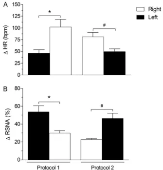
Fig. 4. Maximal changes in cardiovascular parameters from baseline evoked by unilateral microinjection of BMI into right (white bars) and left DMH (black bars) on two experimental protocols. Sequence for injections into unilateral (R or L) DMH followed two different orderli-ness: Protocol 1—First microinjection was done into L-DMH and the second was into R-DMH; Protocol 2—Sequence for injections was reverse of that used in protocol 1, for example, first into R-DMH and second into L-DMH. (A) Heart rate (HR) and (B) renal sympathetic nerve activity (RSNA) sampled from left renal nerve. * P⬍0.05— protocol 1 (L vs. R); # P⬍0.05—protocol 2 (R vs. L). (A) shows
DMH with BMI evokes Fos expression in different nuclei, including the DMH itself, the midline rostral RP, and the lateral septal nucleus, the parvocellular and magnocellular subdivisions of the PVN, the NTS, and the ventrolateral medulla. In the latter bilateral regions, labeling was in-creased on both sides but was markedly greater ipsilateral to the site of DMH stimulation (Zaretskaia et al., 2008). In determining if cardiac sympathoexcitation evoked by acti-vation of neurons on one side of the DMH is transmitted preferentially through ipsilateral relays, we found that dis-inhibition of the right DMH evokes a greater tachycardia than that evoked from the left DMH (Xavier et al., 2009) (Fig. 4). Additionally, disinhibition of the right DMH evokes substantially larger changes in cardiac contractility com-pared to those evoked from left DMH. This effect is inde-pendent of the simultaneous changes in heart rate and afterload and so might be interpreted as a direct positive inotropic effect. Interestingly, in the same study we de-tected a greater number of ectopic beats during the 10 min following injections of BMI into the right DMH (Xavier et al., 2010). This finding prompted us to speculate that recruiting the right DMH during stress exposure might improve the range of cardiac responses and increase the risk of ar-rhythmic episodes.
The possibility that descending input to the DMH from brain structures, such as the insular cortex, may be asym-metric should also be considered (seeFig. 2). First, there is anatomical and functional evidence for connections be-tween the insular cortex and the hypothalamus (Cechetto and Chen, 1990). Second, neural activity in the insular cortex may have an arrythmogenic role according to past findings. In rats, the cardiac effects of stimulation of the insular cortex mimic the repolarization and structural changes that occur with catecholamine-induced cardiomy-opathy seen under certain clinical circumstances, including death following extreme and prolonged stress, and these effects are likely associated with sympathetic neural acti-vation of the ventricular myocardium (Oppenheimer, 2007). Similarly, in humans, insular damage may produce effects on cardiac repolarization (Sander and Klingelhofer, 1994). Third, there is evidence suggesting lateralization and specialization of cardioregulatory function within the insular cortex. The right insular cortex has been primarily implicated with modulation of cardiac sympathetic nerve activity and the left with effects primarily on cardiac vagal activity. Interestingly, patients with stroke lateralized to the left insular cortex reportedly exhibit impaired sympathova-gal balance, with one third of the stroke patients develop-ing sinus tachycardia even in the absence of significant coronary disease (Oppenheimer, 2007). Whether or not insular cortical imbalance might result in consequent asymmetric activity of DMH neurons for triggering adverse cardiac outcomes remains to be determined.
CONCLUSION AND PERSPECTIVES
In conclusion, although many of the details regarding the role of the dorsomedial hypothalamus in the cardiovascu-lar response to emotional stress remain to be determined,
considerable progress has been made in the past few years in determining the central pathways involved. Un-doubtedly, a critical step is to further investigate the impli-cations of the lateralization observed in the descending pathways from the DMH. The role of the DMH in adverse cardiac events observed after cortical stimulation or dam-age deserves extensive investigation. Elucidating the func-tional organization of this network could provide a frame-work for understanding how, in some conditions, stress results in autonomic imbalance resulting in cardiovascular risk.
Acknowledgments—Authors would like to thank the financial sup-port provided by: (1) Conselho Nacional de Desenvolvimento Cientifico e Tecnologico do Brazil (CNPq) and Fundacao de Amparo a Pesquisa do Estado de Minas Gerais (Fapemig-PPM), Brazil; (2) USPHS Grant NS 19883, USA.
REFERENCES
Amat J, Baratta MV, Paul E, Bland ST, Watkins LR, Maier SF (2005) Medial prefrontal cortex determines how stressor controllability affects behavior and dorsal raphe nucleus. Nat Neurosci 8: 365–371.
Amendt K, Czachurski J, Dembowsky K, Seller H (1979) Bulbospinal projections to the intermediolateral cell column: a neuroanatomical study. J Auton Nerv Syst 1:103–107.
Bester H, Besson JM, Bernard JF (1997) Organization of efferent projections from the parabrachial area to the hypothalamus: a Phaseolus vulgaris-leucoagglutinin study in the rat. J Comp Neurol 383:245–281.
Blessing WW (2003) Lower brainstem pathways regulating sympathet-ically mediated changes in cutaneous blood flow. Cell Mol Neuro-biol 23:527–538.
Blessing WW, Goodchild AK, Dampney RA, Chalmers JP (1981) Cell groups in the lower brain stem of the rabbit projecting to the spinal cord, with special reference to catecholamine-containing neurons. Brain Res 221:35–55.
Borelli KG, Ferreira-Netto C, Brandao ML (2006) Distribution of Fos immunoreactivity in the rat brain after freezing or escape elicited by inhibition of glutamic acid decarboxylase or antagonism of GABA-A receptors in the inferior colliculus. Behav Brain Res 170:84 –93. Bush G, Luu P, Posner MI (2000) Cognitive and emotional influences
in anterior cingulate cortex. Trends Cogn Sci 4:215–222. Butcher KS, Cechetto DF (1998) Receptors in lateral hypothalamic
area involved in insular cortex sympathetic responses. Am J Physiol 275:H689 –H696.
Cameron AA, Khan IA, Westlund KN, Cliffer KD, Willis WD (1995) The efferent projections of the periaqueductal gray in the rat: a Phaseo-lus vulgaris-leucoagglutinin study. I. Ascending projections. J Comp Neurol 351:568 –584.
Campos RR, McAllen RM (1999) Cardiac inotropic, chronotropic, and dromotropic actions of subretrofacial neurons of cat RVLM. Am J Physiol 276:R1102–R1111.
Cao WH, Fan W, Morrison SF (2004) Medullary pathways mediating specific sympathetic responses to activation of dorsomedial hypo-thalamus. Neuroscience 126:229 –240.
Cao WH, Morrison SF (2003) Disinhibition of rostral raphe pallidus neurons increases cardiac sympathetic nerve activity and heart rate. Brain Res 980:1–10.
Carrive P (1993) The periaqueductal gray and defensive behavior: functional representation and neuronal organization. Behav Brain Res 58:27– 47.
Cechetto DF, Shoemaker JK (2009) Functional neuroanatomy of au-tonomic regulation. Neuroimage 47:795– 803.
Critchley HD (2005) Neural mechanisms of autonomic, affective, and cognitive integration. J Comp Neurol 493:154 –166.
da Silva LG, de Menezes RC, dos Santos RA, Campagnole-Santos MJ, Fontes MA (2003) Role of periaqueductal gray on the cardio-vascular response evoked by disinhibition of the dorsomedial hy-pothalamus. Brain Res 984:206 –214.
da Silva LG Jr, Menezes RC, Villela DC, Fontes MA (2006) Excitatory amino acid receptors in the periaqueductal gray mediate the car-diovascular response evoked by activation of dorsomedial hypo-thalamic neurons. Neuroscience 139:1129 –1139.
Dampney RA, Horiuchi J, Tagawa T, Fontes MA, Potts PD, Polson JW (2003) Medullary and supramedullary mechanisms regulating sympathetic vasomotor tone. Acta Physiol Scand 177:209 –218. Dampney RA, Tagawa T, Horiuchi J, Potts PD, Fontes M, Polson JW
(2000) What drives the tonic activity of presympathetic neurons in the rostral ventrolateral medulla? Clin Exp Pharmacol Physiol 27:1049 –1053.
de Menezes RC, Zaretsky DV, Fontes MA, Dimicco JA (2006) Micro-injection of muscimol into caudal periaqueductal gray lowers body temperature and attenuates increases in temperature and activity evoked from the dorsomedial hypothalamus. Brain Res 1092: 129 –137.
de Menezes RC, Zaretsky DV, Fontes MA, DiMicco JA (2009) Cardio-vascular and thermal responses evoked from the periaqueductal grey require neuronal activity in the hypothalamus. J Physiol 587:1201–1215.
de Menezes RC, Zaretsky DV, Sarkar S, Fontes MA, Dimicco JA (2008) Microinjection of muscimol into the periaqueductal gray suppresses cardiovascular and neuroendocrine response to air jet stress in conscious rats. Am J Physiol Regul Integr Comp Physiol 295:R881–R890.
De Novellis V, Stotz-Potter EH, Morin SM, Rossi F, DiMicco JA (1995) Hypothalamic sites mediating cardiovascular effects of microin-jected bicuculline and EAAs in rats. Am J Physiol 269:R131–R140. de Oliveira RW, Del Bel EA, Guimaraes FS (2000) Behavioral and c-fos expression changes induced by nitric oxide donors microin-jected into the dorsal periaqueductal gray. Brain Res Bull 51:457– 464.
DiMicco JA, Samuels BC, Zaretskaia MV, Zaretsky DV (2002) The dorsomedial hypothalamus and the response to stress: part renais-sance, part revolution. Pharmacol Biochem Behav 71:469 – 480. Dragunow M, Faull R (1989) The use of c-fos as a metabolic marker in
neuronal pathway tracing. J Neurosci Methods 29:261–265. Fang HS, Wang SC (1962) Cardioaccelerator and cardioaugmentor
points in hypothalamus of the dog. Am J Physiol 203:147–150. Farkas E, Jansen AS, Loewy AD (1998) Periaqueductal gray matter
input to cardiac-related sympathetic premotor neurons. Brain Res 792:179 –192.
Fontes MA, Tagawa T, Polson JW, Cavanagh SJ, Dampney RA (2001) Descending pathways mediating cardiovascular response from dorsomedial hypothalamic nucleus. Am J Physiol Heart Circ Physiol 280:H2891–H2901.
van Ginkel HJ (2008). Urban future. Nature 456:32–33.
Harris JA, Guglielmotti V, Bentivoglio M (1996) Diencephalic asymme-tries. Neurosci Biobehav Rev 20:637– 643.
Hatton DC, Brooks V, Qi Y, McCarron DA (1997) Cardiovascular response to stress: baroreflex resetting and hemodynamics. Am J Physiol 272:R1588 –R1594.
Hoover WB, Vertes RP (2007) Anatomical analysis of afferent projec-tions to the medial prefrontal cortex in the rat. Brain Struct Funct 212:149 –179.
Horiuchi J, Killinger S, Dampney RA (2004a) Contribution to sympa-thetic vasomotor tone of tonic glutamatergic inputs to neurons in the RVLM. Am J Physiol Regul Integr Comp Physiol 287:R1335– R1343.
Horiuchi J, McAllen RM, Allen AM, Killinger S, Fontes MA, Dampney RA (2004b) Descending vasomotor pathways from the dorsome-dial hypothalamic nucleus: role of medullary raphe and RVLM. Am J Physiol Regul Integr Comp Physiol 287:R824 –R832. Horiuchi J, McDowall LM, Dampney RA (2009) Vasomotor and
respi-ratory responses evoked from the dorsolateral periaqueductal grey are mediated by the dorsomedial hypothalamus. J Physiol 587:5149 –5162.
Hosoya Y, Ito R, Kohno K (1987) The topographical organization of neurons in the dorsal hypothalamic area that project to the spinal cord or to the nucleus raphe pallidus in the rat. Exp Brain Res 66:500 –506.
Huang J, Chowhdury SI, Weiss ML (2002) Distribution of sympathetic preganglionic neurons innervating the kidney in the rat: PRV transneuronal tracing and serial reconstruction. Auton Neurosci 95:57–70.
Hudson PM, Lumb BM (1996) Neurones in the midbrain periaqueduc-tal grey send collateral projections to nucleus raphe magnus and the rostral ventrolateral medulla in the rat. Brain Res 733:138 –141. Hunt JL, Zaretsky DV, Sarkar S, Dimicco JA (2010) Dorsomedial hypothalamus mediates autonomic, neuroendocrine, and locomo-tor responses evoked from the medial preoptic area. Am J Physiol Regul Integr Comp Physiol 298:R130 –R140.
Hurley KM, Herbert H, Moga MM, Saper CB (1991) Efferent projec-tions of the infralimbic cortex of the rat. J Comp Neurol 308: 249 –276.
Jansen AS, Wessendorf MW, Loewy AD (1995) Transneuronal label-ing of CNS neuropeptide and monoamine neurons after pseudo-rabies virus injections into the stellate ganglion. Brain Res 683:1–24.
Johnson PL, Shekhar A (2006) Panic-prone state induced in rats with GABA dysfunction in the dorsomedial hypothalamus is mediated by NMDA receptors. J Neurosci 26:7093–7104.
Kanbar R, Orea V, Chapuis B, Barres C, Julien C (2007) A transfer function method for the continuous assessment of baroreflex con-trol of renal sympathetic nerve activity in rats. Am J Physiol Regul Integr Comp Physiol 293:R1938 –R1946.
Kober H, Barrett LF, Joseph J, Bliss-Moreau E, Lindquist K, Wager TD (2008) Functional grouping and cortical-subcortical interactions in emotion: a meta-analysis of neuroimaging studies. Neuroimage 42:998 –1031.
Koutcherov Y, Mai JK, Ashwell KW, Paxinos G (2004) Organisation of the human dorsomedial hypothalamic nucleus. Neuroreport 15: 107–111.
Koutcherov Y, Mai JK, Paxinos G (2003) Hypothalamus of the human fetus. J Chem Neuroanat 26:253–270.
Krout KE, Jansen AS, Loewy AD (1998) Periaqueductal gray matter projection to the parabrachial nucleus in rat. J Comp Neurol 401: 437– 454.
Kunos G, Varga K (1995) The tachycardia associated with the defense reaction involves activation of both GABAA and GABAB receptors in the nucleus tractus solitarii. Clin Exp Hypertens 17:91–100. Lam W, Gundlach AL, Verberne AJ (1997) Neuronal activation in the
forebrain following electrical stimulation of the cuneiform nucleus in the rat: hypothalamic expression of c-fos and NGFI-A messenger RNA. Neuroscience 78:1069 –1085.
Lane JD, Adcock RA, Burnett RE (1992a) Respiratory sinus arrhyth-mia and cardiovascular responses to stress. Psychophysiology 29:461– 470.
Lane RD, Wallace JD, Petrosky PP, Schwartz GE, Gradman AH (1992b) Supraventricular tachycardia in patients with right hemi-sphere strokes. Stroke 23:362–366.
LeDoux J (2007) The amygdala. Curr Biol 17:R868 –R874.
Leor J, Poole WK, Kloner RA (1996) Sudden cardiac death triggered by an earthquake. N Engl J Med 334:413– 419.
cardio-vascular response to experimental stress in rats. Pharmacol Res 21 (Suppl 1):9 –10.
Lisa M, Marmo E, Wible JH Jr, DiMicco JA (1989b) Injection of mus-cimol into posterior hypothalamus blocks stress-induced tachycar-dia. Am J Physiol 257:R246 –R251.
Lloyd-Jones D, Adams R, Carnethon M, De Simone G, Ferguson TB, Flegal K, Ford E, Furie K, Go A, Greenlund K, Haase N, Hailpern S, Ho M, Howard V, Kissela B, Kittner S, Lackland D, Lisabeth L, Marelli A, McDermott M, Meigs J, Mozaffarian D, Nichol G, O’Donnell C, Roger V, Rosamond W, Sacco R, Sorlie P, Stafford R, Steinberger J, Thom T, Wasserthiel-Smoller S, Wong N, Wylie-Rosett J, Hong Y (2009) Heart disease and stroke statistics—2009 update: a report from the American Heart Association Statistics Committee and Stroke Statistics Subcommittee. Circulation 119:480 – 486.
Loewy AD (1981) Raphe pallidus and raphe obscurus projections to the intermediolateral cell column in the rat. Brain Res 222:129 – 133.
Lovallo WR, Gerin W (2003) Psychophysiological reactivity: mecha-nisms and pathways to cardiovascular disease. Psychosom Med 65:36 – 45.
Lumb BM (1990) Hypothalamic influences on viscero-somatic neu-rones in the lower thoracic spinal cord of the anaesthetized rat. J Physiol 424:427– 444.
McAllen RM, May CN, Shafton AD (1995) Functional anatomy of sympathetic premotor cell groups in the medulla. Clin Exp Hyper-tens 17:209 –221.
McDougall SJ, Widdop RE, Lawrence AJ (2004) Medial prefrontal cortical integration of psychological stress in rats. Eur J Neurosci 20:2430 –2440.
McDowall LM, Horiuchi J, Killinger S, Dampney RA (2006) Modulation of the baroreceptor reflex by the dorsomedial hypothalamic nu-cleus and perifornical area. Am J Physiol Regul Integr Comp Physiol 290:R1020 –R1026.
Mittleman MA, Maclure M, Sherwood JB, Mulry RP, Tofler GH, Jacobs SC, Friedman R, Benson H, Muller JE (1995) Triggering of acute myocardial infarction onset by episodes of anger. Determinants of Myocardial Infarction Onset Study Investigators. Circulation 92:1720 –1725.
Morrison SF (2004) Central pathways controlling brown adipose tissue thermogenesis. News Physiol Sci 19:67–74.
Morrison SF, Sved AF, Passerin AM (1999) GABA-mediated inhibition of raphe pallidus neurons regulates sympathetic outflow to brown adipose tissue. Am J Physiol 276:R290 –R297.
Nalivaiko E, Blessing WW (2001) Raphe region mediates changes in cutaneous vascular tone elicited by stimulation of amygdala and hypothalamus in rabbits. Brain Res 891:130 –137.
Nalivaiko E, Ootsuka Y, Blessing WW (2005) Activation of 5-HT1A receptors in the medullary raphe reduces cardiovascular changes elicited by acute psychological and inflammatory stresses in rab-bits. Am J Physiol Regul Integr Comp Physiol 289:R596 –R604. Okamura H, Abitbol M, Julien JF, Dumas S, Berod A, Geffard M,
Kitahama K, Bobillier P, Mallet J, Wiklund L (1990) Neurons con-taining messenger RNA encoding glutamate decarboxylase in rat hypothalamus demonstrated byin situhybridization, with special emphasis on cell groups in medial preoptic area, anterior hypotha-lamic area and dorsomedial hypothahypotha-lamic nucleus. Neuroscience 39:675– 699.
Oppenheimer S (2007) Cortical control of the heart. Cleve Clin J Med 74 (Suppl 1):S27–S29.
Oppenheimer SM, Cechetto DF (1990) Cardiac chronotropic organi-zation of the rat insular cortex. Brain Res 533:66 –72.
Paxinos G, Watson C (1986) The rat brain in stereotaxic coordinates. New York: Academic Press.
Quirk GJ, Gehlert DR (2003) Inhibition of the amygdala: key to path-ological states? Ann N Y Acad Sci 985:263–272.
Quirk GJ, Likhtik E, Pelletier JG, Pare D (2003) Stimulation of medial prefrontal cortex decreases the responsiveness of central amygdala output neurons. J Neurosci 23:8800 – 8807.
Radley JJ, Gosselink KL, Sawchenko PE (2009) A discrete GABAergic relay mediates medial prefrontal cortical inhibition of the neuroen-docrine stress response. J Neurosci 29:7330 –7340.
Samuels BC, Zaretsky DV, DiMicco JA (2002) Tachycardia evoked by disinhibition of the dorsomedial hypothalamus in rats is mediated through medullary raphe. J Physiol 538:941–946.
Samuels BC, Zaretsky DV, DiMicco JA (2004) Dorsomedial hypotha-lamic sites where disinhibition evokes tachycardia correlate with location of raphe-projecting neurons. Am J Physiol Regul Integr Comp Physiol 287:R472–R478.
Sander D, Klingelhofer J (1994) Changes of circadian blood pressure patterns after hemodynamic and thromboembolic brain infarction. Stroke 25:1730 –1737.
Schadt JC, Hasser EM (1998) Hemodynamic effects of acute stres-sors in the conscious rabbit. Am J Physiol 274:R814 –R821. Schadt JC, Hasser EM (2001) Defense reaction alters the response to
blood loss in the conscious rabbit. Am J Physiol Regul Integr Comp Physiol 280:R985–R993.
Sevoz-Couche C, Comet MA, Hamon M, Laguzzi R (2003) Role of nucleus tractus solitarius 5-HT3 receptors in the defense reaction-induced inhibition of the aortic baroreflex in rats. J Neurophysiol 90:2521–2530.
Shaikh MB, Barrett JA, Siegel A (1987) The pathways mediating affective defense and quiet biting attack behavior from the midbrain central gray of the cat: an autoradiographic study. Brain Res 437:9 –25.
Shekhar A, Johnson PL, Sajdyk TJ, Fitz SD, Keim SR, Kelley PE, Gehlert DR, DiMicco JA (2006) Angiotensin-II is a putative neu-rotransmitter in lactate-induced panic-like responses in rats with disruption of GABAergic inhibition in the dorsomedial hypothala-mus. J Neurosci 26:9205–9215.
Siegel A, Schubert KL, Shaikh MB (1997) Neurotransmitters regulating defensive rage behavior in the cat. Neurosci Biobehav Rev 21:733–742.
Smith PA, Graham LN, Mackintosh AF, Stoker JB, Mary DA (2004) Relationship between central sympathetic activity and stages of human hypertension. Am J Hypertens 17:217–222.
Soltis RP, Cook JC, Gregg AE, Stratton JM, Flickinger KA (1998) EAA receptors in the dorsomedial hypothalamic area mediate the car-diovascular response to activation of the amygdala. Am J Physiol 275:R624 –R631.
Soltis RP, DiMicco JA (1991a) GABAA and excitatory amino acid receptors in dorsomedial hypothalamus and heart rate in rats. Am J Physiol 260:R13–R20.
Soltis RP, DiMicco JA (1991b) Interaction of hypothalamic GABAA and excitatory amino acid receptors controlling heart rate in rats. Am J Physiol 261:R427–R433.
Soltis RP, DiMicco JA (1992) Hypothalamic excitatory amino acid receptors mediate stress-induced tachycardia in rats. Am J Physiol 262:R689 –R697.
Stephan KE, Fink GR, Marshall JC (2007) Mechanisms of hemispheric specialization: insights from analyses of connectivity. Neuropsy-chologia 45:209 –228.
Stotz-Potter EH, Morin SM, DiMicco JA (1996a) Effect of microinjec-tion of muscimol into the dorsomedial or paraventricular hypotha-lamic nucleus on air stress-induced neuroendocrine and cardio-vascular changes in rats. Brain Res 742:219 –224.
Taylor RB, Weaver LC (1992) Spinal stimulation to locate pregangli-onic neurons controlling the kidney, spleen, or intestine. Am J Physiol 263:H1026 –H1033.
Ter Horst GJ, Hautvast RW, De Jongste MJ, Korf J (1996) Neuroanat-omy of cardiac activity-regulating circuitry: a transneuronal retro-grade viral labelling study in the rat. Eur J Neurosci 8:2029 –2041. ter Horst GJ, Luiten PG (1986) The projections of the dorsomedial
hypothalamic nucleus in the rat. Brain Res Bull 16:231–248. Thompson RH, Canteras NS, Swanson LW (1996) Organization of
projections from the dorsomedial nucleus of the hypothalamus: a PHA-L study in the rat. J Comp Neurol 376:143–173.
Thompson RH, Swanson LW (1998) Organization of inputs to the dorsomedial nucleus of the hypothalamus: a reexamination with fluorogold and PHAL in the rat. Brain Res Brain Res Rev 27:89 –118.
Toga AW, Thompson PM (2003) Mapping brain asymmetry. Nat Rev Neurosci 4:37– 48.
Vertes RP (2004) Differential projections of the infralimbic and prelim-bic cortex in the rat. Synapse 51:32–58.
Vertes RP (2006) Interactions among the medial prefrontal cortex, hippocampus and midline thalamus in emotional and cognitive processing in the rat. Neuroscience 142:1–20.
Villela DC, da Silva LG Jr, Fontes MA (2009) Activation of 5-HT receptors in the periaqueductal gray attenuates the tachycardia evoked from dorsomedial hypothalamus. Auton Neurosci 148: 36 – 43.
Wittling W, Block A, Genzel S, Schweiger E (1998a) Hemisphere asymmetry in parasympathetic control of the heart. Neuropsycho-logia 36:461– 468.
Wittling W, Block A, Schweiger E, Genzel S (1998b) Hemisphere asymmetry in sympathetic control of the human myocardium. Brain Cogn 38:17–35.
Xavier CH, Beig MI, Ianzer D, Fontes MA, Nalivaiko E (2010) Cardiac chronotropic and inotropic responses evoked from right or left sides of dorsomedial hypothalamus. FASEB J 24:1019 –1020. Xavier CH, Nalivaiko E, Beig MI, Menezes GB, Cara DC,
Campag-nole-Santos MJ, Fontes MA (2009) Functional asymmetry in the descending cardiovascular pathways from dorsomedial hypotha-lamic nucleus. Neuroscience 164:1360 –1368.
Yasui Y, Breder CD, Saper CB, Cechetto DF (1991) Autonomic re-sponses and efferent pathways from the insular cortex in the rat. J Comp Neurol 303:355–374.
Yoshida K, Li X, Cano G, Lazarus M, Saper CB (2009) Parallel preoptic pathways for thermoregulation. J Neurosci 29:11954 – 11964.
Zagon A, Smith AD (1993) Monosynaptic projections from the rostral ventrolateral medulla oblongata to identified sympathetic pregan-glionic neurons. Neuroscience 54:729 –743.
Zaretskaia MV, Zaretsky DV, DiMicco JA (2003) Role of the dorsome-dial hypothalamus in thermogenesis and tachycardia caused by microinjection of prostaglandin E2 into the preoptic area in anes-thetized rats. Neurosci Lett 340:1– 4.
Zaretskaia MV, Zaretsky DV, Sarkar S, Shekhar A, DiMicco JA (2008) Induction of Fos-immunoreactivity in the rat brain follow-ing disinhibition of the dorsomedial hypothalamus. Brain Res 1200:39 –50.
Zaretsky DV, Zaretskaia MV, DiMicco JA (2003a) Stimulation and blockade of GABA(A) receptors in the raphe pallidus: ef-fects on body temperature, heart rate, and blood pressure in conscious rats. Am J Physiol Regul Integr Comp Physiol 285: R110 –R116.
Zaretsky DV, Zaretskaia MV, Samuels BC, Cluxton LK, DiMicco JA (2003b) Microinjection of muscimol into raphe pallidus suppresses tachycardia associated with air stress in conscious rats. J Physiol 546:243–250.