A b s t r a c t A b s t r a c tA b s t r a c t A b s t r a c t A b s t r a c t
Neurochemically distinct systems interact in the regulation of sleep and wakefulness. Wakefulness is promoted by the aminergic, acetylcholinergic brainstem and hypothalamic systems. Each of these arousal systems supports wakefulness, and coordinated activity is required for alertness and electroencephalogram-proven brain activation. Neurons in the pons and preoptic area control rapid eye movement and non-rapid eye movement sleep. Mutual inhibition among these sleep-wake mechanisms generates behavioral states. An updated understanding of these systems should allow clinicians and researchers to better understand the effects of drugs, injury and neurologic disease on sleep and wakefulness.
K KK K
Keywords:eywords:eywords:eywords:eywords: Sleep/physiology; Sleep disorders; Sleep disorders, circadian rhythm; Wakefulness/physiology; Sleep deprivation; Neuropeptides; Anterior hypothalamic nucleus; Brain stem
R e s u m o R e s u m oR e s u m o R e s u m o R e s u m o
Três sub-divisões hipotalâmicas são importantes no ciclo sono-vigília: o hipotálamo anterior (núcleos gabaérgicos e núcleos supraquiasmáticos), o hipotálamo posterior (núcleo túbero-mamilar histaminérgico) e o hipotálamo lateral (sistema hipocretinas). O sistema gabaérgico inibitório do núcleo pré-óptico ventro-lateral (VLPO) do hipotálamo anterior é responsável pelo início e manutenção do sono NREM. Os neurônios supraquiasmáticos (NSQs) do hipotálamo anterior são responsáveis pelo ritmo circadiano do ciclo sono-vigília. Os núcleos aminérgicos, histaminérgicos, as hipocretinas e núcleos colinérgicos do prosencéfalo basal apresentam-se ativos durante a vigília, inibindo o núcleo pré-óptico ventro-lateral, promovendo a vigília. O processo de inibição-estimulação é a base do modelo da interação recíproca entre os grupos de células wake-off-sleep-on e células wake-off-sleep-on reguladores do ciclo sono-vigília. O modelo da interação recíproca também se aplica aos núcleos colinérgicos (células REM-on) e aminérgicos (células REM-off) do tronco cerebral no controle temporal do sono REM-NREM.
Descritores: Descritores: Descritores: Descritores:
Descritores: Sono/fisiologia; Transtornos do sono, do ritmo circadiano; Vigília/fisiologia; Privação do sono; Neuropeptídeos; Núcleo hipotálamico anterior; Tronco encefálico
1Interdepartmental Center for Sleep Studies at the Universidade de São Paulo School of Medicine Hospital das Clínicas, São Paulo, Brazil 2Clinical Neurology Department of the Universidade de São Paulo School of Medicine Hospital das Clínicas, São Paulo, Brazil
Correspondence Flávio Alóe
Rua Bergamota 326 #172 05468-000 São Paulo, SP Phone: (11) 3071-0972 E-mail: [email protected]
Sleep-wake cycle mechanisms
Mecanismos do ciclo sono-vigília
Flávio Alóe,
1Alexandre Pinto de Azevedo,
1I n t r o d u c t i o n I n t r o d u c t i o n I n t r o d u c t i o n I n t r o d u c t i o n I n t r o d u c t i o n
Sleep is a complex behavioral state and one of the great mysteries of modern neuroscience.1 Sleep began to be understood when it was associated with rapid eye movement (REM) by Aserinsky and Kleitman2 in 1953. In 1998, the discovery of the hypothalamic peptides known as orexins (or hypocretins), together with the identification of their role in the sleep-wake cycle and in the physiopathology of narcolepsy-cataplexy, contradicted conventional wisdom regarding the hypothalamus and the sleep-wake cycle, the control of which had previously been exclusively attributed to structures located in the brain stem and thalamus.3-6 Control of this cycle is now attributed to the hypothalamic systems and to their respective functional interactions with the circadian timing system.6-7 In the present study, we describe recent advances in the neurobiology of sleep.
C h o l i n e r g i c a n d m o n o a m i n e r g i c s y s t e m s C h o l i n e r g i c a n d m o n o a m i n e r g i c s y s t e m s C h o l i n e r g i c a n d m o n o a m i n e r g i c s y s t e m s C h o l i n e r g i c a n d m o n o a m i n e r g i c s y s t e m s C h o l i n e r g i c a n d m o n o a m i n e r g i c s y s t e m s
Normal sleep consists of alternation between REM and NREM stages. Characterized by the presence of synchronized waves in the electroencephalogram (EEG), NREM sleep can be subdivided into four phases (phases 3 and 4 correspond to slow-wave sleep or delta sleep). The REM sleep stage is characterized by EEG desynchronization and low-amplitude waves.
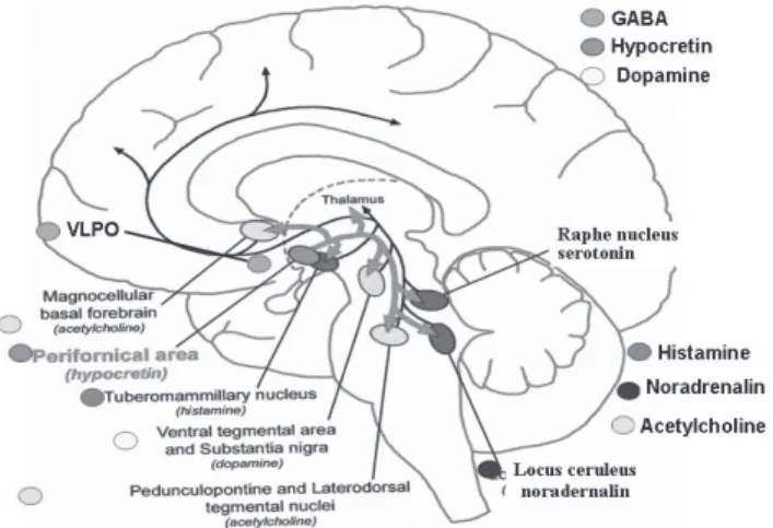
The synchronization-desynchronization of EEG waves during NREM-REM sleep and wakefulness is a consequence of neural activity in the thalamocortical circuits (reticular nuclei in the thalamus and cerebral cortex), derived from the interaction between monoaminergic and cholinergic nuclei in the brain stem.7-8 The monoaminergic system of ascending reticular activation is composed of the serotoninergic dorsal raphe nuclei (DRN), noradrenergic locus coeruleus (LC) of the brain stem and the histaminergic tuberomammillary nucleus (TMN) in the posterior hypothalamus, which are diffusely projected to the cortex and reticular nuclei of the thalamus (Figure 1).8
The thalamocortical circuit and the aminergic-cholinergic
projections are responsible for the desynchronization of the EEG during wakefulness. High aminergic activity during active wakefulness activates the thalamocortical circuits but is reduced during NREM sleep, and is absent during REM sleep. Aminergic neurons are called wake-on-and-sleep-off cells. The cerebral cortex is aminergically demodulated during REM sleep due to the lack of hypocretin tone.6,8-9
Aminergic systems project to the anterior hypothalamus, inhibiting GABAergic and galaninergic cells in the ventrolateral preoptic area (VLPO).7 Cholinergic neurons in the dorsolateral pontine nucleus, pedunculopontine tegmental nucleus and nucleus basalis of the forebrain, under the inhibitory control of the DRN-LC system, make excitatory connections in the thalamic reticular nuclei and project to the thalamo-limbic region (cortex and amygdala) as well as directly into the cortex (Figure 1).7
These cholinergic thalamocortical and limbic thalamocortical projections are fundamental for desynchronization of the EEG during wakefulness and during REM sleep.9 In contrast with aminergic activity, which is absent during REM sleep, the cholinergic activity of the dorsolateral pontine and pedunculopontine tegmental nuclei, as well as that in the nucleus basalis of the forebrain, is at a maximum during REM sleep and wakefulness but is minimal or absent during NREM sleep.6,8-9 Therefore, cholinergic nuclei are activated during wakefulness and during REM sleep with EEG desynchronization. Cholinergic cells are known as "REM-and-wake-on" cells.7
H o w e v e r, t h e r e i s a d i f f e r e n c e b e t w e e n t h e E E G desynchronization occurring during REM sleep and that seen in a state of wakefulness. During REM sleep, the aminergic systems are not active and cholinergic activation activates the cortex directly. During wakefulness, aminergic, dopaminergic, hypocretin and cholinergic systems are active (cortical aminergic modulation).7-9 The difference in the thalamocortical activation process between REM sleep and wakefulness provides an opportunity to understand certain sleep alterations. For example, depressive disorders (reduced REM-sleep latency/ cholinergic hyperactivity) and Alzheimer's disease (reduced
Figure 1 - Aminergic and cholinergic pathways Figure 1 - Aminergic and cholinergic pathways Figure 1 - Aminergic and cholinergic pathways Figure 1 - Aminergic and cholinergic pathways Figure 1 - Aminergic and cholinergic pathways
Ascending projections from the brain stem via the thalamus, Ascending projections from the brain stem via the thalamus, Ascending projections from the brain stem via the thalamus, Ascending projections from the brain stem via the thalamus, Ascending projections from the brain stem via the thalamus, pos-terior hypothalamus and basal forebrain (BF). Neurons in the terior hypothalamus and basal forebrain (BF). Neurons in the terior hypothalamus and basal forebrain (BF). Neurons in the terior hypothalamus and basal forebrain (BF). Neurons in the terior hypothalamus and basal forebrain (BF). Neurons in the dorsolateral nucleus (DLN) and pedunculopontine tegmental (PPT) dorsolateral nucleus (DLN) and pedunculopontine tegmental (PPT) dorsolateral nucleus (DLN) and pedunculopontine tegmental (PPT) dorsolateral nucleus (DLN) and pedunculopontine tegmental (PPT) dorsolateral nucleus (DLN) and pedunculopontine tegmental (PPT) nucleus (blue circles) send cholinergic fibers to the thalamus and nucleus (blue circles) send cholinergic fibers to the thalamus and nucleus (blue circles) send cholinergic fibers to the thalamus and nucleus (blue circles) send cholinergic fibers to the thalamus and nucleus (blue circles) send cholinergic fibers to the thalamus and directly to the cortex. Aminergic nuclei (green circles) are diffusely directly to the cortex. Aminergic nuclei (green circles) are diffusely directly to the cortex. Aminergic nuclei (green circles) are diffusely directly to the cortex. Aminergic nuclei (green circles) are diffusely directly to the cortex. Aminergic nuclei (green circles) are diffusely projected straight to the cor
projected straight to the cor projected straight to the cor projected straight to the cor
projected straight to the cortex. Ttex. Ttex. Ttex. Ttex. Tuberomamillaruberomamillaruberomamillaruberomamillar y nuclei (TMN):uberomamillary nuclei (TMN):y nuclei (TMN):y nuclei (TMN):y nuclei (TMN): histamine. Dorsal raphe nucleus (DRN): serotonin (5-HT). Locus histamine. Dorsal raphe nucleus (DRN): serotonin (5-HT). Locus histamine. Dorsal raphe nucleus (DRN): serotonin (5-HT). Locus histamine. Dorsal raphe nucleus (DRN): serotonin (5-HT). Locus histamine. Dorsal raphe nucleus (DRN): serotonin (5-HT). Locus coeruleus (LC): noradrenalin (NA). Ventrolateral preoptic area coeruleus (LC): noradrenalin (NA). Ventrolateral preoptic area coeruleus (LC): noradrenalin (NA). Ventrolateral preoptic area coeruleus (LC): noradrenalin (NA). Ventrolateral preoptic area coeruleus (LC): noradrenalin (NA). Ventrolateral preoptic area (VLPO): GABA and galanin.
(VLPO): GABA and galanin. (VLPO): GABA and galanin. (VLPO): GABA and galanin. (VLPO): GABA and galanin.
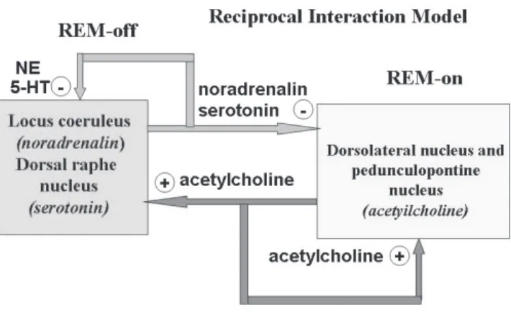
Figure 2 - Reciprocal Interaction Model Figure 2 - Reciprocal Interaction Model Figure 2 - Reciprocal Interaction Model Figure 2 - Reciprocal Interaction Model Figure 2 - Reciprocal Interaction Model
Cholinergic on cells and serotoninergic-noradrenergic Cholinergic on cells and serotoninergic-noradrenergic Cholinergic on cells and serotoninergic-noradrenergic Cholinergic on cells and serotoninergic-noradrenergic Cholinergic on cells and serotoninergic-noradrenergic REM-off cells. During wakefulness, aminergic REM-REM-off system is tonically off cells. During wakefulness, aminergic REM-off system is tonically off cells. During wakefulness, aminergic REM-off system is tonically off cells. During wakefulness, aminergic REM-off system is tonically off cells. During wakefulness, aminergic REM-off system is tonically activated, causing EEG desynchronization and inhibiting cholinergic activated, causing EEG desynchronization and inhibiting cholinergic activated, causing EEG desynchronization and inhibiting cholinergic activated, causing EEG desynchronization and inhibiting cholinergic activated, causing EEG desynchronization and inhibiting cholinergic REM-on cells. During REM sleep, aminergic REM-off cells are REM-on cells. During REM sleep, aminergic REM-off cells are REM-on cells. During REM sleep, aminergic REM-off cells are REM-on cells. During REM sleep, aminergic REM-off cells are REM-on cells. During REM sleep, aminergic REM-off cells are silenced and the cholinergic system, free from inhibitory influences, silenced and the cholinergic system, free from inhibitory influences, silenced and the cholinergic system, free from inhibitory influences, silenced and the cholinergic system, free from inhibitory influences, silenced and the cholinergic system, free from inhibitory influences, reaches its peak.
REM-sleep time/cholinergic hypoactivity) are accompanied by specific alterations in sleep architecture.10-11 Studies involving 18-fluoro-2-deoxyglucose positron emission tomography (18-FDG PET) of patients suffering from major depression have shown cholinergic hyperactivity affecting the metabolism in the limbic and paralimbic areas.12-17 This hyperactivity is compatible with the hypothesis of unbalanced aminergic-cholinergic limbic dysfunction.
R e c i p r o c a l i n t e r a c t i o n m o d e l R e c i p r o c a l i n t e r a c t i o n m o d e l R e c i p r o c a l i n t e r a c t i o n m o d e l R e c i p r o c a l i n t e r a c t i o n m o d e l R e c i p r o c a l i n t e r a c t i o n m o d e l
The reciprocal interaction model is a functional model that establishes wakefulness as a predominantly aminergic state, REM sleep as a predominantly muscarinic cholinergic state and NREM sleep as an intermediate state.18 The model proposes two types of cell groups located in the reticular formation: the cholinergic REM-on cells and the serotoninergic-noradrenergic REM-off cells (Figure 2).
During wakefulness, the aminergic REM-off system, which is tonically activated, generating EEG desynchronization, inhibits the cholinergic REM-on system, suppressing REM sleep.7,18 During REM sleep, aminergic REM-off cells, as well as the cholinergic system, are free from inhibitory influences and reach their peak. Therefore, REM sleep only occurs when the aminergic system suspends its inhibitory effect on cholinergic activity (Figure 2).7,18
The histaminergic system of the posterior hypothalamus (TMN) and the dopaminergic system in the ventral tegmental area join the serotoninergic and noradrenergic systems in order to inhibit cholinergic REM-on cells. The excitatory stimulation caused by lateral hypothalamus hypocretins in the REM-on aminergic system is similarly affected (Figure 3).6-9
Glutamatergic excitatory interneurons of the pontine reticular formation, interposed between REM-on cells, become active as aminergic inhibition decreases during NREM sleep. These neurons, working in self-excitatory loops, activate REM-on cells, exponentially increasing the discharge frequency and triggering the initiation and maintenance of REM sleep.7,19-20
D o p a m i n e r g i c m e c h a n i s m s o f s l e e p r e g u l a t i o n D o p a m i n e r g i c m e c h a n i s m s o f s l e e p r e g u l a t i o n D o p a m i n e r g i c m e c h a n i s m s o f s l e e p r e g u l a t i o n D o p a m i n e r g i c m e c h a n i s m s o f s l e e p r e g u l a t i o n D o p a m i n e r g i c m e c h a n i s m s o f s l e e p r e g u l a t i o n Neurons in the mesencephalic ventral tegmental area (VTA), situated next to the substantia nigra, project to the cerebral cortex via the mesocortical limbic pathway.7 Excitatory axons
of the VTA are projected to the LC and the limbic thalamic nuclei, connecting the mesostriatal dopaminergic system directly with the ascending activating system responsible for wakefulness.7 Spinal cord dopaminergic neurons from the diencephalic tract, originating in the motor nuclei of the thalamus, project to the lower motor neurons and are involved in the genesis of the restless legs syndrome and periodic limb m o v e m e n t d i s o r d e r, p a t h o l o g i e s t h a t r e s p o n d t o D - 2 dopaminergic agonists.21
Neurons in the mesencephalic ventral tegmental area receive excitatory synapses from hypocretinergic cells in the lateral hypothalamus which, together with the excitatory activity of aminergic, cholinergic and hypocretin systems, promote EEG desynchronization during wakefulness.6-7 For example, dopaminergic deficiency in Parkinson's disease causes sleepiness, restless legs syndrome and involuntary movements in the lower limbs.21
The effect of dopamine agonists during the sleep-wake cycle depends on the type of dopaminergic receptors (D1, D2, etc.) that are stimulated (pre- or postsynaptically), doses and types of agonists.21 In patients with Parkinson's disease, low doses of D2 dopamine agonists cause sleepiness, as well as increasing the duration of REM sleep and the frequency of daytime "sleep attacks".21 However, higher doses actually suppress NREM and REM sleep due to the stimulation of D1 receptors.21 The soporific effects of D2 agonists are mediated by inhibitory D2 autoreceptors located in the cytoplasm of cells in the mesocortical limbic pathway. The stimulation of these autoreceptors inhibits the activity of the mesocortical limbic pathway, inducing REM sleep.21
Psychomotor stimulants used for the treatment of excessive sleepiness, such as amphetamines, methylphenidate and pemoline, generate an increase in noradrenalin, dopamine and serotonin neurotransmission.21-25 Modafinil acts in a distinct way, increasing noradrenergic inhibition of the LC over the GABAergic VLPO of the anterior hypothalamus. Modafinil inhibits the dopamine transporting protein, increasing dopaminergic neurotransmission in the dopaminergic circuits during wakefulness.21-25 Since modafinil does not potentiate the neurotransmission of catecholamines, it has no undesirable peripheral autonomic effects and does not cause intensified motor activity, insomnia, anxiety, stimulation of the
hypophyseal-Figure 3 - Hypocretin system and connections Figure 3 - Hypocretin system and connections Figure 3 - Hypocretin system and connections Figure 3 - Hypocretin system and connections Figure 3 - Hypocretin system and connections
Hypocretin I and II excitatory neurons from the lateral hypothalamus Hypocretin I and II excitatory neurons from the lateral hypothalamus Hypocretin I and II excitatory neurons from the lateral hypothalamus Hypocretin I and II excitatory neurons from the lateral hypothalamus Hypocretin I and II excitatory neurons from the lateral hypothalamus innervate the ascending activating system and cerebral cortex. innervate the ascending activating system and cerebral cortex. innervate the ascending activating system and cerebral cortex. innervate the ascending activating system and cerebral cortex. innervate the ascending activating system and cerebral cortex. GABA: gamma-hydroxybutyric acid.
GABA: gamma-hydroxybutyric acid. GABA: gamma-hydroxybutyric acid. GABA: gamma-hydroxybutyric acid. GABA: gamma-hydroxybutyric acid.
Figure 4 - VLPO projections Figure 4 - VLPO projections Figure 4 - VLPO projections Figure 4 - VLPO projections Figure 4 - VLPO projections
adrenal axis, tolerance or dependence.24-25 Therefore, modafinil is known as an atypical wake-promoting agent.24-25
Gamma-hydroxybutyrate (GHB) is a CNS neuropeptide that i n h i b i t s m e s o c o r t i c a l l i m b i c D 1 d o p a m i n e r g i c neurotransmission and increases hypothalamic GABAergic transmission (GABA-B receptors) of the VLPO.26-29 In normal individuals, GHB produces an increase in delta sleep without creating high-amplitude EEG activity.27 In patients suffering from narcolepsy, GHB reduces the number of cataplexy attacks during wakefulness, sleep instability, awakenings and REM sleep periods without atonia, as well as increasing the amount of REM sleep.29 Since it increases the quantity of delta sleep, as well as reducing insomnia, superficial sleep and muscular pain, GHB also has great therapeutic potential for use in the treatment of fibromyalgia.30
A n t e r i o r h y p o t h a l a m u s A n t e r i o r h y p o t h a l a m u s A n t e r i o r h y p o t h a l a m u s A n t e r i o r h y p o t h a l a m u s A n t e r i o r h y p o t h a l a m u s
Galaninergic and GABAergic inhibitory neurons of the VLPO of the anterior hypothalamus are exclusively activated during NREM and REM sleep (sleep-on).6-8 The VLPO is related to slow-wave sleep, and anatomic lesions reduce the quantity of such sleep.6-8 Cells are directly projected from the VLPO to the T M N , D R N , L C , c h o l i n e r g i c d o r s o l a t e r a l p o n t i n e a n d pedunculopontine tegmental nuclei, as well as to the hypocretin system, inhibiting the wake-promoting excitatory effect of these nuclei (Figure 4).6-8
The VLPO remains active, inhibiting the hypocretin, aminergic and cholinergic systems and, since it inhibits REM-off cells, allows REM sleep to begin.7 The VLPO receives inhibitory synapses from the TMN, DRN and LC, as well as from the limbic system nuclei and suprachiasmatic nuclei (SCN), but not from the lateral hypothalamus (hypocretins), using other pathways for the control of the sleep-wake cycle.7 Therefore, the VLPO and the aminergic system have a functional relationship of reciprocal inhibition.8
When the VLPO is active during sleep, it inhibits cells in the cholinergic system. Likewise, when the aminergic-cholinergic neurons are active during wakefulness, they inhibit the VLPO. This reciprocity model, proposed by Saper et al,8 presupposes that sleep or wakefulness remain stable when one of the balancing components is sufficiently activated. In experi-mental rat models of acute and chronic stress caused by insomnia stress causes sleep discontinuity through anatomic connections of the amygdalae, hippocampus and anterior cingulate with the VLPO, inhibiting VLPO activity at the expense of a higher degree of activity in the aminergic nuclei.8
More recent data indicate that GABAergic inhibition of the DRN and LC is the final synaptic stage for REM-off cell deactivation, initiating REM sleep in accordance with the reciprocal interaction model.7
C i r c a d i a n p a c e m a k e r C i r c a d i a n p a c e m a k e r C i r c a d i a n p a c e m a k e r C i r c a d i a n p a c e m a k e r C i r c a d i a n p a c e m a k e r
The SCN are anatomic structures that are located in the anterior hypothalamus over the optic chiasm and contain approximately ten thousand cells. Collectively, the SCN are the biological clock, able to generate its own endogenous rhythm that can be synchronized by signals from internal or environmental sources (sunlight).31-32
The initial stage of SCN synchronization occurs in the photoreceptive retinal ganglion cells. These cells possess type-1 melatonin (ML-type-1) receptors and two specific photoreceptors, cryptochrome and melanopsin, which are responsible for the photoreception and transduction of the luminous stimulus
transmitted via glutamate by the retinohypothalamic tract up to the SCN.31-36 SCN cells transmit the synchronized light-dark rhythm information to other adjacent hypothalamic nuclei responsible for hormone secretion periodicity, temperature changes in the CNS, food ingestion, tendency and duration of sleep-wake cycle and melatonin secretion.7 The SCN signal can also be synchronized by other nonphotic stimuli, stimuli from the limbic system and other social rhythms, such as meal timing.34,36
The SCN efferent projections to the VLPO, lateral hypothalamus and LC are those that are the most important for the sleep-wake cycle. The functional role of SCN efferent projection to the VLPO is to block its inhibition at the end of wakefulness (when the SCN signal decreases), thereby allowing the initiation of NREM sleep.7 The functional relationship between SCN and the lateral hypothalamus (hypocretins) is excitatory. The SCN present no direct efferent projection to the excitatory aminergic system other than that projected to the LC. The SCN receive afferents from the cholinergic nuclei of basal forebrain (excitatory), serotoninergic nuclei of the DRN and amygdaloid complex of the limbic system.31,37
Some of the advancements in the understanding of how SCN work have been achieved through the elucidation of genetic mechanisms in the generation of circadian rhythms.36 For example, it has been reported that cloning the clock gene in mutant mice lengthens the circadian period in those animals.38 In a recent clinical study, it was reported that, in a family comprising 32 individuals, 20 were diagnosed with advanced sleep-phase syndrome as an autosomal dominant inheritance.39 This study represents an important step in the identification of genes that are responsible for sleep and circadian rhythm regulation in humans, confirming that sleep pattern is a phenotype determined by genetic inheritance.32,40-42
The SCN photic synchronization from cells in the retinohypothalamic tract involves glutaminergic excitation of N-methyl-d-aspartate receptors in the SCN, followed by the calcium-dependent release of nitrous oxide.31,36 The in vitro administration of glutamate in SCN cells causes a phase delay in the firing pattern of these cells. These findings have partially revealed the biochemical mechanisms of SCN synchronization to light-dark cycles, allowing the development of pharmacological models with calcium channel blocking agents for SCN phase changes.43 The photic synchronized signal from the SCN cells is multisynaptically transmitted to the pineal gland, which is responsible for serum melatonin secretion during the night sleep period. There are two specific melatonin sub-receptors (ML-1 and ML-2) exerting inhibitory effects in glutamatergic SCN cells in the retinal ganglion cells.44-45
The existence of ML-1 subreceptors in the retinal ganglion c e l l s a n d i n t h e g l u t a m a t e r g i c S C N c e l l s p e r m i t s t h e development of ML-1 agonists such as TAK-375, which has been being tested in clinical trials for the treatment of insomnia and circadian disorders.46
H o m e o s t a t i c c o n t r o l o f s l e e p - a d e n o s i n e H o m e o s t a t i c c o n t r o l o f s l e e p - a d e n o s i n e H o m e o s t a t i c c o n t r o l o f s l e e p - a d e n o s i n e H o m e o s t a t i c c o n t r o l o f s l e e p - a d e n o s i n e H o m e o s t a t i c c o n t r o l o f s l e e p - a d e n o s i n e
basal forebrain. The decrease in the activity of these cholinergic cells blocks inhibition of GABAergic cells in the VLPO as well as blocking stimulation of the hypocretin system, initiating NREM sleep at the end of the period of activity or wakefulness.47 The reduction in cholinergic activity in the basal forebrain caused by the accumulation of adenosine blocks inhibition of the VLPO, which, in conjunction with the SCN effect, induces NREM sleep. This is the double trigger for sleep onset.7-8 The antagonistic effects of caffeine, aminophylline and theophylline on the adenosine-1 receptors are responsible for stimulating or inhibiting sleep.47
P P P P
Po s t e r i o r h y p o t h a l a m u so s t e r i o r h y p o t h a l a m u so s t e r i o r h y p o t h a l a m u so s t e r i o r h y p o t h a l a m u so s t e r i o r h y p o t h a l a m u s
Hypocretins were initially described in 1998 and were designated either (due to their hypothalamic origin) as hypocretins I and II or (due to their appetite-stimulating effect) as orexins A and B.4-5 The molecular structure and function of hypocretins are similar in all mammals. Hypocretins I and II have 33 and 28 amino acids, respectively. Both have a consistently excitatory effect.4-5,48-49
The approximately 1100 neurons that produce hypocretins I and II are located in the perifornical region of the posterior hypothalamus and project excitatory axons to various areas in the CNS, including the cortex, brain stem and spinal medulla but not the cerebellum (Figure 3).5 These neurons regulate the sleep-wake cycle, energy balance and appetite, autonomous ner vous system activity, neuroendocrine secretion and locomotor activity.48 Hypocretins I and II present excitatory projections to the reticular thalamic nuclei (thalamocortical circuits), reticular activating system, direct projections to the cerebral cortex, limbic system (amygdala complex) and spinal medulla. Denser projections of hypocretinergic neurons are directed to the LC, TMN, DRN, VTA and substantia nigra,48 as well as to cholinergic nuclei in the bridge (dorsolateral and pedunculopontine tegmental nuclei) and to the basal forebrain
(Figure 3), although there are no synaptic projections to the VLPO. However, the VLPO inhibits hypocretinergic cells.48 The hypocretin system receives excitatory afferents from the limbic system, basal forebrain (cholinergic-adenosinergic nucleus) and SCN (Figure 5).7,48 The excitatory afferents projected from the SCN to the posterior hypothalamus confirm that the circadian signal is transmitted to the hypocretin system, indicating that hypocretin activity has a circadian rhythm.50 In diurnal animals, the level of hypocretin activity is higher at the end of the photoperiod, whereas, in nocturnal animals, it is higher at the end of periods of locomotor activity, when the homeostatic pressure to sleep (adenosine accumulation) reaches its peak.51 Hypocretins play a central role in maintaining alertness during sleep deprivation. It has been shown that hypocretin levels increase in laboratory animals submitted to sleep deprivation.48 The limbic system is responsible for hypocretin stimulation in sleep deprivation in order to compensate for the reduced circadian signal in the SCN at the end of the light-dark activity period.51
Hypocretins play a key role in stabilizing aminergic and cholinergic systems during the sleep-wake cycle. The effect of hypocretins is highest during wakefulness, stimulating all the excitatory circuits responsible for wakefulness (wake-on-sleep-off cells), and are absent during NREM and REM sleep ( F i g u r e 5 ) . H y p o c r e t i n s i n c r e a s e m o n o a m i n e t o n u s , maintaining VLPO inhibition and preventing sleep onset.18 However, the suspension of excitatory stimuli from the SCN and from the basal forebrain (adenosine accumulation), together with the inhibition of the hypocretin system by the VLPO, are responsible for initiating NREM sleep8 (Figure 6). The liberation of dorsolateral cholinergic and pedunculopontine nuclei by the suspension of inhibitory stimuli from hypocretins and amines allows cholinergic activity and REM sleep expression in accordance with the reciprocal interaction model (Figure 7).
Hypocretin deficiency is the cause of sleep symptoms related to the systemic instability observed in narcolepsy-cataplexy in animals and humans,52-53 evidenced by excessive sleepiness and the intrusion of REM phenomena, such as cataplexy, into wakefulness.53 There is intrusion of wakefulness into sleep with multiple awakenings (fragmented sleep) and intrusion of REM phenomena with sleep paralysis and hypnagogic hallucinations.52 Recent studies in humans have revealed that
F FF F
Figure 5 - Wigure 5 - Wigure 5 - Wigure 5 - Wigure 5 - Wakefulnessakefulnessakefulnessakefulnessakefulness Hypocretin activity Hypocretin activityHypocretin activity Hypocretin activity
Hypocretin activity, aminergic and cholinergic nuclei are responsible, aminergic and cholinergic nuclei are responsible, aminergic and cholinergic nuclei are responsible, aminergic and cholinergic nuclei are responsible, aminergic and cholinergic nuclei are responsible for EEG desynchronization. The hypocretin system receives excitatory for EEG desynchronization. The hypocretin system receives excitatoryfor EEG desynchronization. The hypocretin system receives excitatory for EEG desynchronization. The hypocretin system receives excitatory for EEG desynchronization. The hypocretin system receives excitatory afferents (red arrows). The VLPO presents inhibitory projections (white afferents (red arrows). The VLPO presents inhibitory projections (whiteafferents (red arrows). The VLPO presents inhibitory projections (white afferents (red arrows). The VLPO presents inhibitory projections (white afferents (red arrows). The VLPO presents inhibitory projections (white arrows) forming a reciprocal relationship between excitatory aminergic-arrows) forming a reciprocal relationship between excitatory aminergic-arrows) forming a reciprocal relationship between excitatory arrows) forming a reciprocal relationship between excitatory arrows) forming a reciprocal relationship between excitatory aminergic-cholinergic nuclei and the VLPO. The VLPO cells are active exclusively cholinergic nuclei and the VLPO. The VLPO cells are active exclusivelycholinergic nuclei and the VLPO. The VLPO cells are active exclusively cholinergic nuclei and the VLPO. The VLPO cells are active exclusively cholinergic nuclei and the VLPO. The VLPO cells are active exclusively during NREM and REM sleep. VT
during NREM and REM sleep. VTduring NREM and REM sleep. VT during NREM and REM sleep. VT
during NREM and REM sleep. VTA: ventral tegmental area (dopamine).A: ventral tegmental area (dopamine).A: ventral tegmental area (dopamine).A: ventral tegmental area (dopamine).A: ventral tegmental area (dopamine). SCN: suprachiasmatic nucleus. (+) excitatory synapse (-) inhibitory SCN: suprachiasmatic nucleus. (+) excitatory synapse (-) inhibitorySCN: suprachiasmatic nucleus. (+) excitatory synapse (-) inhibitory SCN: suprachiasmatic nucleus. (+) excitatory synapse (-) inhibitory SCN: suprachiasmatic nucleus. (+) excitatory synapse (-) inhibitory synapse. T
synapse. Tsynapse. T synapse. T
synapse. Transparent arrowransparent arrowransparent arrowransparent arrow: non-active circuit. Blue arrowransparent arrow: non-active circuit. Blue arrow: non-active circuit. Blue arrow: non-active circuit. Blue arrow: non-active circuit. Blue arrow: active: active: active: active: active inhibitory circuit. Red arrow: active excitatory circuit.
inhibitory circuit. Red arrow: active excitatory circuit.inhibitory circuit. Red arrow: active excitatory circuit. inhibitory circuit. Red arrow: active excitatory circuit. inhibitory circuit. Red arrow: active excitatory circuit.
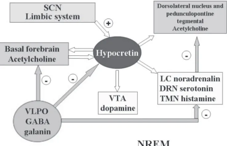
Figure 6 - NREM sleep Figure 6 - NREM sleep Figure 6 - NREM sleep Figure 6 - NREM sleep Figure 6 - NREM sleep
The absence of excitatory stimuli from the suprachiasmatic nucleus The absence of excitatory stimuli from the suprachiasmatic nucleus The absence of excitatory stimuli from the suprachiasmatic nucleus The absence of excitatory stimuli from the suprachiasmatic nucleus The absence of excitatory stimuli from the suprachiasmatic nucleus (SCN), basal forebrain and limbic system (white arrows), as well (SCN), basal forebrain and limbic system (white arrows), as well (SCN), basal forebrain and limbic system (white arrows), as well (SCN), basal forebrain and limbic system (white arrows), as well (SCN), basal forebrain and limbic system (white arrows), as well as the inhibitory projections (blue arrows) from the VLPO to the as the inhibitory projections (blue arrows) from the VLPO to the as the inhibitory projections (blue arrows) from the VLPO to the as the inhibitory projections (blue arrows) from the VLPO to the as the inhibitory projections (blue arrows) from the VLPO to the hypocretin system, induce NREM sleep.
narcoleptic individuals suffer hypocretin I deficiency, and autopsy results have revealed that such individuals also present a decrease in the number of hypocretin cells in the brain.53
The TMN is the only histaminergic nucleus of the CNS and is located in the posterior hypothalamus. As previously stated, the TMN is related to the maintenance of wakefulness, being the main inhibitor of the VLPO nucleus.7 Histaminergic neurons i n n e r v a t e p r a c t i c a l l y t h e e n t i r e b r a i n , i n c l u d i n g t h e mesopontine junction region, which is responsible for REM sleep. Histaminergic activity promotes wakefulness, and injury to the TMN results in excessive sleepiness. However, histaminergic activity is severely inhibited by the VLPO during NREM and REM sleep.7
P P P P
Po t e n t i a l t h e r a p e u t i c t a r g e t s f o r t h e d e v e l o p m e n to t e n t i a l t h e r a p e u t i c t a r g e t s f o r t h e d e v e l o p m e n to t e n t i a l t h e r a p e u t i c t a r g e t s f o r t h e d e v e l o p m e n to t e n t i a l t h e r a p e u t i c t a r g e t s f o r t h e d e v e l o p m e n to t e n t i a l t h e r a p e u t i c t a r g e t s f o r t h e d e v e l o p m e n t o f n e w t r e a t m e n t s
o f n e w t r e a t m e n t s o f n e w t r e a t m e n t s o f n e w t r e a t m e n t s o f n e w t r e a t m e n t s
The understanding of sleep neurobiology is potentially c o n d u c i v e t o n e w f u n c t i o n a l , e t i o l o g i c a n d pharmacotherapeutic models of sleep-wake cycles and men-tal disorders.10-18 Benzodiazepine and non-benzodiazepine hypnotics increase GABAergic transmission and they bind with a specific region of the GABA-A receptor protein complex. However, these agents can cause various degrees of tolerance, d e p e n d e n c e a n d u n d e s i r a b l e a l t e r a t i o n s i n t h e s l e e p architecture. Agents that specifically act to increase the action of endogenous GABA, more specifically in the GABAergic-g a l a n i n e r GABAergic-g i c s y s t e m o f t h e V L P O , s h o u l d p r o d u c e satisfactory therapeutic effects with fewer side effects. A direct GABA-A agonist called gaboxadol, which is still in development, has hypnotic effects with the increase of del-ta sleep. Pregabaline belongs to a new class of anxiolytic a g e n t s w i t h a m e c h a n i s m o f a c t i o n d i f f e r e n t f r o m benzodiazepines since they do not act on A and GABA-B receptors. Acting on the mechanisms of calcium channel activity, pregabaline prevents the presynaptic release of excitatory neurotransmitters such as glutamate, aspartate, and P substance in regions of the limbic system such as the hippocampus, amygdala and cingulate. Pregabaline has
anxiolytic effects, increases delta and is also a potential hypnotic agent for patients suffering from fibromyalgia.
The same can be said for the more selective anti-H-1 agents, which can represent an alternative as hypnotic agents with fewer side effects.
Traditional stimulating agents, such as amphetamines, methylphenidate, pemoline and drugs such as cocaine primarily act as alpha-adrenergic agonists and mesocortical-limbic dopaminergic agonists, and they respectively present peripheral autonomous effects, central stimulating effects with tolerance and abstinence. Hypocretin system or histaminergic agonists as wakefulness promoters can be therapeutic alternatives with no central and peripheral limiting side effects. Another model of wakefulness-promoting mechanism is the inhibition of the GABAergic system of the VLPO, facilitating or blocking the inhibition of wakefulness mechanisms without blocking the mechanisms responsible for the beginning and maintenance of NREM and REM sleep, as traditional stimulants do. An example of a promoting (or wakefulness-unblocking) agent is modafinil, which mainly acts as a noradrenergic agonist by inhibiting GABAergic activity in the VLPO. Other potential therapeutic targets are the biochemical cascade steps in the process of signal generation of SCN, which could be affected by pharmacological agents that act on components of the temporizing (SCN) system, such as the melatonin ML-1 sub-receptors.
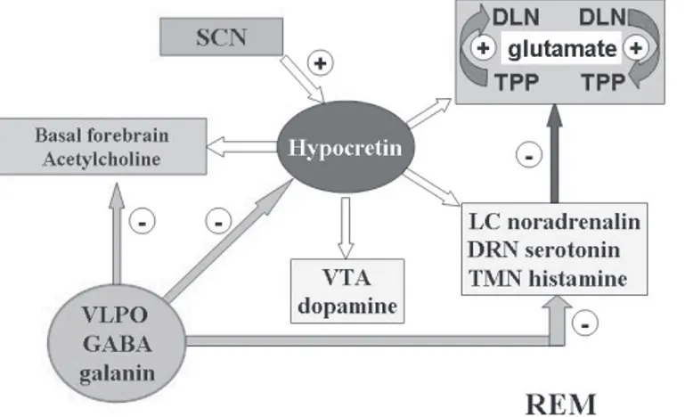
Figure 7 - REM sleep Figure 7 - REM sleep Figure 7 - REM sleep Figure 7 - REM sleep Figure 7 - REM sleep
The activity of cholinergic dorsolateral nucleus (DLN) and The activity of cholinergic dorsolateral nucleus (DLN) and The activity of cholinergic dorsolateral nucleus (DLN) and The activity of cholinergic dorsolateral nucleus (DLN) and The activity of cholinergic dorsolateral nucleus (DLN) and pedunculopontine tegmental (PPT) nuclei (REM-on cells) is pedunculopontine tegmental (PPT) nuclei (REM-on cells) is pedunculopontine tegmental (PPT) nuclei (REM-on cells) is pedunculopontine tegmental (PPT) nuclei (REM-on cells) is pedunculopontine tegmental (PPT) nuclei (REM-on cells) is liberated by the restriction of inhibitory stimuli (light blue arrow) liberated by the restriction of inhibitory stimuli (light blue arrow) liberated by the restriction of inhibitory stimuli (light blue arrow) liberated by the restriction of inhibitory stimuli (light blue arrow) liberated by the restriction of inhibitory stimuli (light blue arrow) from the aminergic system - REM-off cells (dark blue arrow), thereby from the aminergic system - REM-off cells (dark blue arrow), thereby from the aminergic system - REM-off cells (dark blue arrow), thereby from the aminergic system - REM-off cells (dark blue arrow), thereby from the aminergic system - REM-off cells (dark blue arrow), thereby increasing cholinergic activity (REM sleep) in accordance with increasing cholinergic activity (REM sleep) in accordance with increasing cholinergic activity (REM sleep) in accordance with increasing cholinergic activity (REM sleep) in accordance with increasing cholinergic activity (REM sleep) in accordance with the reciprocal integration model.11 The hypocretin and amine the reciprocal integration model.11 The hypocretin and amine the reciprocal integration model.11 The hypocretin and amine the reciprocal integration model.11 The hypocretin and amine the reciprocal integration model.11 The hypocretin and amine systems remain inhibited by the VLPO. White arrows represent systems remain inhibited by the VLPO. White arrows represent systems remain inhibited by the VLPO. White arrows represent systems remain inhibited by the VLPO. White arrows represent systems remain inhibited by the VLPO. White arrows represent lack of activity
lack of activity lack of activity lack of activity lack of activity...
R e f e r e n c e s R e f e r e n c e sR e f e r e n c e s R e f e r e n c e s R e f e r e n c e s
1 . 1 .1 . 1 .
1 . Rechtschaffen A, Bergmann BM. Sleep deprivation in the rat: an update of the 1989 paper. Sleep. 2002;25(1):18-24.
2 . 2 .2 . 2 .
2 . Aserinsky E, Kleitman N. Regularly occurring periods of eye motility, and concomitant phenomena during sleep. J Neuropsychiatry Clin Neurosci. 2003;15(4):454-5.
3 . 3 .3 . 3 .
3 . Hobson JA. Sleep. New York: Scientific American Library; 1989.
4 . 4 .4 . 4 .
4 . de Lecea L, Kilduff TS, Peyron C, Gao X, Foye PE, Danielson PE, et al. The hypocretins: hypothalamus-specific peptides with neuroexcitatory activity. Proc Natl Acad USA. 1998;95(1):322-7.
5 . 5 .5 . 5 .
5 . Sakurai T, Amemiya A, Ishii M, Matsuzaki I, Chemelli RM, Tanaka H, et al. Orexins and orexin receptors: a family of hypothalamic neuropeptides and G protein- coupled receptors that regulate feeding behavior. Cell. 1998,92(5):573-85. Comment in: Cell. 1998;92(5):696.
6 . 6 .6 . 6 .
6 . Mignot E, Taheri S, Nishino S. Sleeping with the hypothalamus: emerging therapeutic targes for sleep disorders. Nat Neurosci. 2002;5(suppl):1071-5.
7 . 7 .7 . 7 .
7 . Pace-Schott EF, Hobson JA. The neurobiology of sleep: genetics, cellular physiology and subcortical networks. Nat Rev Neurosci. 2002;3(9):591-605.
8 . 8 .8 . 8 .
8 . Saper C, Chou T C, Scammell TE. The sleep switch: hypothalamic control of sleep and wakefulness. Trends Neurosci. 2001;24(12):726-31.
9 . 9 .9 . 9 .
9 . Taheri S, Zeitzer J M, Mignot E. The role of hypocretins (orexins) in sleep regulation and narcolepsy. Annu Rev Neurosci. 2002;25:283-313.
1 0 . 1 0 .1 0 . 1 0 .
1 0 . Petit D, Montplaisir J, Lorrain D, Gauthier S. Spectral analysis of the rapid eye movement sleep electroencephalogram in right and left temporal regions: a biological marker of Alzheimer's disease. Ann Neurol. 1992;32(2):172-6.
1 1 . 1 1 .1 1 . 1 1 .
1 1 . Montplaisir J, Petit D, Gauthier S, Gaudreau H, Decary A. Sleep disturbances and EEG slowing in Alzheimer Disease. Sleep Res Online 1998;1(4):147-51.
1 2 . 1 2 .1 2 . 1 2 .
1 2 . Nofzinger EA, Buysse DJ, Germain A, Carter C, Luna B, Price JC, et al. Increased activation of anterior paralimbic and executive cortex from waking to REM sleep in depression. Arch Gen Psychiatry. 2004;61(7):695-702.
1 3 . 1 3 .1 3 . 1 3 .
regio-nal cerebral glucose metabolism during NREM sleep. Psychiatry Res. 2000;98(2):71-91.
1 4 . 1 4 .1 4 . 1 4 .
1 4 . Wu J, Buchsbaum M, Bunney WE Jr. Clinical neurochemical implications of sleep deprivation's effects on the anterior cingulate of depressed responders. Neuropsychopharmacology. 2001;25(5 Suppl):S74-8.
1 5 . 1 5 .1 5 . 1 5 .
1 5 . Buysse DJ, Hall ME, Begley A, Cherry CR, Houck PR, Land S, et al. Sleep and treatment response in depression: new findings using power spectral analysis. Psychiatry Res. 2001;103(1):51-67.
1 6 . 1 6 .1 6 . 1 6 .
1 6 . Jindal RD, Thase ME, Fasiczka AL, Friedman ES, Buysse DJ, Frank E, et al. Electroencephalographic sleep profiles in single-episode and recurrent unipolar forms of major depression: II. Comparison during remission. Biol Psychiatry. 2002;51(3):230-6.
1 7 . 1 7 .1 7 . 1 7 .
1 7 . Cook IA, Leuchter AF, Morgan M, Witte E, Stubbeman WF, Abrams M, et al. Early changes in prefrontal activity characterize clinical responders to antidepressants. Neuropsychopharmacology. 2002;27(1):120-31.
1 8 . 1 8 .1 8 . 1 8 .
1 8 . Hobson JA, McCarkey RW, Wyzinki PW. Sleep cycle oscillation: reciprocal discharge by two brain stem neuronal groups. Science. 1975,189(4196):55-8.
1 9 . 1 9 .1 9 . 1 9 .
1 9 . Nitz D, Siegel JM. GABA release in the locus ceruleus as a function of sleep/wake state. Neuroscience. 1997;78(3):795-801.
2 0 . 2 0 .2 0 . 2 0 .
2 0 . Hobson JA, Pace Schott EF, Stickgold R. Dreaming and the brain: towards a cognitive neuroscience of conscious states. Behav Brain Sci. 2000;23(6):793-842; discussion 904-1121.
2 1 . 2 1 .2 1 . 2 1 .
2 1 . Rye DB, Jankovic J. Emerging views of dopamine in modulating sleep/wake state from an unlikely source: PD. Neurology. 2002;58(3):341-6.
2 2 . 2 2 .2 2 . 2 2 .
2 2 . Randomized trial of modafinil as a treatment for the excessive daytime somnolence of narcolepsy: US Modafinil in Narcolepsy Multicenter Study Group. Neurology. 2000;54(5):1166-75.
2 3 . 2 3 .2 3 . 2 3 .
2 3 . Högl B, Saletu M, Brandauer E, Glatzl S, Frauscher B, Seppi K, et al.Modafinil for the treatment of daytime sleepiness in Parkinson's Disease: a double-blind, randomized, crossover, placebo-controlled polygraphic trial. Sleep. 2002;25(8):905-9.
2 4 . 2 4 .2 4 . 2 4 .
2 4 . Boutrel B, Koob GF. What keeps us awake: the neuropharmacology of stimulants and wakefulness-promoting medications. Sleep. 2004;27(6):1181-94.
2 5 . 2 5 .2 5 . 2 5 .
2 5 . Scammell TE, Estabrooke IV, McCar thy MT, Chemelli RM, Yanagisawa M, Miller MS, et al. Hypothalamic arousal regions are activated during modafinil-induced wakefulness. J Neurosci. 2000;20(22):8620-8.
2 6 . 2 6 .2 6 . 2 6 .
2 6 . Erhardt S, Andersson B, Nissbrandt H, Engberg G. Inhibition of firing rate and changes in the firing pattern of nigral dopamine neurons by gamma-hydroxybutyric acid (GHBA) are specifically induced by activation of GABA(B) receptors. Naunyn Schmiedebergs Arch Pharmacol. 1998;357(6):611-9.
2 7 . 2 7 .2 7 . 2 7 .
2 7 . Carai MAM, Colombo G, Brunetti G, Melis S, Serra S, Vacca G, et al. Role of GABA(B) receptors in the sedative/hypnotic effect of gamma-hydroxybutyric acid. Eur J Pharmacol. 2001;428(3):315-21.
2 8 . 2 8 .2 8 . 2 8 .
2 8 . Nicholson KL, Balster RL. GHB: a new and novel drug of abuse. Drug Alcohol Depend. 2001;63(1):1-22.
2 9 . 2 9 .2 9 . 2 9 .
2 9 . A randomized, double blind, placebo-controlled multicenter trial comparing the effects of three doses of orally administered sodium oxybate with placebo for the treatment of narcolepsy. Sleep. 2002;25(1):42-9.
3 0 . 3 0 .3 0 . 3 0 .
3 0 . Scharf M, Baumann M, Berkowitz DV. The effects of sodium oxybate on clinical symptoms and sleep patterns in patients with fibromyalgia. J Rheumatol. 2003;30(5):1070-4.
3 1 . 3 1 .3 1 . 3 1 .
3 1 . Van Gelder RN. Recent insights into mammalian circadian rhythms.Sleep. 2004;27(1):166-71.
3 2 . 3 2 .3 2 . 3 2 .
3 2 . Albrecht U. Invited review: regulation of mammalian circadian clock genes. J Appl Physiol. 2002;92(3):1348-55.
3 3 . 3 3 .3 3 . 3 3 .
3 3 . Moore RY, Speh JC, Leak RK. Suprachiasmatic nucleus organization. Cell Tissue Res. 2002;309(1):89-98.
3 4 . 3 4 .3 4 . 3 4 .
3 4 . Mrozovsky N. Beyond the suprachiasmatic nucleus. Chronobiol Int. 2003;20(1):201-8.
3 5 . 3 5 .3 5 . 3 5 .
3 5 . Golley JJ, Lu J, Fischer D, Saper CB. A broad role for melanopsin in nonvisual photoreception. J Neurosci. 2003;23(18):7093-106.
3 6 . 3 6 .3 6 . 3 6 .
3 6 . Miller JD, Morin LP, Schartz WP, Moore RY. New insights into the mammalian circadian clock. Sleep. 1996;19(8):641-67.
3 7 . 3 7 .3 7 . 3 7 .
3 7 . Krout KE, Kawano J, Mettenleiter TC, Loewy AD. CNS inputs to the
suprachiasmatic nucleus of the rat. Neuroscience. 2002;110(1):73-92.
3 8 . 3 8 . 3 8 . 3 8 .
3 8 . Vitaterna MH, King DP, Chang AM, Kornhauser JM, Lowrey PL, McDonald JD, et al. Mutagenesis and mapping of a mouse gene, Clock, essential for circadian behavior. Science. 1994;264(5159):719-25.
3 9 . 3 9 . 3 9 . 3 9 .
3 9 . Reid KJ, Chang AM, Dubocovich ML, Turek FW, Takahashi JS, Zee PC. Familial advanced sleep phase syndrome. Arch Neurol. 2001;58(7):1089-94.
4 0 . 4 0 . 4 0 . 4 0 .
4 0 . Katzenberg D, Young T, Finn L, Lin L, King DP,Takahashi JS, et al. A CLOCK polymorphism associated with human diurnal preference. Sleep. 1998;21(6):569-76.
4 1 . 4 1 . 4 1 . 4 1 .
4 1 . Toh KL, Jones CR, He Y, Eide EJ, Hinz WA, Virshup DM, et al. An hPer2 phosphorylation site mutation in familial advanced sleep phase syndrome. Science. 2001;291(5506):1040-3.
4 2 . 4 2 . 4 2 . 4 2 .
4 2 . Lowrey PL, Shimomura K, Antoch MP, Yamazaki S, Zemenides PD, Ralph MR, et al. Positional cloning and functional characterization of the mammalian circadian mutation tau. Science. 2000;288(5465):483-91.
4 3 . 4 3 . 4 3 . 4 3 .
4 3 . Weber ET, Gannon RL, Michel AM, Gillette MU, Rea MA. Nitric oxide synthase inhibitor blocks light induced phase shifts of the circadian activity rhythm, but not c-fos expression in the suprachiasmatic nucleus of the Syrian hamster. Brain Res. 1995;692(1-2):137-42 .
4 4 . 4 4 . 4 4 . 4 4 .
4 4 . Gillette MU, Buchanan GF, Artinian L, Hamilton SE, Nathanson NM, Liu C. Role of the M1 receptor in regulating circadian rhythms. Life Sci. 2001;68(22-23):2467-72.
4 4 4 4
4 5 .5 .5 .5 .5 . Hirai K, Kato K, Nishiyama K. TAK-375 and its metabolites are selective agonists at MLs1s receptors [abstract]. Sleep. 2003;26(suppl):0193C. [Presented of the Associated Professional Sleep Societies 17th Annual Meeting. June 3-8, 2003. Chicago, Illinois, USA].
4 6 . 4 6 . 4 6 . 4 6 .
4 6 . Roth T, Walsh JK, Rogowski R. Efficacy and tolerability of indiplon (NBI-34060) solution in healthy adults in a model of transient insomnia [abstract]. Sleep. 2003;26(suppl):0214C. (Presented of the Associated Professional Sleep Societies 17th Annual Meeting. June 3-8, 2003. Chicago, Illinois, USA].
4 7 . 4 7 . 4 7 . 4 7 .
4 7 . Porkka-Heiskanen T, Alanko L, Kalinchuk A, Stenberg D. Adenosine and sleep. Sleep. Med Rev. 2002;6(4):321-32.
4 8 . 4 8 . 4 8 . 4 8 .
4 8 . Beukmann CT, Yanagisawa M. Orexins: from neuropeptides to energy homeostasis and sleep-wake regulation. J Mol Med. 2002;80(6):329-42.
4 9 . 4 9 . 4 9 . 4 9 .
4 9 . Peyron C, Tighe DK, van den Pol AN, de Lecea L, Heller HC, Sutcliffe J, et al. Neurons containing hypocretin (orexin) project to multiple neuronal systems. J Neurosci. 2002;18(23):9996-10015.
5 0 . 5 0 . 5 0 . 5 0 .
5 0 . Aston-Jones G, Chen S, Zhu Y, Oshinsky ML. A neural circuit for circadian regulation of arousal. Nat Neurosci. 2001;4(7):732-8.
5 1 . 5 1 . 5 1 . 5 1 .
5 1 . Zhang S, Zeitzer JM, Yoshida Y, Wisor JP, Nishino S, Edgar DM, et al. Lesions of the suprachiasmatic nucleus eliminate the daily rhythm of hypocretin release. Sleep. 2004;2(4)7:619-27.
5 2 . 5 2 . 5 2 . 5 2 .
5 2 . Thannickal TC, Moore RY, Niehius R, Ramanathan L, Gulyani S, Aldrich M, et al. Reduced number of hypocretin neurons in human narcolepsy. Neuron. 2000;27(3):469-74.
5 3 . 5 3 . 5 3 . 5 3 .