○ ○ ○ ○ ○ ○ ○ ○ ○ ○ ○ ○ ○ ○ ○ ○ ○ ○ ○
ABSTRACT
○ ○ ○ ○ ○ ○ ○ ○ ○ ○ ○ ○ ○ ○ ○ ○ ○ ○ ○ ○
INTRODUCTION
Electron microscopy has been used for the morphological diagnosis of glomerular diseases for more than three decades and its value has been widely emphasized.1 However, recent
re-ports have analyzed the routine use of electron microscopy critically. Its use in other areas of diagnosis such as tumor diseases has declined considerably; in addition, in view of the una-voidable financial pressure for the reduction of costs due to investigations and diagnostic rou-tines, the selection of cases for electron micros-copy has been quite rigorous.2-4
Some investigators have observed that about 85% of renal biopsies had an indica-tion of electron microscopy for diagnostic confirmation.5 Routine diagnostic electron
microscopy has proved to be of high value in the differential diagnosis of nephrotic syndrome, especially in early membranous glomerulonephritis and in cases of minimal lesion glomerulopathy. The use of electron microscopy for the classification of glome-rular diseases has been well established, and the technique can also be used for thera-peutic monitoring.6
Some lesions detected via light microscopy and immunofluorescence can be better char-acterized by electron microscopy, as is the case for the localization of immune deposits and structural changes in the glomerular basement membrane. The use of electron microscopy has allowed the recognition of changes not observed under light microscopy, thereby con-tributing to the understanding of the pathogenesis of renal diseases.7 This is the
tech-nique used for the determination of glomeru-• Marcello Fabiano Franco
for the diagnosis of
glomerulopathies
Departments of Pathology, Universidade Metropolitana de Santos and
Universidade Federal de São Paulo – Escola Paulista de Medicina, São
Paulo, Brazil
CONTEXT: Electron microscopy has been used for the morphological diagnosis of glomerular diseases for more than three decades and its value has been widely emphasized. However, recent reports have analyzed the routine use of electron microscopy critically. Its use in other areas of diagnosis such as tumor diseases has declined considerably; in addition, in view of the unavoidable financial pres-sure for the reduction of costs due to investiga-tions and diagnostic routines, the selection of cases for electron microscopy has been quite rigorous.
OBJECTIVE: To identify the glomerular diseases that depend on electron microscopy for a final diag-nosis, by means of reviewing renal biopsies per-formed over a 12-year period.
DESIGN: Prospective
SETTING: Hospital Ana Costa, Hospital Guilherme Álvaro and Serviço de Anatomia Patológica de Santos, Santos, São Paulo, Brazil.
PARTICIPANTS: 200 consecutive renal biopsies ob-tained from private hospitals and the teaching hospital from 1979 to 1991 were studied.
MAIN MEASUREMENTS: All cases were analyzed via light microscopy, immunofluorescence and elec-tron microscopy. The diagnosis was first made via light microscopy plus immunofluorescence and then via electron microscopy.
RESULTS: Electron microscopy was diagnostic or essen-tial for diagnosis in 10.0% of the cases, correspond-ing to 3.4% of primary glomerulopathies and 100% of hereditary glomerulopathies. Electron microscopy was contributory (useful) to the diagnosis in 5.5% of the cases, confirming the preliminary diagnosis formulated on the basis of clinical and laboratory data and light microscopy plus immunofluorescence findings. We obtained a 7.5% rate of discordant immunofluorescence, which was considered as such when negative immunofluorescence findings were not confirmed by electron microscopy. The final di-agnosis with the use of light microscopy plus im-munofluorescence alone was 77.0%.
CONCLUSIONS: It was possible to diagnose with cer-tainty a great percentage of glomerulopathies (82.5-90% of the cases) based on the light microscopy and immunofluorescence findings alone. Electron microscopy was essential for the diagnosis of hereditary nephropathies.
KEY WORDS: Electron microscopy. Kidney. Biopsy. Glomerulonephritis. Membranous glomerulonephritis.
lar basement membrane damage in non-im-mune glomerulopathies such as Alport syn-drome, thin basement membrane disease and nephrotic syndrome with minimal lesion glomerulopathy.8
In thin basement membrane disease, light microscopy only reveals the presence of blood casts in the tubular lumen. The early thicken-ing of the glomerular basement membrane, which may occur in diabetic nephropathy, hypertension and glomerulonephritis, can also be seen only via electron microscopy. The lo-calization of immunocomplexes is important for defining the type of glomerulonephritis. Finally, ultrastructural evaluation is essential for adequate characterization of fibrillar glomerulonephritis such as microfibrillar and immunotactoid glomerulonephritis.9,10
The objective of the present study was to analyze the role of electron microscopy exami-nation for the diagnosis of glomerular disease in a consecutive series of biopsies analyzed by the same pathologist with the systematic use of light microscopy, immunofluorescence and electron microscopy.
○ ○ ○ ○ ○ ○ ○ ○ ○ ○ ○ ○ ○ ○ ○ ○ ○ ○ ○ ○
METHODS
A total of 200 consecutive renal biopsies obtained from private hospitals and the teach-ing hospital of the Santos School of Medical Sciences, State of São Paulo, from 1979 to 1991 were studied via light microscopy, im-munofluorescence and electron microscopy. Most of the biopsies were obtained using a needle and about 15% were obtained via open surgery, especially the biopsies from children. The renal fragments were received for
Original Ar
C
ase Repor
t
analysis without prior fixing. They were di-vided into three portions: I) the extremities were fixed in glutaraldehyde and reserved for electron microscopy; II) the central portion was frozen at –20º C and used for immun-ofluorescence; and III) the remaining portions were fixed in 10% formalin for paraffin sec-tions and examination via light microscopy. The material was processed for electron microscopy by resin embedding and cutting into 750 Å sections using a diamond knife. The sections were placed on a net for obser-vation under a Philips electron microscope.
Immunofluorescence involved the search for immunoglobulins A, G and M (IgA, IgG and IgM), C3 and C1q complement compo-nents, and also albumin and fibrinogen. Sec-tions of 3 to 4 µm were obtained using a
cryo-stat and fixed in cold acetone. After incubation and washing in phosphate-buffered saline so-lution (PBS, pH 7.2), readings were taken us-ing a Zeiss fluorescence microscope. For light microscopy analysis, the paraffin blocks were cut into 3 to 5 µm sections, which were stained using hematoxylin-eosin, Masson trichrome, periodic acid Schiff, and silver impregnation.
The light microscopy, immunofluores-cence and electron microscopy findings as a whole were reviewed by two pathologists for definition of the final diagnosis. Each case was first analyzed using light microscopy and im-munofluorescence findings together with the clinical and laboratory data for the morpho-logical and nosomorpho-logical interpretation of the glomerulopathy. These findings were then reevaluated together with the ultrastructural study in order to determine the impact of elec-tron microscopy on the diagnosis of the glomerular disease.
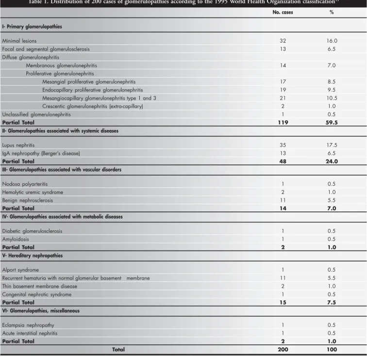
Table 1. Distribution of 200 cases of glomerulopathies according to the 1995 World Health Organization classification11
No. cases %
I- Primary glomerulopathies
Minimal lesions 32 16.0
Focal and segmental glomerulosclerosis 13 6.5
Diffuse glomerulonephritis
Membranous glomerulonephritis 14 7.0
Proliferative glomerulonephritis
Mesangial proliferative glomerulonephritis 17 8.5
Endocapillary proliferative glomerulonephritis 19 9.5
Mesangiocapillary glomerulonephritis type 1 and 3 21 10.5
Crescentic glomerulonephritis (extra-capillary) 2 1.0
Unclassified glomerulonephritis 1 0.5
Partial Total 119 59.5
II- Glomerulopathies associated with systemic diseases
Lupus nephritis 35 17.5
IgA nephropathy (Berger’s disease) 13 6.5
Partial Total 48 24.0
III- Glomerulopathies associated with vascular disorders
Nodosa polyarteritis 1 0.5
Hemolytic uremic syndrome 2 1.0
Benign nephrosclerosis 11 5.5
Partial Total 14 7.0
IV- Glomerulopathies associated with metabolic diseases
Diabetic glomerulosclerosis 1 0.5
Amyloidosis 1 0.5
Partial Total 2 1.0
V- Hereditary nephropathies
Alport syndrome 1 0.5
Recurrent hematuria with normal glomerular basement membrane 11 5.5
Thin basement membrane disease 2 1.0
Congenital nephrotic syndrome 1 0.5
Partial Total 15 7.5
VI- Glomerulopathies, miscellaneous
Eclampsia nephropathy 1 0.5
Acute interstitial nephritis 1 0.5
Partial Total 2 1.0
○ ○ ○ ○ ○ ○ ○ ○ ○ ○ ○ ○ ○ ○ ○ ○ ○ ○ ○ ○
RESULTS
The distribution of the 200 cases studied according to the World Health Organization classification of glomerulonephritis11 is
pre-sented in Table 1. Primary glomerulonephri-tis was observed in 59.5% of the cases, sys-temic diseases with glomerulonephritis in 24%, vascular disease with glomerulonephri-tis in 7.0%, metabolic disease with glomeru-lonephritis in 1.0%, hereditary nephropathies in 7.5%, and diverse glomerular diseases in 1.0%. There was sharp predominance of mini-mal lesion glomerulopathy in the primary glomerulonephritis group and of lupus nephri-tis in the glomerulonephrinephri-tis associated with systemic diseases.
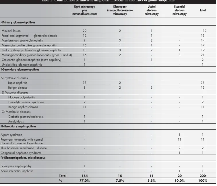
The case series was evaluated according
to: 1) number of cases in which the diagnosis was reached via light microscopy plus immun-ofluorescence alone; 2) number of cases in which immunofluorescence was considered discordant from the electron microscopy find-ings; and 3) number of cases in which elec-tron microscopy was interpreted as essential for the diagnosis (Table 2).
In 154 biopsies (77.0%), the morphologi-cal diagnosis of glomerular disease was based on light microscopy and immunofluorescence findings alone. In 15 cases (7.5%), immun-ofluorescence yielded results that were not confirmed by electron microscopy (discrep-ant immunofluorescence). In 11 cases (5.5%), electron microscopy confirmed the light microscopy and fluorescence findings (con-tributory electron microscopy). In 20 cases
(10.0%), the electron microscopy was essen-tial for the final diagnosis.
Electron microscopy was mainly contribu-tory or confirmacontribu-tory for diagnosis when it revealed or confirmed important morphologi-cal elements that had not been clearly observed via light microscopy or immunofluorescence, as follows: cases of membranous glomerulonephri-tis in the early state with negative immunofluo-rescence, in which electron microscopy revealed electron-dense subepithelial deposits; cases of proliferative endocapillary glomerulonephritis with negative immunofluorescence in which electron microscopy revealed electron-dense sub-epithelial “hump” type deposits; cases of IgA ne-phropathy with doubtful immunofluorescence examination, in which electron microscopy revealed electron-dense paramesangial deposits.
Table 2. Contribution of different diagnostic methods in 200 cases of glomerulopathies
Light microscopy Discrepant Useful Essential
plus ímmunofluorescence electron electron Total
immunofluorescence microscopy microscopy microscopy
I-Primary glomerulopathies
Minimal lesion 29 2 1 - 32
Focal and segmental glomerulosclerosis 12 - 1 - 13
Membranous glomerulonephritis 9 3 2 - 14
Mesangial proliferative glomerulonephritis 15 1 1 - 17
Endocapillary proliferative glomerulonephritis 13 3 2 1 19
Mesangiocapillary glomerulonephritis (types 1 and 3) 16 2 - 3 21
Crescentic glomerulonephritis (extra-capillary) 1 - 1 - 2
Unclassified glomerulonephritis 1 - - - 1
II-Secondary glomerulopathies
A) Systemic diseases
Lupus nephritis 33 2 - - 35
Berger disease 8 2 3 - 13
B) Vascular diseases
Nodosa polyarteritis 1 - - - 1
Hemolytic uremic syndrome 2 - - - 2
Benign nephrosclerosis 11 - - - 11
C) Metabolic diseases
Diabetic glomerulosclerosis 1 - - - 1
Amyloidosis 1 - - - 1
III-Hereditary nephropathies
Alport syndrome - - - 1 1
Recurrent hematuria with normal - - - 11 11
glomerular basement membrane
Thin basement membrane disease - - - 2 2
Congenital nephrotic syndrome - - - 1 1
IV-Glomerulopathies, miscellaneous
Eclampsia nephropathy 1 - - - 1
Acute interstitial nephritis - - - 1 1
Total 154 15 11 20 200
In the four cases of proliferative glomerulone-phritis (one case of endocapillary proliferative glomerulonephritis and three cases of mesangiocapillary glomerulonephritis), electron microscopy was diagnostic by demonstrating subepithelial and subendothelial electron-dense deposits, respectively.
Within the group in which electron microscopy was essential to the diagnosis, the highest percentage of cases consisted of heredi-tary nephropathies (15 cases), divided into Alport syndrome1, benign recurrent
hema-turia11, thin basement membrane disease2, and
congenital nephrotic syndrome1.
○ ○ ○ ○ ○ ○ ○ ○ ○ ○ ○ ○ ○ ○ ○ ○ ○ ○ ○ ○
DISCUSSION
Although renal biopsies started to be per-formed in medical practice in 1951, pioneered by Iversen & Brun,12 it was only 20 years later
that the importance of immunofluorescence became emphasized in relation to complemen-tary assessment of the anatomopathological evaluation.
Subsequently, Habib & Gluber,13 in 1983,
tried to correlate light microscopy and elec-tron microscopy findings and define the pathogenesis via immunofluorescence. No attempt was made in any of these reports to systematically quantify the importance of each procedure or determine what conditions would not be diagnosed in the absence of one of the stains or methods used.
The importance of such assessment resides in the fact that most histopathology laborato-ries in Brazil do not have immunofluorescence capability, and only a minority can use elec-tron microscopy, which is usually performed in the laboratories of the major university hospitals. For countries with fewer resources, the diagnosis of glomerular disease needs to be made possible by first using less expensive methods before employing electron microscopy.2-4
In the presence of minimal lesion glome-rulopathy, although the primary lesion is ul-trastructural, with podocyte effacement, normal light microscopy and negative immunofluores-cence findings in combination with clinical data are indicative for the diagnosis. In focal and seg-mental glomerulosclerosis, immunofluorescence can reveal trapping of IgM and/or C3 in the sclerosed glomeruli, and thus electron microscopy is important for the diagnosis since it reveals fusion of the foot processes in the glomeruli, which appear normal via light microscopy and without electron-dense depos-its. In membranous glomerulonephritis, the di-agnosis may be difficult in stage I of the disease
since, at the beginning, the changes are not evi-dent via special staining, with the absence of spikes in silver impregnation.
Some diagnosis can be made only via im-munofluorescence, but when this procedure is considered erroneously negative, as in cases of weak or irregular staining due to previous treatment or technical error, the final diagno-sis will depend on electron microscopy exami-nation finding electron-dense deposits.9
Endocapillary proliferative glomerulone-phritis usually presents no diagnostic difficul-ties when it shows endocapillary proliferation and neutrophilic exudation via light microscopy and granular deposits predominantly of C3 via immunofluorescence. If immunofluorescence is inconclusive, it is important to make a dif-ferential diagnosis with other entities in which complement activation occurs and neutrophils are present. In such cases, the differential diag-nosis is made via electron microscopy when epimembranous deposits with a “hump” pat-tern are detected. Mesangiocapillary lonephritis is frequently recognized by glomeru-lar basement membrane duplication, which is well demonstrated in most cases by silver stain-ing. In immunofluorescence, the detection of granular and peripheral C3 deposits contrib-utes to the diagnosis.
Only three cases in the present series were defined by electron microscopy. One case had glomerular changes similar to mesangio-capillary glomerulonephritis, in which fibril-lar deposits were detected via electron mi-croscopy. The other two cases were in the early phases of mesangiocapillary glomerulonephri-tis, with the detection of submembranous deposits via electron microscopy. Such ul-trastructural finding allowed differentiation between mesangiocapillary glomerulonephri-tis and acute diffuse glomerulonephriglomerulonephri-tis, which would not have occurred if only light microscopy and immunofluorescence had been used. None of our cases was assigned to mesangiocapillary glomerulonephritis type II. Among the systemic diseases, lupus nephri-tis was the most frequent. Light microscopy and immunofluorescence are sufficient for ad-equate definition of the various types of lupus lesion and for identifying active or chronic le-sion.14,15 In Berger’s disease, light microscopy
can have various presentations, ranging from normal glomeruli to glomeruli with focal le-sion or global sclerosis. The definitive diagno-sis of IgA nephropathy is obtained via immun-ofluorescence with the detection of IgA of mesangial location. Electron microscopy is complementary, revealing electron-dense de-posits in paramesangial regions. Although not
specific, such findings strongly support the di-agnosis of IgA nephropathy.
When immunofluorescence does not demonstrate these deposits, the electron mi-croscopy findings can be indicative of the en-tity, especially in the presence of predominant hematuria. In the group of metabolic and vas-cular diseases, the diagnostic conclusion is normally reached via light microscopy, since immunofluorescence is negative.16 In fibrillar
glomerulonephritis, special stains such as Congo red for the demonstration of amyloid are useful for the diagnostic procedures, ex-cept in the early stage. None of our cases showed scarce amyloid deposits via electron microscopy, which would not have shown positivity using Congo red staining. In non-amyloid cases, examination via electron microscopy and other clinical and laboratory data are essential for the diagnosis.
In the hereditary nephropathies group, electron microscopy makes the definitive di-agnosis. Light microscopy can be normal at first and immunofluorescence is always nega-tive, but electron microscopy allows documen-tation of alterations at the glomerular base-ment membrane level. When the glomerular basement membrane is delaminated and of variable thickness, Alport syndrome is char-acterized. When the membrane thickness is very reduced, usually to about one-third of normal or approximately 200 nm, thin base-ment membrane disease is characterized.17
When nephrotic syndrome is observed in newborn infants and the clinical data is remi-niscent of minimal lesion glomerulopathy, congenital nephrotic syndrome is diagnosed. Nephritic cases with normal findings from light microscopy, immunofluorescence and electron microscopy are classified as recurrent hematuria with normal glomerular basement membrane. A large part of our group was as-signed to this category because recurrent hematuria is a clinical indication for biopsy, and this is useful for ruling out more severe disease of poor prognosis.
With regard to the two cases of diverse glomerular diseases, light microscopy defined the case of eclampsia nephropathy and elec-tron microscopy was essential for the diag-nosis in the case of acute interstitial nephritis, in order to rule out other conditions accom-panied by hematuria, such as hereditary ne-phropathy.
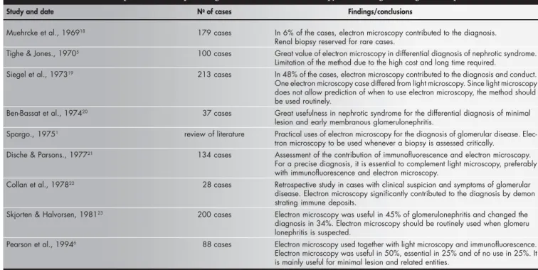
Table 3. List of publications emphasizing the use of electron microscopy for the diagnosis of glomerulopathies
Study and date No of cases Findings/conclusions
Muehrcke et al., 196918 179 cases In 6% of the cases, electron microscopy contributed to the diagnosis.
Renal biopsy reserved for rare cases.
Tighe & Jones., 19705 100 cases Great value of electron microscopy in differential diagnosis of nephrotic syndrome.
Limitation of the method due to the high cost and long time required.
Siegel et al., 197319 213 cases In 48% of the cases, electron microscopy contributed to the diagnosis and conduct.
One electron microscopy case differed from light microscopy. Since light microscopy does not allow prediction of when to use electron microscopy, the method should be used routinely.
Ben-Bassat et al., 197420 37 cases Great usefulness in nephrotic syndrome for the differential diagnosis of minimal
lesion and early membranous glomerulonephritis.
Spargo., 19751 review of literature Practical uses of electron microscopy for the diagnosis of glomerular disease.
Elec-tron microscopy to be used whenever a biopsy is assessed critically.
Dische & Parsons., 197721 134 cases Assessment of the contribution of immunofluorescence and electron microscopy.
For a precise diagnosis, it is essential to complement light microscopy, preferably with immunofluorescence and electron microscopy.
Collan et al., 197822 28 cases Retrospective study in cases with clinical suspicion and symptoms of glomerular
disease. Electron microscopy significantly contributed to the diagnosis by demon strating immune deposits.
Skjorten & Halvorsen, 198123 200 cases Electron microscopy was useful in 45% of glomerulonephritis and changed the
diagnosis in 34%. Electron microscopy should be routinely used when glomeru lonephritis is suspected.
Pearson et al., 19946 88 cases Electron microscopy used together with light microscopy and immunofluorescence.
Electron microscopy was useful in 50%, essential in 25% and of no use in 25%. It is mainly useful for minimal lesion and related entities.
studies only mention the generic importance of its use or its importance for groups of glomerulopathies, as can be seen in Table 3.
Pearson et al.,6 in 1994, concluded that
elec-tron microscopy plays an important role in the diagnosis of renal disease and therefore renal tis-sue should be submitted to electron microscopy whenever possible. In some selected cases, when light microscopy and immunofluorescence re-sults are already known, the ultrastructural ex-amination could be predicted. Electron microscopy would be particularly useful for the differential diagnosis of glomerulopathies that progress with nephrotic syndrome.
In our series, electron microscopy was es-sential for the diagnosis in 10% of the cases, i.e. the correct diagnosis would not have been
possible without it. It was contributory in 5.5% of the cases, in situations in which it confirmed the light microscopy plus immunofluorescence findings. If we succeed in reducing the percentage of immunofluorescence that is considered discrepant (7.5%), by means of improved controls, reduction of technical errors and avoiding post-treatment biopsies (false-negative results), we would reach 90% accuracy in the diagnosis of biopsied glomerulopathies, using only light microscopy plus immunofluorescence.
○ ○ ○ ○ ○ ○ ○ ○ ○ ○ ○ ○ ○ ○ ○ ○ ○ ○ ○ ○
CONCLUSION
1. The frequencies of glomerulopathies di-agnosed by biopsy, in the city of Santos, Brazil, not including cases of renal
trans-plantation, were as follows: predominance of primary glomerulopathies (59.5%); lower frequency for glomerulopathies as-sociated with systemic diseases (24%); similar low frequencies for glomeru-lopathies associated with vascular diseases (7%) and hereditary nephropathies (7.5%); very low frequency for glomeru-lopathies associated with metabolic dis-eases (1.0%) and miscellaneous (1.0%). 2. It was possible to diagnose with certainty
O valor da microscopia eletrônica no diagnósti-co das glomerulopatias
CONTEXTO: A microscopia eletrônica tem sido usada há mais de três décadas para o diag-nóstico morfológico das doenças glomerulares e seu valor tem sido amplamente enfatizado. Entretanto, relatos recentes têm analisado cri-ticamente o uso rotineiro da microscopia ele-trônica. O seu uso em outras áreas de diag-nóstico como doenças tumorais tem declina-do consideravelmente. Além disso, em virtu-de da inevitável pressão financeira para redu-ção dos custos da investigaredu-ção na rotina diagnóstica, a seleção dos casos para micros-copia eletrônica tem sido rigorosa.
OBJETIVO: Com o intuito de se identificarem as doenças glomerulares que dependem da microscopia eletrônica para o diagnóstico fi-nal, foram revisadas biópsias renais recebidas no período de 12 anos.
TIPO DE ESTUDO: Prospectivo
LOCAL: Hospital Ana Costa, Hospital Guilher-me Álvaro e Serviço de Anatomia Patológica de Santos, São Paulo, Brasil.
PARTICIPANTES: 200 biópsias renais consecu-tivas, obtidas de hospital privado e hospital-escola de 1979 a 1991.
PRINCIPAIS VARIÁVEIS: Todos os casos foram analisados por microscopia óptica,
imuno-○ ○ ○ ○ ○ ○ ○ ○ ○ ○ ○ ○ ○ ○ ○ ○ ○ ○ ○ ○ ○ ○ ○ ○ ○ ○ ○ ○ ○ ○ ○ ○ ○ ○ ○ ○ ○ ○ ○ ○ ○ ○
RESUMO
Acknowledgements: The authors thank Dr. Tatiana Antonovych, Armed Forces Institute of Pathology, United States, for the electron microscopy analysis.
Angelo Sementilli, MD. Departments of Pathology, Universidade Metropolitana de Santos (Unimes) and Centro Universitário Lusíada (Unilus), Santos, São Paulo, Brazil.
Luiz Antonio Moura, MD, PhD. Department of Pathol-ogy, Escola Paulista de Medicina, Universidade Federal de São Paulo, São Paulo, Brazil.
Marcello Fabiano Franco, MD, PhD. Department of Pathology, Escola Paulista de Medicina, Universidade Fed-eral de São Paulo, São Paulo, Brazil.
Sources of funding: CNPq master’s degree scholarship
Conflict of interest: Not declared
Date of first submission: January 6, 2003
Last received: July 28, 2003
Accepted: October 29, 2003
Address for correspondence:
Angelo Sementilli
Serviço de Anatomia Patológica de Santos R. Monsenhor Paula Rodrigues, 197
Vila Belmiro – Santos/SP – Brasil – CEP 11075-350 Tel. (+55 13) 3234-9328
Fax (+55 13) 3222-2150 E-mail: [email protected]
COPYRIGHT © 2004, Associação Paulista de Medicina
○ ○ ○Publishing information○ ○ ○ ○ ○ ○ ○ ○ ○ ○ ○ ○ ○ ○ ○ ○ ○
fluorescência e microscopia eletrônica. O di-agnóstico foi inicialmente feito por microscopia óptica e de imunofluorescência, e posteriormente pela microscopia eletrônica.
RESULTADOS: A microscopia eletrônica foi diagnóstica ou essencial para o diagnóstico em 10,0% dos casos, correspondendo a 3,4% de glomerulopatias primárias e 100% das glomerulopatias hereditárias. A microscopia eletrônica foi contributiva para o diagnósti-co em 5,5% dos casos, diagnósti-confirmando os diag-nósticos formulados com base em dados clí-nicos e laboratoriais, e achados de microscopia óptica e de imunofluorescência. Obtivemos 7,5% de imunofluorescências discordantes, assim consideradas quando os achados de imunofluorescência não foram confirmados pela microscopia eletrônica. Em 77,0% dos casos, o diagnóstico final pôde ser estabeleci-do exclusivamente com base nos achaestabeleci-dos de microscopia óptica e de imunofluorescência.
CONCLUSÕES: Foi possível diagnosticar com exatidão grande porcentagem (82,5 - 90,0%) dos casos com base nos achados isolados de microscopia óptica e de imunofluorescência. A microscopia eletrônica foi essencial para o diagnóstico das nefropatias hereditárias.
PALAVRAS-CHAVE: Glomerulopatia mem-branosa. Microscopia eletrônica. Biópsia.. Glomerulonefrite.
1. Spargo BH. Practical use of electron microscopy for the diag-nosis of glomerular disease. Hum Pathol. 1975;6(4):405-20. 2. Tucker JA. The continuing value of electron microscopy in
sur-gical pathology. Ultrastruct Pathol. 2000;24(6):383-9. 3. Mierau GW. Electron microscopy for tumour diagnosis: is it
redundant? Histopathology. 1999;35(2):99-101. 4. Kovacs K, Scheithauer BW, Horvath E, Lloyd RV. The World
Health Organization classification of adenohypophysial neoplasms. A proposed five-tier scheme. Cancer. 1996;78(3):502-10. 5. Tighe JR, Jones NF. The diagnostic value of routine electron
microscopy of renal biopsies. Proc R Soc Med. 1970;63(5):475-7. 6. Pearson JM, McWilliam LJ, Coyne JD, Curry A. Value of
elec-tron microscopy in diagnosis of renal disease. J Clin Pathol. 1994;47(2):126-8.
7. Ordonez NG. The use of electron microscopy in Kidney bi-opsy interpretation In: Baur SP, Mackay B, Editors. Diagnostic Electron Microscopy. New York: Appleton Century Crofts; 1981. p.75-129.
8. Danilewicz M, Wagrowska-Danilewicz M. Glomerular base-ment membrane thickness in primary diffuse IgA nephropa-thy: ultrastructural morphometric analysis. Int Urol Nephrol. 1998;30(4):513-9.
○ ○ ○ ○ ○ ○ ○ ○ ○ ○ ○ ○ ○ ○ ○ ○ ○ ○ ○ ○ ○ ○ ○ ○ ○ ○ ○ ○ ○ ○ ○ ○ ○ ○ ○ ○ ○ ○ ○ ○ ○ ○ ○ ○ ○ ○ ○ ○ ○ ○ ○ ○ ○ ○ ○ ○ ○ ○ ○ ○ ○ ○ ○ ○
REFERENCES
9. Radford MG, Donadio JV, Holley KE, Björnsson J, Grande JP. Renal biopsy in clinical practice. Mayo Clin Proc. 1994; 69(10):983-4.
10. Choi YJ, Lee JD, Yang RH, et al. Immunotactoid glome-rulopathy associated with idiopathic hypereosinophilic syn-drome. Am J Nephrol. 1998;18(4):337-43.
11. Schwartz MM. The pathologic diagnosis of renal disease. In: Jennette JC, Olson JL, Schwartz MM, Silva FG, editors. Heptinstall’s Pathology of the kidney. 5th edition. Philadelphia:
Lippincott-Raven Publishers; 1998. p. 169-80.
12. Iversen P, Brun C. Aspiration biopsy of the kidney. Am J Med. 1951;11(3):324-30.
13. Habib R, Gonzalès-Burchard G. Glomérulonéphrite aiguë post-infectieuse. In: Royer P, Habib R, Mathieu H, Broyer M, edi-tors. Nephrologie pediatrique. 3rd edition. Paris: Flammarion
Medicine-Sciences; 1983. p. 294-305.
14. Raine AE. Renal biopsy and the prognosis of lupus nephritis. Q J Med. 1991;81(295):879-81.
15. Grande JP, Balow JE. Renal biopsy in lupus nephritis. Lupus. 1998;7(9):611-7.
16. Osterby R. Lessons from kidney biopsies. Diabetes Metab Rew. 1996;12(3):151-74.
17. Dische FE. Measurement of glomerular basement membrane thickness and its application to the diagnosis of thin-membrane nephropathy. Arch Pathol Lab Med. 1992;116(1):43-9. 18. Muehrcke RC, Mandal AK, Gotoff SP, Isaacs EW, Volini FI.
The clinical value of electron microscopy in renal disease. Arch Intern Med. 1969;124(2):170-6.
19. Siegel NJ, Spargo BH, Kashgarian M, Hayslett JP. An evalua-tion of routine electron microscopy in the examinaevalua-tion of renal biopsies. Nephron. 1973;10(4):209-15.
20. Ben-Bassat M, Stark H, Robson M, Rosenfeld J. Value of rou-tine electron microscopy in the differential diagnosis of the nephrotic syndrome. Pathol Microbiol. 1974;41(1):26-40. 21. Dische FE, Parsons V. Experience in the diagnosis of
glomeru-lonephritis using combined light microscopical, ultrastructural and immunofluorescence techniques: an analysis of 134 cases. Histopathology. 1977;1(5):331-62.
22. Collan Y, Klockars M, Heino M. Revision of light microscopic kidney biopsy diagnosis in glomerular disease. Nephron. 1978;20(1):24-31.