M E D iili
O U R N A L
A ch i/(ja L isb o a B itte n co u rt, K le b e r C h a g a s, V a n ia A yre s T e ixe ira C a la b rick
L a r g e p la c e n ta l h e m a n g io m a d ia g n o s e d
b y u ltr a s o n o g r a p h y
- a c a s e r e p o r t
O b s te tr ic s S e r v ic e , H o s p ita l S a n to A m a r o (F u n d a fiio J o s e S ilv e ir a ); P a th o lo g ic a l A n a to m y D e p a r tm e n t, M e d ic a lS c h o o l, U n iv e r s id a d e F e d e r a l d a B a h ia , S a lv a d o r - B A , B r a z il
We present a case of large placental hemangioma comprising more than half of the organ and not causing feto-maternal complica-tions. It appeared after 29 weeks of gestation. At 29 weeks an ultrasonography disclosed a normal placenta. At 35 weeks of gestation it measured 60 x 57 mm and appeared as a well delineated hypoechoic image. Delivery took place at 38 weeks by cesarean section and the child was normal.
UNITERMS: Chorioangiomas. Placental ultrasound. Placental pathology.
INTRODUCTION
P
l a c e n t a la b o u t 1%h e m a n g i o m a so f p l a c e n t a s , ( c h o r i o a n g i o m a s )b u t t h e m a j o r i t y o c c u ro f t h e s ei n t u m o r s m e a s u r e l e s s t h a n o n e c e n t i m e t e r a n d a r en o t o b s e r v e d a t i n s p e c t i o n o f t h e o r g a n ( 3 ) .
L a r g e t u m o r s a r e r a r e a n d m a y c a u s e v a r i o u s c l i n i c a l
c o m p l i c a t i o n s i n t h e m o t h e r a n d f e t u s / n e w b o r n . F e t a l
c a r d i o m e g a l y , c o n g e s t i v e h e a r t f a i l u r e a n d h y d r o p s y h a v e
b e e n a t t r i b u t e d t o s h u n t i n g o f b l o o d t h r o u g h t h e
h e m a n g i o m a .
A d d re ss fo r co rre sp o n d e n ce : A ch ile a L isb o a B itte n co u rt
H o sp ita l P ro f. E d g a rd S a n to s - U n ive rs. F e d e ra l d a B a h ia S e rvir;o d e A n a to m ia P a to l6 g ica
R u a Jo a o d a s B o ta s sin , C a n e la S a lva d o rlB A - B ra sil - C E P 4 0 1 1 0 -1 6 0
O t h e r f e t a l c o m p l i c a t i o n s a s s o c i a t e d w i t h t u m o r s ,
l a r g e r t h a n 5 c m , a r e f e t a l g r o w t h r e t a r d a t i o n , a n e m i a ,
t h r o m b o c y t o p e n i a , d i s s e m i n a t e d i n t r a v a s c u l a r c o a g u l a t i o n
a n d h e m a n g i o m a s i n o t h e r f e t a l a r e a s . T h e s e l a r g e t u m o r s
a r e g e n e r a l l y a c c o m p a n i e d b y h y d r a m n i o s , p r e - e c l a m p s i a
a n d p r e m a t u r e d e l i v e r y ( 2 ) .
H e m a n g i o m a s i n o t h e r t i s s u e s o f t h e b o d y a r e
c h a r a c t e r i z e d b y a v e r y s l o w ' g r o w t h a n d l a c k o f
c a p s u l a t i o n . H o w e v e r , i n t h e p l a c e n t a , t h e s e t u m o r s a r e
c a p s u l a t e d a n d g r o w f a s t e r . I n t h i s p a p e r , a c a s e o f a l a r g e
p l a c e n t a l h e m a n g i o m a d i a g n o s e d b y u l t r a s o n o g r a p h y ( U S )
t h a t c a u s e d n o d i s t u r b a n c e s i n t h e m o t h e r a n d f e t u s i s
d i s c u s s e d .
CASE REPORT
A 2 8 - y e a r - o l d w o m a n , 0 3 P I , b e g a n t o h a v e p r e n a t a l
c a r e a t e i g h t w e e k s o f g e s t a t i o n . I n t h e 1 7 t h w e e k o f
CHAGAS, K.; BITTENCOURT, A.; SANTANA, A.M.; TEIXEIRA, V. - Large placental hemangioma diagnosed by ultraspnography - a case report
1034
D IS C U S S IO N
S he w as re-evaluated
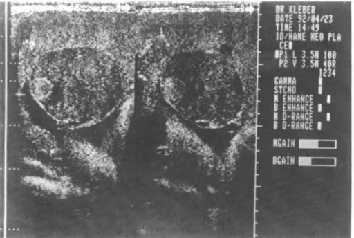
ultrasonographically at 35 w eeks
w hen a 60 x 57 m m subchorionic
lesion w as visualized in an anterior
placenta. It consisted of a w ell
delineated hypoechoic im age w ith
a sm all echogenic round a.rea
(F ig. I ). T he echographic diagnosis
w as hem angiom a. U
ltrasono-graphically, the fetus presented
norm al biom etrical param eters for
the gestational age w ithout
cardiom egaly or hepatom egaly.
D elivery took place at 38 w eeks of gestation by cesarean section
and w as uneventful. T he m ale
child w as born w eighing 3.420 g.
E chocardiography and abdom inal
U S of the new born did not disclose
cardiac or hepatic alterations and
a hem atological evaluation w as
norm al.
T he placenta w eighed 500g and m easured 18 x 13
cm . A large tum or m easuring 6 x 6 cm bulged on the
surface. It w as subchorionic and presented a fleshy and
red cut surface, w ell delineated by a thin capsule (F ig.2).
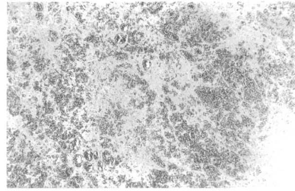
T he um bilical cord w as norm al. M icroscopically, the tum or
consisted of sm all vessels w ithin a m yxedem atous strom a (F ig.3). It w as delineated by a fibrous capsule and exhibited
the aspect of a capillary hem angiom a. T he placental villi
w ere norm al for the gestational age, although they presented m ore capi Ilaries than
is norm ally observed
(chorio-angiosis).
A ccording to P hilippe (5),
placental hem angiom as gene-rally appear after 30 w eeks
gestation. H ow ever, there are
tw o reports of chorionangiom as
diagnosed at 22 and 25 w eeks by ultrasonography (1,4). In the
present case, the tum or appeared
after 29 w eeks of gestation and show ed a rapid grow th, reaching
. 60 x 57 m m in less than six FIGURE 2 -C ut surface of the tum or show ing a large and tum or surrounded by a thin capsule. w eeks.'
gestation, a routine ultasonography (U S ) yielded norm al
results. In the her first pregnancy she had provoked
abortion. H er second pregnancy, three years ago, resulted
in a term vaginal deli very of a healthy child. T he course
of the present pregnancy w as uneventful until the 29th
w eek, w hen she began to present uterine contractions. A t
this tim e, the U S w as norm al.
S he w as recom m ended to rest and w as treated w ith
betam im etics (B rycanil).
FIGURE 1 - U ltrasonography show s a w ell-delineated hypoechoic im age w ith a sm all echogenic round area at the left.
1035
FIGURE 3 - M icroscopical section of the tum or show any sm all blood w ithin a m yxom atous strom a. H E,40x.
The tumor was
subchorionic,
close
to
the
umbilical
cord, and comprised
more than half ofthe
placental
paren-chyma.
In spite of
this, the chi ld was
born without
com-plications.
It was
submitted
to
an
echocardiograph
ic
study
and
to
an
abdominal
ultra-sonography
in order
to"search for cardiac
enlargement
and
abdominal
tumors,
respectively,
but no
abnormalities
were
observed.
In
addi-tion, no
hemangio-mas were seen in the
newborn's
skin. Probably,
the chorioangiosis
observed
in the placental
villi resulted
from
a compensatory
mechanism
because
of the reduction
of the functional
parenchyma.
The present case is unusual because the very large
placental hemangioma
did not cause abnormalities
in the
conceptus, and the pregnancy
was uneventful.
The ultrasonographic
and placental
examinations
indicated a rapid growth of the hemangioma.
RESUMO
Introduyao: Apresenta-se um caso de volum oso hem angiom a placentario envolvendo m ais de m etade do 6rgao sem causar com plica~oes feto-m aternas. R esultados: C om 29 sem anas a placenta era norm al ao ultra-som e com 35 sem anas ja exibia im agem hipoec6ica com 60 x 57 cm .0parto ocorreu com 38 sem anas sem intercorrencias e0recem -nascidd nao apresentou alterayoes.
REFERENCES
I. ACHIRON, R; SHAIA, M; SHIMMED, M et al.
-Choriongioma with hydrops in twins. The Fetus 2: I,1992.
2. BENIRSCHKE, K.& KAUFMAN, P. -Pathology of the Human Placenta, New York, Springer-Verlag, 1990, p 841. 3. BITrE COURT, A.L. - Tumores Primarios da Placenta.
Nao Trofoblasticos, in Garcia, A; Azoubel, R. (eds): A
placenta humana. Morfologia e Patologia Fetal e Perinatal. Rio de Janeiro, Athenell, 1986, p 187.
4. DAO, A.H.; ROGERS, W. & WONG, S.W.
-Chorioangioma of the placenta: Report of two cases with
1Iitasound study. Obstet Gynecol 57:46S, 1981.
5. PHILIPPE, E. - Histopathologie Placentaire, Paris,
Masson & Cie Editeurs, 1974, p 101.
C H AG AS, K.; BITIEN C O U R T, A.; SAN TAN A, A.M .; TEIXEIR A, V. - Large placental hem angiom a diagnosed by ultrasonography - a case report