Application of BRED technology to construct recombinant D29
reporter phage expressing EGFP
Joas L. da Silva1, Mariana Piuri2, Gregory Broussard3, Laura J. Marinelli3, Gisele M. Bastos1, Rosario D.C. Hirata1, Graham F. Hatfull3& Mario H. Hirata1
1Laboratory of Applied Molecular Biology and Pharmacogenomics, School of Pharmaceutical Sciences, University of Sao Paulo, Sao Paulo, Brazil; 2Departamento de Quımica Biologica, Facultad de Ciencias Exactas y Naturales, Universidad de Buenos Aires, IQUIBICEN-CONICET, Buenos Aires,
Argentina; and3Department of Biological Sciences, Pittsburgh Bacteriophage Institute, University of Pittsburgh, Pittsburgh, PA, USA
Correspondence:Joas L. da Silva, Laboratory of Applied Molecular Biology and Pharmacogenomics, School of Pharmaceutical Sciences, University of Sao Paulo, Av. Lineu Prestes 580, B-17, 05508-900, S~ao Paulo, SP, Brazil. Tel./fax: +55 11 3091 3660; e-mail: [email protected]
Received 26 March 2013; revised 19 April 2013; accepted 3 May 2013. Final version published online 3 June 2013.
DOI: 10.1111/1574-6968.12171
Editor: Wolfgang Schumann
Keywords
Mycobacterium; recombineering; bacteriophage; green fluorescent protein.
Abstract
Bacteriophage Recombineering of Electroporated DNA (BRED) has been described for construction of gene deletion and point mutations in
mycobacte-riophages. Using BRED, we inserted a Phsp60-egfp cassette (1143 bp) into the
mycobacteriophage D29 genome to construct a new reporter phage, which was used for detection of mycobacterial cells. The cassette was successfully inserted and recombinant mycobacteriophage purified. DNA sequencing of the cassette
did not show any mutations even after several phage generations.
Mycobacte-rium smegmatis mc2155 cells were infected with D29::Phsp60-egfp (MOI of 10)
and evaluated for EGFP expression by microscopy. Fluorescence was observed at around 2 h after infection, but dissipated in later times because of cell lysis.
We attempted to construct a lysis-defective mutant by deleting the lysA gene,
although we were unable to purify the mutant to homogeneity even with com-plementation. These observations demonstrate the ability of BRED to insert
c. 1 kbp-sized DNA segments into mycobacteriophage genomes as a strategy
for constructing new diagnostic reporter phages.
Introduction
Recombinant bacteriophages are useful tools for studying gene function and construction of phage reporters for
detecting viable Mycobacterium tuberculosis in clinical
specimens. The insertion of gene cassettes into mycobacte-riophages was first accomplished with shuttle phasmids
based on the lytic mycobacteriophage TM4 (Jacobset al.,
1993). Shuttle phasmids have a combination of phage and plasmid features, can be genetically manipulated in vitro,
amplified in Escherichia coli as plasmids, and used for
infecting Mycobacteria where they can be propagated as bacteriophages. Since then, a variety of luciferase phage reporters have been constructed for detection of active and nonreplicatingM. tuberculosis (Sarkiset al., 1995; Pearson
et al., 1996; Carriereet al., 1997; Dusthackeeret al., 2008). Based on the same technology, a new group of reporter phages (phAE87::Phsp60-egfp and phAE87::Phsp60-ZsYellow)
expressing fluorescent proteins were constructed (Piuri
et al., 2009). These fluoromycobacteriophages allowed the
detection of viable mycobacteria in fluorescent microcopy and by flow cytometry. The phage expressing the EGFP (enhanced green fluorescent protein) was further evalu-ated to detect drug resistance to isoniazid, rifampicin and
streptomycin in a total of 155 strains of M. tuberculosis
showing a low cost per sample and rapid results if compared with standard phenotypic assays; sensitivity for detecting isoniazid and rifampicin resistance was simi-lar to that determined using a resazurin assay, but slightly lower for streptomycin resistance (Rondon et al., 2011).
The expression rate of GFP by fluorophages was recently increased 100-fold by Jain et al. (2012), using a more efficient promoter and a new reporter phage vector. This high-level expression of GFP overcame the influence of background fluorescence in the assay and permitted
the detection of M. tuberculosisin sputum in preliminary
results. The performance of this fluoromycobacteriophage in a high-burden tuberculosis setting remains to be fully evaluated.
MICR
Genetic manipulation to obtain gene replacement,
point mutations,in vivocloning, and unmarked deletions
in mycobacteria became less complex with the advent of a recombineering system by Van Kessel & Hatfull (2007). Analysis of bacteriophage Che9c genome revealed genes encoding an exonuclease (gp60) and a recombinase
(gp61), and homologs to RecE and RecT fromEscherichia
coliRac prophage, respectively. These genes were used for constructing pJV62 containing only gp61 to generate tar-get point mutations with ssDNA as substrate and pJV53 containing both genes for dsDNA recombineering.
Recombineering technology has also been applied for generating mutations, deletions, gene replacement, and insertions in mycobacteriophages with high efficiency. Using Bacteriophage Recombineering of Electroporated
DNA (BRED), M. smegmatis mc2155 with
recombineer-ing functions is simultaneously electroporated with phage genomic DNA and a targeting substrate (Van Kessel & Hatfull, 2008; Van Kessel et al., 2008; Marinel-li et al., 2012). Phage particles can then be recovered by plating the electroporated cells. In general, 1–15% of the screened plaques contain the expected mutant, which can be identified by PCR. These primary plaques are mixed, containing nonrecombinant mycobacterio-phages as well as the mutant. Several rounds of plating and PCR are needed to isolate recombinants (Marinelli
et al., 2008).
There are many advantages in using BRED; there is no need for constructing complex cloning systems, no requirement for a selectable marker, mutations can be made in any part of a mycobacteriophage genome, the recombineering process takes place in vivo, and detection of phage recombinants is rapidly accomplished by PCR. Although BRED is faster and less complex than
alterna-tive cloning systems, insertion or deletion of large DNA segments is anticipated to be less efficient. This has to be considered for constructing reporter phages containing gene cassettes that are usually larger than 400 bp.
As every phage reporter constructed to date took advantage of shuttle phasmid technology, here we attempted to use the BRED approach for constructing a recombinant D29 mycobacteriophage expressing EGFP
under the control of thehsp60 promoter and applying it
to the detection of mycobacteria.
Materials and methods
Electrocompetent cells
Mycobacterium smegmatismc2155 cells containing plasmid
pJV53 were grown for 48 h at 37°C in middlebrook 7H9
supplemented with 10% ADC (albumin, dextrose, catalase) (Becton Dickinson GmbH, Heidelberg, GE), 0.05% Tween
80, and kanamycin at 30lg mL 1 (Sigma-Aldrich, St.
Louis, MO). A volume of 500–1000lL was transferred
from the culture to 100 mL of 7H9 broth with 1 mM CaCl2,
30lg mL 1 kanamycin, and 0.2% succinic acid
(Sigma-Aldrich) to obtain a culture with an optical density (OD600 nm) of 0.02. The culture was incubated at 37°C,
250 rpm until it reached an OD600 of 0.4. Acetamide
(Sigma-Aldrich) was added to a final concentration of 0.2%, and the culture was further incubated for 3 h, at
37°C, 250 rpm. The culture was maintained on ice for
30 min and washed four times with ice cold 10% glycerol (Sigma-Aldrich). The bacterial pellet was suspended in 2 mL of ice cold 10% glycerol. Aliquots were kept
on 80°C until the electroporation or up to 6 months
(Piuriet al., 2009).
Table 1. Oligonucleotides used for D29 phage recombinants construction and detection
Oligonucleotide Sequence
100 bp 82/71 5′-CGTGTTAGTTCAGGAGTTCCTCGATGTCGGGTGGCCAGCACCAGATCATGTGCAGGTTCTTGTAGACGAAGACGCGAATTGGCT TGGGGTTCATGCGATC-3′
D29-p1 5′-GCGGTTCCTTACTGCGTGGGCTCCGCTGGCTACGGAGAACGCACGCGCGTCGTGTTAGTTCAGGAGTTCCTCGAT-3′ D29-p2 5′-TTGAGTCGGACCCGACGCTTCAGCGTCAAAGACAACTACCTGGATGACTGGATCGCATGAACCCCAAGCCAATTC-3′ D29-p3 5′-CGTGTTAGTTCAGGAGTTCCTCGATGTCGGGTGGCCAGCACCAGATCATTTACTTGTACAGCTCGTCCATGCC-3′ D29-p4 5′-GATCGCATGAACCCCAAGCCAATTCGCGTCTTCGTCTACAAGAACCTGCACTCTAGAGGTGACCACAACGAC-3′
100 bp LysA 5‘-CATGACGCTCATAGTCACACGCGACCACGCGCAGTGGGTCCACGACATGAACCCCGAGTACCTACAGGCGTACATCGCCAG GAATGGAGCCCTATGAG- 3‘
LysA-1 5‘-GCAGGTTCGAGTCCTGCTCTCGCGACTTGACAGCCACCACGAAAGGAACCCATGACGCTCATAGTCACACGCGA-3‘ LysA-2 5‘-TGCCGGGGACGAGAGTGCCGACGTAGTAGAGCGTTTCACGGATCTTGGGGCTCATAGGGCTCCATTCCTGGCGAT-3 D1 5′-TACGAAGGTATCGGCGAGCCATC-3′
D2 5′-GCTAGTGAGCGGCATTGCGG-3′ D3 5‘-TCGTTGTGGTCACCTCTAGAGTG-3‘ D4 5‘-GTTCGAGTCCTGCTCTCGCGA-3‘
D5 5‘-ACGAGAGTGCCGACGTAGTAGA-3‘
Construction of a 200-bp recombineering substrate
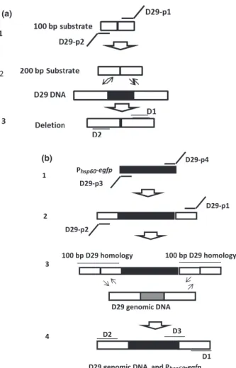
A 100-base oligonucleotide (Integrated DNA Technolo-gies, San Diego, CA) named 100 bp 82/71 (Table 1), with 50-base upstream and downstream homology to the D29 genome segment to be deleted, was amplified by PCR. Two 75-base primers, D29-p1and D29-p2 (Integrated DNA Technologies), with 50-base overlaps on each end of the 100-mer, were used to generate a 200-bp recombi-neering substrate with 100-bp homology on either side of the D29 DNA segment selected for being deleted (Fig. 1).
The PCR was performed on a 2720 thermal cycler (Applied Biosystems, Foster City, CA) in a 0.2-mL tube
containing 1 U of platinum Taq, 0.8lL of dNTPs at
25 mM each, 4 lL of 2 mM MgCl2, 1lL of each primer at
25 pmol L 1, 10lL of 109 buffer (600 mM Tris-SO4,
180 mM (NH4)2SO4), 4lL of DMSO, and PCR-graded
water to a total reaction volume of 100lL. PCR cycling
parameters were set to 2 min at 95°C followed by 30 cycles at 95°C for 30 s, 56°C for 30 s, and 68°C for 1 min, and a final 10-min step at 68°C. The PCR products were puri-fied from agarose gel with QIAquick Gel Extraction Kit (Qiagen, Hilden, GE) following manufacturer‘s protocol.
Construction of thePhsp60-egfprecombineering
substrate
The construction of recombineering substrate was per-formed in two independent PCR. Firstly, the cassette Phsp60-egfp was amplified from plasmid pYL37-egfp. A
50-bp homology flanking the target D29 DNA segment was added upstream and downstream of the cassette (Fig. 1b). Primers D29-p3 and D29-p4 (Table 1) with 50-base (IDT) overlaps on each end of the cassette were used in this first step, as previously described (Marinelliet al., 2008).
The PCR was performed on a 2720 thermal cycler (Applied Biosystems) in a reaction mixture containing
1 U of PFU DNA polymerase, 0.8ll of dNTPs at 25 mM
each, 1lL of each primer at 25 pmol L 1, 10lL of
109 buffer (750 mM Tris-HCl (pH 9.0); 500 mM KCl;
200 mM, (NH4)2SO4), 4 lL of DMSO, 10 ng of plasmid
DNA and PCR-graded water to a total reaction volume
of 100lL. The cycling conditions were set to 1 cycle at
95°C for 5 min, 30 cycles (95 °C for 1 min, 56 °C for
30 s, 72°C for 1.5 min), and a final cycle at 72 °C for
10 min. The PCR product were gel purified using QIAquick Gel Extraction Kit.
A second PCR was carried out to add more 50 bp on each end of the cassette and obtain a final recombineering substrate with 100-bp homology on either side of the target sequence. The PCR assay was performed on a 2720
thermal cycler (Applied Biosystems) with primers
D29-p1and D29-p2 (Table 1) in a reaction containing
0.2lL (1 U) of Platinum Taq high-fidelity DNA
polymer-ase (Invitrogen Corporation, Carlsbad, CA), 0.8lL of
dNTPs at 25 mM each, 4lL of 2 mM MgCl2, 1lL of
each primer at 25 pmol L 1, 10lL of 109 buffer
(600 mM Tris-SO4, 180 mM (NH4)2SO4), 4 lL of DMSO,
10 ng of the first PCR product, and PCR-graded water to
a total reaction volume of 100lL. Cycling parameters
were set to 1 cycle at 95°C for 2 min, 30 cycles (95°C for 1 min, 57°C for 30 s, 68°C for 1.5 min), and a final cycle
at 68°C for 10 min. The PCR product was gel purified
using QIAquick Gel Extraction Kit. Phsp60-egfp
D29-p3
D29-p4
D29-p1
D29-p2
100 bp D29 homology 100 bp D29 homology
D29 genomic DNA
D29 genomic DNA and Phsp60-egfp (b)
(a)
1
2
3
4 D3
D1 D2
Electroporation of recombineering substrates
Aliquots (100lL) of electrocompetent M. smegmatis
mc2155 carrying pJV53 were mixed with 100 ng of phage
DNA and recombineering substrate at concentrations
varying over the range 50–700 ng. The solution was
maintained on ice for 15 min, loaded into 2-cm cuvettes, and immediately electroporated using Gene Pulser Xcell Electroporation Total System (Bio-Rad, Hercules, CA)
using a pulse of 2500 V, 25lF, and 1000 Ω (Goude &
Parish, 2008). Electroporated cells were kept in 7H9
sup-plemented with 10% ADC and 1 mM CaCl2 at 37°C/
250 rpm/1 h, gently mixed with 4 mL of molten 7H9 soft
agar (10% ADC, 1 mM CaCl2, 0,75% agar), and plated
onto 7H10 with 500lL of M. smegmatis mc2 155. After
16-h incubation at 37°C, each primary plaque was
transferred to 100lL of phage buffer (10 mM Tris-HCl,
pH 7.5; 10 mM MgSO4; 68.5 mM NaCl; 1 mM CaCl2).
Detection of 472-bp deletion
Recombinant phages from primary plaques were detected
in 1lL of phages by PCR using PFU DNA polymerase.
Primers D1 and D2 (Integrated DNA Technologies) (Table 1) flanking the deleted region were used to amplify a 1002-bp or a 530-bp segment of wild-type and recombinant phage, respectively. The concentrations of each PCR component were the same as described above.
The PCR was set to 1 cycle at 95°C for 5 min, 30 cycles
(95°C for 1 min, 59°C for 30 s, 72 °C for 1 min), and a final step at 72°C for 10 min.
Primary plaques containing recombinant and wild-type phages were transferred to 100lL phage buffer and serially
diluted from 10 1to 10 6. Dilutions were incubated with
500 lL of M. smegmatis mc2155 at 37°C/15 min, mixed
with 5 mL of molten 7H9 soft agar and replated onto 7H10
supplemented with 10% ADC and 1 mM CaCl2. New
pla-ques were screened by PCR for mutant detection as described above. This process was performed until only pure recombinant phages were detected in plaques.
To confirm the deletion, DNA sequencing of the recombinant phages was carried out on ABI 3730 DNA Analyzer using BigDye Terminator v3.1 Cycle Sequencing Kit (Applied Biosystem) with the same set of primers (D1 and D2) previously used for detecting phage mutants. The data obtained were analyzed with BioEdit software v7.0.9 (Ibis Bioscience, Carlsbad, CA).
Detection of thePhsp60-egfpinsertion/
replacement
Primary plaques were first screened by PCR using primers D1 and D3 (Table 1). The PCR was performed on a 2720
thermal Cycler (Applied Biosystems) in a reaction
con-taining 0.2lL of Platinum Taq high-fidelity DNA
poly-merase (Invitrogen Corporation), 0.8lL of dNTPs at
25 mM each, 4lL of 2 mM MgCl2, 0.1lL of each
pri-mer at 25 pmol L 1, 2.5lL of 109 buffer (600 mM
Tris-SO4, 180 mM (NH4)2SO4), 1 lL of DMSO, 1lL of
phage, and PCR-graded water to a total reaction volume of 25lL. Cycling parameters were set to 1 cycle at 95°C
for 2 min, 30 cycles (95°C for 1 min, 58°C for 1 min),
and a final cycle at 68°C for 10 min.
A second PCR was carried out with primers D1 and D2 (Table 1) flanking the recombineering site to confirm the presence of samples containing only recombinant
phages. The PCR was performed in a 25lL reaction
con-taining 1 U of PFU DNA polymerase, 0.4lL of dNTPs at
25 mM each, 0.1lL of each primer at 25 pmol L 1,
2.5lL of 109 buffer (750 mM Tris-HCl (pH 9.0);
500 mM KCl; 200 mM, (NH4)2SO4), and 1lL of sample.
The cycling conditions were set to 1 cycle at 95°C for
5 min, 30 cycles (95°C for 1 min, 59°C for 30 s, 72 °C
for 2 min), and a final cycle at 72°C for 10 min.
Deletion of lysA
A 200-bp substrate containing 100-bp homology on either
side of the lysA gene targeting sequence was constructed
as previously described using the oligonucleotide 100-bp LysA and primers LysA1 and LysA2 (Integrated DNA Technologies) (Table 1).
Mutant phages were detected by PCR (Marinelli et al.,
2008) using platinum Taq (Invitrogen Corporation) with primers D4 and D6, which fully hybridizes only with recombinant phages. The amplification was performed
under the following cycling conditions: 2 min at 95°C,
30 cycles (30 s at 95°C, 30 s at 59°C), and a 10 min at
68°C.
A PCR with PFU DNA polymerase was also performed with primers D4 and D7. This set of primer amplifies the targeting DNA segment of both recombinant and wild-type phage D29, and it was used to confirm the presence of only phage recombinants after several steps of phage purification. The following cycling parameters were per-formed: 5 min at 95°C, 35 cycles (30 s at 95 °C, 30 s at 59°C, and 45 s at 72°C), and 10 min at 72°C.
Evaluation of EGFP expression
Three aliquots (500lL) of M. smegmatis mc2155 were
infected with recombinant D29 (MOI of 10) and incubated
at 30, 37, and 42°C. A volume of 100lL was recovered
0.22-lm filter and immediately evaluated by fluorescence
microscopy (Axiostar Plus, Carl Zeiss, Gottingen, GE)
with a 1009 objective and oil immersion, using the REL.
4.6 software. Filters 42002-HQ 479/30X, HQ 520/40 m, and Q495LP (Chroma Technology Corporation, Bellows Falls, VT) were used for detection of EGFP. No software was used for enhancing image quality.
Results and discussion
Construction of a D29::Phsp60-egfpreporter
phage
Mycobacteriophage D29 is a lytic phage related to the
temperate phage L5 (Ford et al., 1998). It carries two
lysogeny-related genes partially deleted in its right arm
(Ford et al., 1998). Additionally, a nonessential region
near the cohesive end in the right arm of D29 genome was previously described using phasmid-based technology (Pearsonet al., 1996).
To verify that the region encompassing gene 82/71
(Sarkiset al., 1995; Fordet al., 1998) was nonessential for lytic growth of D29, a segment of 472 bp was deleted
(D29 genome coordinates 45.387–45.859). This deletion
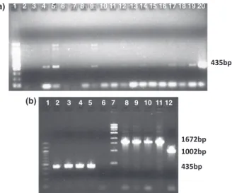
was accomplished in a single co-electroporation of 100 ng D29 genomic DNA and targeting substrate (Table 1). In a total of 100 plaques screened, over 20% contained the deletion mutant, as identified by PCR of the flanking sequences (Fig. 2). Recombinant bacteriophages were
pla-que purified and shown to grow with normal plapla-que morphology and production of phage particles (data not shown).
The region encompassing the 472-bp sequence was than replaced by a Phsp60-egfpcassette (Fig. 3). Differently from
the previous segment deletion, we started co-electroporating 200 ng of D29 genomic DNA and targeting DNA substrate at concentrations varying from 100 to 700 ng. No plaques containing phage recombinants were found using flanking PCR or mismatched PCR with concentrations of targeting substrate below 600 ng. In a total of 600 primary plaques screened, we found only around 1% of individual plaques containing phage recombinants.
Several steps of serial dilutions followed by recovery and replating mixed populations were needed to obtain pure phage recombinants, indicating that they represented only a minor portion of particles in the primary plaques. This relatively poor recovery of recombinants may be due to the size of the insert, because the previous deletion of the same region with a 200-bp substrate generated a high proportion of mutant phages. DNA sequencing confirmed insertion of the Phsp60-egfp cassette into D29 genome, and
mutations were not found even after production of sev-eral phage stocks. Although mutant detection required multiple rounds of PCR, it was not more laborious than using shuttle phasmid technology for the construction. Although the number of phage recombinants containing
(a)
(b)
Fig. 2. Gel electrophoresis of PCR products using flanking primers D1 and D2 for detection of plaques containing phage D29 with a deletion in the right arm. (a) 1. 1-kb ladder, 2–16. Primary plaques containing a mixed population of wild-type D29 phages (901 bp) and recombinant D29 phages (529 bp). (b) 1. 100-bp ladder, 2. Negative control, DNA band amplified from phage D29 genomic DNA, 3. Positive control, DNA bands generated from a primary mixed plaque, 4–12. Production of a single 529-pb DNA band from pure plaques of D29 phage recombinants.
435bp
435bp 1672bp
1002bp
1 2 3 4 5 6 7 8 9 10 11 12 1 2 3 4 5 6 7 8 9 10 11 12 13 14 15 16 17 18 19 20 (a)
(b)
the Phsp60-egfpcassette was lower than when compared to
the cloning of DNA segments under 400 bp, there was no need of carrying a serial of recombineerings to insert the whole segment in parts, what significantly reduced the time and cost of the procedure.
Detection ofM. smegmatisusing the D29::Phsp60-egfpreporter phage
To evaluate if EGFP is expressed from the D29:: Phsp60-egfp
phage at levels visible by fluorescent microscopy,
M. smegmatis mc2155 was grown to mid-log phase and infected with mycobacteriophage D29::Phsp60-egfpat a
mul-tiplicity of infection of 10. Samples collected every 30 min during 6 h, washed in PBS buffer, and concentrated in a
0.22lm membrane were immediately analyzed for
detecting fluorescent cells. Fluorescence was not visualized in the first 1 h 30 min of infection. Instead, a growing number of lysed cells were observed in the course of infection. Fluorescent cells were detected in the 2-h
infec-tion at 37°C (Fig. 4b), but phage D29::Phsp60-egfp had
lysed the majority of cells at this time as observed on Fig. 4a. Infection time over 3 h reduced the living cells to a number difficult to visualize under microscopy.
Infections carried out at 30 and 42°C did not provide
fluorescence cells.
Attempts to construct a lysis-defective mutant of D29
We reasoned that we could extend the period of fluores-cence and increase its intensity by constructing a lysis defect mutant of the fluorophage. One approach is to
delete the lysA gene encoding the endolysin, which is
known to be required for lysis (Payneet al., 2009). Using
BRED to construct this deletion, a lysA mutant was
detected by PCR in 5% of the recovered primary plaques, showing recombination was successful. Attempts to purify the mutant phage away from the wild-type helper phage particles in the mixed primary plaque were unsuccessful, consistent with lysin A being an essential function for plaque formation. We thus constructed a complementing
plasmid encoding the Corndog lysin A, which has been
shown previously to be tolerated byM. smegmatisand to
complement a lysin A mutant of mycobacteriophage Giles
(Marinelli et al., 2008). However, we were not able to
purify the lysA mutant by complementation even after
screening over 1600 plaques.
With some reporter phages already developed, the next main steps into improving phenotypic assays based on bacteriophage technology are to reduce the time needed
to detect M. tuberculosis and increase the sensibility of
phage-based assays. The lytic phage machinery may be valuable for construction of recombinant phages present-ing high mRNA transcription rate, which can improve mRNA transcription of reporter genes. The advent of BRED technology makes possible the construction of phages containing reporter genes virtually in any portion of a bacteriophage genome under the control of any pro-moter. Using BRED technology, it was possible to insert the whole Phsp60-egfp cassette into the genome of
myco-bacteriophage D29. This approach may be useful to con-struct novel reporter phages and improve phasmids expressing reporter proteins already developed.
Acknowledgement
This research was supported by grant from FAPESP (Project 2008/05076-7), Brazil.
Competing interests
The authors declare no competing interests.
References
Carriere C, Riska PF, Zimhony O, Kriakov J, Bardarov S, Burns J, Chan J & Jacobs WR Jr (1997) Conditionally replicating luciferase reporter phages: improved sensitivity for rapid detection and assessment of drug susceptibility of
Mycobacterium tuberculosis.J Clin Microbiol35: 3232–3239. Dusthackeer A, Kumar V, Subbian S, Sivaramakrishnan G,
Zhu G, Subramanyam B, Hassan S, Nagamaiah S, Chan J & Paranji Rama N (2008) Construction and evaluation of
(a) (b)
Fig. 4. Mycobacterium smegmatismc2155
luciferase reporter phages for the detection of active and non-replicating tubercle bacilli.J Microbiol Methods73: 18–25.
Ford ME, Sarkis GJ, Belanger AE, Hendrix RW & Hatfull GF (1998) Genome structure of mycobacteriophage D29: implications for phage evolution.J Mol Biol279: 143–164. Goude R & Parish T (2008) Electroporation of mycobacteria.
J Vis Exp23: 15. Pii. 761.
Jacobs WR Jr, Barletta RG, Udani R, Chan J, Kalkut G, Sosne G, Kieser T, Sarkis GJ, Hatfull GF & Bloom BR (1993) Rapid assessment of drug susceptibilities ofMycobacterium tuberculosisby means of luciferase reporter phages.Science
260: 819–822.
Jain P, Hartman TE, Eisenberg Net al.(2012) (2GFP10, a high-intensity fluorophage, enables detection and rapid drug susceptibility testing ofMycobacterium tuberculosisdirectly from sputum samples.J Clin Microbiol50: 1362–1369. Marinelli LJ, Piuri M, Swigonov a Z, Balachandran A, Oldfield
LM, Van Kessel JC & Hatfull GF (2008) BRED: a simple and powerful tool for constructing mutant and recombinant bacteriophage genomes.PLoS ONE3: e3957.
Marinelli LJ, Hatfull GF & Piuri M (2012) Recombineering: a powerful tool for modification of bacteriophage genomes.
Bacteriophages2: 5–14.
Payne K, Sun Q, Sacchettini J & Hatfull GF (2009)
Mycobacteriophage Lysin B is a novel mycolylarabinogactan esterase.Mol Microbiol73: 367–381.
Pearson RE, Jurgensen S, Sarkis GJ, Hatfull GF & Jacobs WR Jr (1996) Construction of D29 shuttle phasmids and luciferase reporter phages for detection of mycobacteria.
Gene183: 129–136.
Piuri M, Jacobs WR Jr & Hatfull GF (2009)
Fluoromycobacteriophages for rapid, specific, and sensitive antibiotic susceptibility testing ofMycobacterium
tuberculosis.PLoS ONE4: e4870.
Rondon L, Piuri M, Jacobs WR Jr, De Ward J, Hatfull GF & Takiff HE (2011) Evaluation of fluoromycobacteriophages for detecting drug resistance inMycobacterium tuberculosis.
J Clin Microbiol49: 1838–1842.
Sarkis GJ, Jacobs WR Jr & Hatfull GF (1995) L5 luciferase reporter mycobacteriophages: a sensitive tool for the detection and assay of live mycobacteria.Mol Microbiol15: 1055–1067.
Van Kessel JC & Hatfull GF (2007) Recombineering in
Mycobacterium tuberculosis.Nat Methods4: 147–152. Van Kessel JC & Hatfull GF (2008) Efficient point mutagenesis
in mycobacteria using single-stranded DNA recombineering: characterization of antimycobacterial drug targets.Mol Microbiol67: 1094–1107.
Van Kessel JC, Marinelli LJ & Hatfull GF (2008)