Effects of
N
-Glycosylation Site Removal in
Archaellins on the Assembly and Function of
Archaella in
Methanococcus maripaludis
Yan Ding1, Kaoru Uchida2, Shin-Ichi Aizawa2, Kathleen Murphy3, Alison Berezuk3, Cezar M. Khursigara3, James P. J. Chong4, Ken F. Jarrell1*
1Department of Biomedical and Molecular Sciences, Queen’s University, Kingston, Ontario, Canada,
2Department of Life Sciences, Prefectural University of Hiroshima, 562 Nanatsuka, Shobara, Hiroshima, Japan,3Department of Molecular and Cellular Biology, University of Guelph, Guelph, Ontario, Canada,
4Department of Biology, University of York, Heslington, York, United Kingdom
Abstract
InMethanococcus maripaludisS2, the swimming organelle, the archaellum, is composed of three archaellins, FlaB1S2, FlaB2S2and FlaB3S2. All three are modified with anN-linked
tetrasaccharide at multiple sites. Disruption of theN-linked glycosylation pathway is known to cause defects in archaella assembly or function. Here, we explored the potential require-ment ofN-glycosylation of archaellins on archaellation by investigating the effects of elimi-nating the 4N-glycosylation sites in the wildtype FlaB2S2protein in all possible
combinations either by Asn to Glu (N to Q) substitution or Asn to Asp (N to D) substitutions of theN-glycosylation sequon asparagine. The ability of these mutant derivatives to comple-ment a non-archaellatedΔflaB2S2strain was examined by electron microscopy (for archae-lla assembly) and swarm plates (for analysis of swimming). Western blot results showed that all mutated FlaB2S2proteins were expressed and of smaller apparent molecular mass
compared to wildtype FlaB2S2, consistent with the loss of glycosylation sites. In the 8
sin-gle-site mutant complements, archaella were observed on the surface of Q2, D2 and D4 (numbers after N or Q refer to the 1stto 4thglycosylation site). Of the 6 double-site mutation
complementations all were archaellated except D1,3. Of the 4 triple-site mutation comple-ments, only D2,3,4 was archaellated. Elimination of all 4N-glycosylation sites resulted in non-archaellated cells, indicating some minimum amount of archaellin glycosylation was necessary for their incorporation into stable archaella. All complementations that led to a re-turn of archaella also resulted in motile cells with the exception of the D4 version. In addi-tion, a series of FlaB2S2scanning deletions each missing 10 amino acids was also
generated and tested for their ability to complement theΔflaB2S2strain. While most variants were expressed, none of them restored archaellation, although FlaB2S2harbouring a
smaller 3-amino acid deletion was able to partially restore archaellation. OPEN ACCESS
Citation:Ding Y, Uchida K, Aizawa S-I, Murphy K, Berezuk A, Khursigara CM, et al. (2015) Effects of N-Glycosylation Site Removal in Archaellins on the Assembly and Function of Archaella in Methanococcus maripaludis. PLoS ONE 10(2): e0116402. doi:10.1371/journal.pone.0116402
Academic Editor:Frederique Lisacek, Swiss Institute of Bioinformatics, SWITZERLAND
Received:October 8, 2014
Accepted:December 9, 2014
Published:February 20, 2015
Copyright:© 2015 Ding et al. This is an open access article distributed under the terms of the Creative Commons Attribution License, which permits unrestricted use, distribution, and reproduction in any medium, provided the original author and source are credited.
Data Availability Statement:All relevant data are within the paper.
Introduction
N-glycosylation is a prevalent protein modification found in all three domains of life in which the attachment of the glycan is via the nitrogen atom in asparagine residues located in the accep-tor glycoprotein [1–6]. General features of theN-glycosylation pathways are shared among the three domains [1–3,5,7,8]. TheN-glycan precursor is first synthesized on a phosphorylated iso-prene-based lipid carrier, either a dolichol derivative in Eukarya and Archaea, or an undecapre-nol derivative in Bacteria, via the activities of glycosyltransferases. The assembled lipid-linked glycan is then flipped across a membrane, i.e. to face into the lumen of the endoplasmic reticu-lum in Eukarya or to the external face of the cytoplasmic membrane in Archaea and Bacteria. A signature enzyme of the pathway, the oligosaccharyltransferase (OST), transfers the completed glycanen blocfrom its lipid carrier to select Asn residues in target proteins, although further sugars can still be added to the protein-bound glycan. The Asn residue to which theN-glycan is attached is usually located in an Asn-Xaa-Ser/Thr sequon (Xaa cannot be Pro), although in some Bacteria, i.e.Campylobacterspp., a negatively charged amino acid residue is also needed at the-2 position. Between the two prokaryotic domains, Bacteria and Archaea,N-glycosylation appears to be much more widespread among Archaea, where 166 of 168 sequenced genomes ex-amined contained at least one copy of a gene encoding the archaeal OST AglB [9]. A variety of archaeal proteins, mainly S-layer proteins and the subunits of surface structures including both pilins and archaellins (formerly archaeal flagellins [10]), have been shown to be modified with N-glycans [11–18]. Recent work on archaealN-glycosylation systems has combined both struc-tural and genetic methods, typically using archaellins and S-layer proteins as reporter proteins. Since the first archaeal glycosylation (agl) genes were identified in 2006 [14,19], this combined approach has focused on a few key archaeal model organisms [20]:Methanococcus maripaludis S2,Methanococcus voltaePS,Haloferax volcaniiH53 andSulfolobus acidocaldariusMW001 [1,6,11,12,15,16,21]. Interestingly, theN-glycosylation pathway is not essential for any of the three studied euryarchaeotes (M. maripaludisS2,M. voltaePS andHfx. volcaniiH53), as mu-tants carrying a deletion or insertional inactivation ofaglBwere readily obtained [14,19,22]. However, in the crenarchaeoteS. acidocaldariusMW001, repeated attempts to delete or inter-ruptaglBwere unsuccessful and only the integration of a second copy ofaglBinto the genome allowed for the deletion of the originalaglB[23]. The key reported effects that result from per-turbation or complete abolition of theN-glycosylation pathway in Archaea are on S-layer stabil-ity, growth of cells at high salinities and on archaellation and motility [18,19,22,24–30].
The archaellum (formerly archaeal flagellum) is the major motility apparatus found in Ar-chaea [10]. It is a rotating appendage unrelated to the bacterial flagellum but bearing instead similarities to the bacterial type IV pilus. These similarities include homologous ATPase and membrane platform proteins involved in assembly of the structure as well as similarities in their major structural proteins [31–35]. The structural proteins, called archaellins, are made as preproteins with class III (type IV pilin-like) signal peptides which are removed by a specific prepilin peptidase-like enzyme (termed FlaK in methanogens or PibD in Sulfolobales and halo-philes) [36–39]. Signal peptide removal is critical for incorporation of archaellins into the fila-ment [37,39]. In addition, it appears that archaellins are commonly modified withN-linked glycans and interruption of the normalN-glycosylation pathway leads to defects in archaella assembly or function [1,40]. In species whereaglBhas been deleted or insertionally inactivated (M. voltaePS,M. maripaludisS2,Hfx. volcaniiH53), the cells are unable to make archaella. If the pathway is interrupted at steps that lead to a truncated glycan, there is an impairment in motility, although archaella are still made unless the truncation is too great [18,19,22]. InM. maripaludisS2 strain, archaella contain 3 archaellins: the major archaellins FlaB1S2and
FlaB2S2form the filament while the minor archaellin FlaB3S2comprises the hook region [41].
The three archaellins share sequence similarities including a class III signal peptide cleaved by FlaK, conserved N-terminal and C-terminal regions, and a hypervariable region in the middle [41]. The hypervariable regions of FlaB1S2, FlaB2S2and FlaB3S2are decorated at multiple
posi-tions with a unique tetrasaccharide [11]. Cells are archaellated if they carry deletions inagl genes that result in aN-glycan of at least two sugars, but non-archaellated if the deletedagl gene results in a glycan of only a single sugar or preventsN-glycosylation totally (as in anaglB deletion) [22,42,43]. These results could mean that the archaellins must be glycosylated at some or all of its glycosylation sequons by at least a two sugar glycan for those archaellins to be assembled into a structure. However, an alternative explanation is that the necessity for the gly-cosylation lies at a different step in archaella assembly.
A major goal of this study was to examine the requirement ofN-glycosylation of the major archaellin FlaB2S2for archaellation inM. maripaludisS2. FlaB2S2has fiveN-glycosylation
sequons,26NTS28,66NIT68,110NLT112,119NTT121and124NWS126. The first sequon26NTS28 lo-cated in the N-terminal conserved region was previously reported to be unoccupied with glycan while the remaining four, located in the hypervariable region, were modified with tetrasacchar-ide (Fig. 1, [11]). For these experiments, we eliminated the 4 occupied sequons (66NIT68,
110NLT112,119NTT121and124NWS126, designated as the 1st, 2nd, 3rdand 4thN-glycosylation
site, respectively) in all possible combinations (creating single-, double-, triple- and quadruple-site mutations in FlaB2S2). We also generated a series of FlaB2S2scanning deletions in an
at-tempt to determine regions of the molecule that were essential for assembly of archaella.
Materials and Methods
Strains and growth conditions
M. maripaludisS2Δhpt(Mm900) [44],M. maripaludisS2ΔhptΔflaB2S2(ΔflaB2S2in short
hereafter) [41] and all complemented strains, as well asM. maripaludisΔRC (formerly Metha-nococcus deltaeΔRC [45,46]) were routinely cultured anaerobically in 125 mL sealed serum bottles containing 10 mL Balch medium III under an atmosphere of CO2:H2(20:80) at 37°C
with shaking [47]. Cells carrying a complementation plasmid were cultured in the presence of 2.5μg/mL puromycin for plasmid selection [48]. For swarming assays, cells were inoculated
onto plates of Balch medium III containing 0.25% (w/v) agar in the presence of 2.5μg/mL
pu-romycin [22].Escherichia coliTOP 10 cells (Invitrogen Inc.), used for molecular cloning steps, were cultured at 37°C in Luria Broth (LB) medium with shaking or on LB plates (containing 1.5% w/v agar) in the presence of 100μg/mL ampicillin for plasmid selection. Strains and
plas-mids used in this study are listed inTable 1.
Construction of mutant
flaB2
S2genes using site-directed mutagenesis
(SDM)
To generate mutantflaB2S2genes, the wildtypeflaB2S2gene was first cloned into the
pCR2.1-TOPO-TA vector (Invitrogen Inc.) to create pKJ902. This pCR2.1-TOPO-flaB2S2and its
deriv-atives were used as template to generate the mutants listed inTable 2. The wildtypeflaB2S2
gene used in cloning was generated by PCR using the complementation primers listed in
Table 3and genomic DNA from Mm900 as template.
To generate the mutantflaB2S2genes that would encode proteins in whichN-glycosylation
transformed intoE. coliTOP10 competent cells. Plasmids extracted from the transformants were sequenced to confirm the mutation. Using this method, 8 single-site mutantflaB2S2genes
were generated that resulted in 4 N to Q single-site mutations and 4 N to D single-site muta-tions in their protein products. Double-site mutantflaB2S2genes were then generated using the
plasmids with the single-site changes inflaB2S2as template. The same strategy was used to
cre-ate the triple and quadruple glycosylation site mutantflaB2S2genes. The multi-site mutant
pro-teins all contained N to D changes only.
TheG6(flaB2ΔRC) gene was amplified by PCR usingM. maripaludisΔRC whole cells as
template with the complementation primers inTable 3and subsequently cloned into pCR2.1-TOPO. TheG10gene whose protein product contains additional glycosylation sites was chemi-cally synthesized by GenScript USA Inc. (Piscataway, NJ).
Fig 1. Protein sequence alignment of FlaB2S2, G6 (FlaB2ΔRC) and G10.Signal peptide is shown in grey; the first sequon26NTS28that is not occupied with
N-glycan is shown in orange; the 4 occupiedN-glycosylation sequons are shown in red; the 3-amino acid61GTA63deletion in 3AA is shown in green; extra
sequons in the G6 and G10 are also shown in orange; sequences differences from FlaB2S2in G6 and G10 that do not introduce new sequons are shown
in blue.
doi:10.1371/journal.pone.0116402.g001
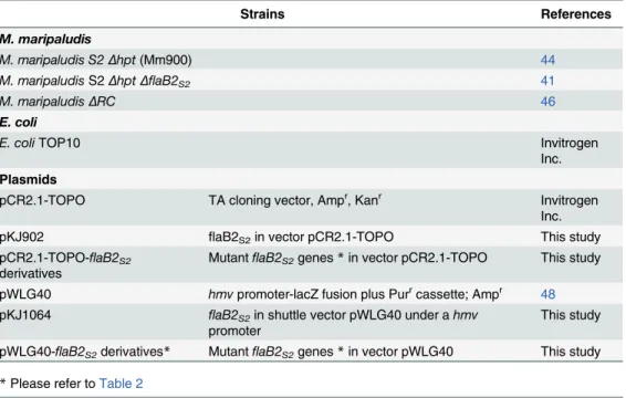
Table 1. Strains and plasmids used in this study.
Strains References
M. maripaludis
M. maripaludis S2Δhpt(Mm900) 44
M. maripaludisS2ΔhptΔflaB2S2 41
M. maripaludisΔRC 46
E. coli
E. coliTOP10 Invitrogen
Inc.
Plasmids
pCR2.1-TOPO TA cloning vector, Ampr, Kanr Invitrogen
Inc.
pKJ902 flaB2S2in vector pCR2.1-TOPO This study
pCR2.1-TOPO-flaB2S2
derivatives Mutantfl
aB2S2genes*in vector pCR2.1-TOPO This study pWLG40 hmvpromoter-lacZ fusion plus Purrcassette; Ampr 48
pKJ1064 flaB2S2in shuttle vector pWLG40 under ahmv
promoter This study
pWLG40-flaB2S2derivatives* MutantflaB2S2genes*in vector pWLG40 This study *Please refer toTable 2
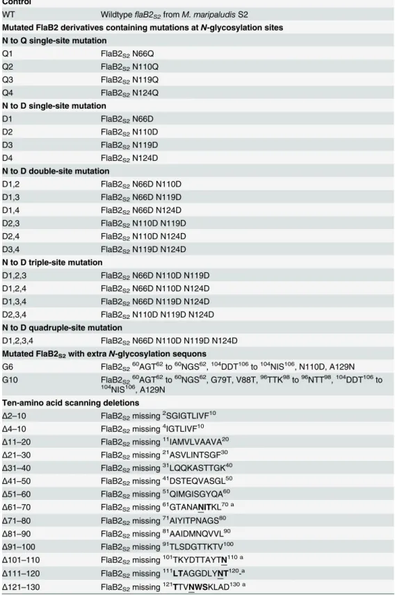
Table 2. Mutated FlaB2S2 derivatives generated in this study.
Mutated FlaB2 derivatives
Description
Control
WT WildtypeflaB2S2fromM. maripaludisS2
Mutated FlaB2 derivatives containing mutations atN-glycosylation sites
N to Q single-site mutation
Q1 FlaB2S2N66Q
Q2 FlaB2S2N110Q
Q3 FlaB2S2N119Q
Q4 FlaB2S2N124Q
N to D single-site mutation
D1 FlaB2S2N66D
D2 FlaB2S2N110D
D3 FlaB2S2N119D
D4 FlaB2S2N124D
N to D double-site mutation
D1,2 FlaB2S2N66D N110D
D1,3 FlaB2S2N66D N119D
D1,4 FlaB2S2N66D N124D
D2,3 FlaB2S2N110D N119D
D2,4 FlaB2S2N110D N124D
D3,4 FlaB2S2N119D N124D
N to D triple-site mutation
D1,2,3 FlaB2S2N66D N110D N119D
D1,2,4 FlaB2S2N66D N110D N124D
D1,3,4 FlaB2S2N66D N119D N124D
D2,3,4 FlaB2S2N110D N119D N124D
N to D quadruple-site mutation
D1,2,3,4 FlaB2S2N66D N110D N119D N124D Mutated FlaB2S2with extraN-glycosylation sequons
G6 FlaB2S260AGT62to60NGS62,104DDT106to104NIS106, N110D, A129N
G10 FlaB2S260AGT62to60NGS62, G79T, V88T,96TTK98to96NTT98,104DDT106to 104NIS106, A129N
Ten-amino acid scanning deletions
Δ2–10 FlaB2S2missing2SGIGTLIVF10 Δ4–10 FlaB2S2missing4IGTLIVF10 Δ11–20 FlaB2S2missing11IAMVLVAAVA20 Δ21–30 FlaB2S2missing21ASVLINTSGF30 Δ31–40 FlaB2S2missing31LQQKASTTGK40 Δ41–50 FlaB2S2missing41DSTEQVASGL50 Δ51–60 FlaB2S2missing51QIMGISGYQA60 Δ61–70 FlaB2S2missing61GTANANITKL70 a Δ71–80 FlaB2S2missing71AIYITPNAGS80 Δ81–90 FlaB2S2missing81AAIDMNQVVL90 Δ91–100 FlaB2S2missing91TLSDGTTKTV100 Δ101–110 FlaB2S2missing101TKYDTTAYTN110 a Δ111–120 FlaB2S2missing111LTAGGDLYNT120-a Δ121–130 FlaB2S2missing121TTVNWSKLAD130 a
For construction of the mutant versions offlaB2S2whose protein products would contain
scanning deletions, inverse PCR and overlapping primers was employed, again with pKJ902 as template. The forward primer was designed to contain theflaB2S2gene sequence flanking the
desired in-frame 30 bp deletion. The reverse primer was complementary to the forward primer sequence upstream of the deletion (Table 2). AfterDpnI digestion, purified PCR products were transformed intoE. coliTOP10 competent cells. Recombinant plasmids were extracted from the transformants and used as template for amplifying theflaB2S2mutant genes by PCR with
the complementation primers listed inTable 2. The smallerflaB2S2mutant genes were
identi-fied by agarose gel electrophoresis of the PCR products which were also sequenced to confirm the deletion. The same protocol was used to generate the 3 amino acid deletion,61GTA63, where the deletion of the 9bp resulted in the removal of anRsaI restriction site. This allowed for the screening of theflaB2S2gene in plasmids carried by transformants for the small deletion
by digestion of subsequentflaB2S2PCR products withRsaI. Lastly, a mutantflaB2S2gene
en-coding a mutated FlaB2S2protein with a 10-amino acid deletion at91TLSDGTTKTV100had
those amino acids replaced with a copy of the 10 amino acids at161IIVSGVSFDT170, thereby generating a substitution mutant version of FlaB2S2(SUB) that was still the same length as the
wildtype version. All the mutant versions offlaB2S2generated were sequenced to confirm
the mutations.
Construction of complementation vectors
To generate complementation plasmids forM. maripaludisS2, mutantflaB2S2genes in the
pCR2.1-TOPO vector were PCR amplified using complementation primers with anNsiI re-striction site incorporated into the forward primer and anXbaI site into the reverse primer (Table 2). AfterNsiI andXbaI digestion, the PCR product was cloned into the shuttle vector pWLG40 where transcription of the cloned gene is under the control of the strong constitutive hmvpromoter [48]. MutantflaB2S2genes in pWLG40 were sequenced to confirm the insert
gene sequence. As a further control, plasmids were re-isolated from the complemented cells and re-sequenced.
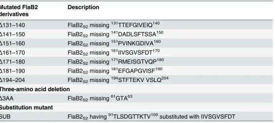
Table 2. (Continued)
Mutated FlaB2 derivatives
Description
Δ131–140 FlaB2S2missing131TTEFGIVEIQ140 Δ141–150 FlaB2S2missing141DADLSFTSSA150 Δ151–160 FlaB2S2missing151PVINKGDIVA160 Δ161–170 FlaB2S2missing161IIVSGVSFDT170 Δ171–180 FlaB2S2missing171RMEISGTVQP180 Δ181–190 FlaB2S2missing181EFGAPGVISF190 Δ194–204 FlaB2S2missing194STFTEKV VSLQ204 Three-amino acid deletion
Δ3AA FlaB2S2missing61GTA63 Substitution mutant
SUB FlaB2S2having91TLSDGTTKTV100substituted with IIVSGVSFDT
The mutantflaB2S2genes werefirst generated in pCR2.1-TOPO and then cloned into theM. maripaludis expression vector pWLG40.
abold letters:N-glycosylation sequons with the asparagine underlined
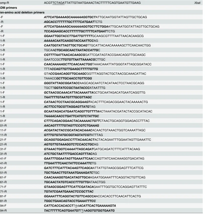
Table 3. Primers used in this studya.
Complementation primers Notes
comp-F CTAGATGCATGAAAATAACAGAATTCATGAAAAGCAAAAAAGGTGCTTC NsiI
comp-R ACGTTCTAGATTATTGTAATGAAACTACTTTTTCAGTGAATGTTGAAG XbaI
SDM primers
Ten-amino acid deletion primers
2-F ATTCATGAAAAGCAAAAAAGGTGCTATTGCAATGGTATTAGTTGCTGCAG 2-R AGCACCTTTTTTGCTTTTCATGAATTCTG
4-F ATTCATGAAAAGCAAAAAAGGTGCTTCTGGAATTGCAATGGTATTAGTTGCTGCAG 4-R TCCAGAAGCACCTTTTTTGCTTTTCATGAATTCTG
11-F GGAATTGGTACCTTGATTGTTTTTGCAAGCGTTTTAATTAACACAAGCG 11-R AAAAACAATCAAGGTACCAATTCCAG
21-F CAATGGTATTAGTTGCTGCAGTTGCATTACAACAAAAAGCTTCAACAACTGG
21-R TGCAACTGCAGCAACTAATACCATTGC
31-F CGTTTTAATTAACACAAGCGGATTCGATAGTACCGAACAGGTTGCAAGC
31-R GAATCCGCTTGTGTTAATTAAAACGCTTGC
41-F CAACAAAAAGCTTCAACAACTGGTAAACAAATTATGGGTATTAGCGGATACC 41-R TTTACCAGTTGTTGAAGCTTTTTGTTG
51-F GTACCGAACAGGTTGCAAGCGGTTTAGGTACTGCTAACGCAAACATTAC
51-R TAAACCGCTTGCAACCTGTTCGG
61-F GGGTATTAGCGGATACCAAGCAGCAATCTACATAACTCCTAACGCAGG
61-R TGCTTGGTATCCGCTAATACCCATAATTTG
71-F GCTAACGCAAACATTACAAAATTAGCTGCAATAGACATGAATCAGGTTG
71-R TAATTTTGTAATGTTTGCGTTAGC
81-F CATAACTCCTAACGCAGGAAGTACACTTTCAGACGGAACTACAAAAACTG
81-R ACTTCCTGCGTTAGGAGTTATGTAG
91-F GCAATAGACATGAATCAGGTTGTTTTAACTAAATACGATACTACCGCATACAC 91-R TAAAACAACCTGATTCATGTCTATTGC
101-F CTTTCAGACGGAACTACAAAAACTGTTCTAACTGCAGGTGGAGACCTTTAC
101-R AACAGTTTTTGTAGTTCCGTCTGAAAG
111-F ACGATACTACCGCATACACAAACACAACTGTAAACTGGTCAAAATTAGC
111-R GTTTGTGTATGCGGTAGTATCGTATTTAG
121-F GCAGGTGGAGACCTTTACAACACTACTACAGAATTTGGAATAGTTGAAATTC
121-R AGTGTTGTAAAGGTCTCCACCTGCAG
131-F GTAAACTGGTCAAAATTAGCAGATGATGCAGATCTTTCATTTACAAG
131-R ATCTGCTAATTTTGACCAGTTTACAG
141-F GAATTTGGAATAGTTGAAATTCAACCAGTTATCAACAAAGGTGACATAG
141-R TTGAATTTCAACTATTCCAAATTCTG
151-F GATCTTTCATTTACAAGTTCAGCAATTATTGTAAGCGGAGTTTCATTCG
151-R TGCTGAACTTGTAAATGAAAGATCTGC
161-F CAACAAAGGTGACATAGTTGCAAGAATGGAAATTTCAGGTACTGTTCAG
161-R TGCAACTATGTCACCTTTGTTGATAACTGG
171-F GTAAGCGGAGTTTCATTCGATACAGAATTTGGTGCTCCAGGAGTTATTTC
171-R TGTATCGAATGAAACTCCGCTTAC
181-F GGAAATTTCAGGTACTGTTCAGCCAACCACACCTTCAACATTCACTG
181-R TGGCTGAACAGTACCTGAAATTTCC
194-F CATTCACCACACCTTAAACATTCACTGAAAAAGTA
194-R TACTTTTTCAGTGAATGTTTAAGGTGTGGTGAATG
Complementation of a
Δ
flaB2
S2mutant using mutant
flaB2
S2derivatives
To determine if the mutant FlaB2S2proteins generated above could restore archaellation and
motility in theΔflaB2S2mutant, recombinant pWLG40 plasmids carrying the various mutant
flaB2S2derivatives were transformed individually into theΔflaB2S2mutant using a PEG-based
method [41,50]. Transformants were cultured in Balch medium III containing 0.25μg/mL
pu-romycin for plasmid selection [48].
Western blot analysis of the
Δ
flaB2
S2mutant complemented with mutant
flaB2
S2derivatives
Whole cell lysates of complemented cells carrying the various mutantflaB2S2genes were
sepa-rated by SDS-PAGE (15% gels) and then transferred onto an Immobilon-P membrane (Milli-pore Inc.) [51]. Mutant FlaB2S2proteins were detected using chicken anti-FlaB2S2specific
primary antibody [41]. Horseradish peroxidase-conjugated rabbit anti-chicken immunoglobu-lin Y (Jackson Immuno Research Laboratories) was used as secondary antibody, and the blots were developed using Immobilon Western Chemiluminescent HRP Substrate (Millipore Inc.).
Table 3. (Continued)
Complementation primers Notes
Three-amino acid deletion primers
3aa-F ATGGGTATTAGCGGATACCAAGCAAACGCAAACATTACAAAATTAGC
3aa-R TGCTTGGTATCCGCTAATACCCATAATTTG
Ten-amino acid substitution (sub) primers
sub-F TGTAAGCGGAGTTTCATTCGATACAACTAAATACGATACTACCGC
sub-R CGAATGAAACTCCGCTTACAATAATTAAAACAACCTGATTCATGTC
N-glycosylation site mutation primers
Q1-F GTACTGCTAACGCACAAATTACAAAATTAGC
Q1-R GCTAATTTTGTAATTTGTGCGTTAGCAGTAC
Q2-F CTACCGCATACACACAACTAACTGCAGGTGGAG
Q2-R CTCCACCTGCAGTTAGTTGTGTGTATGCGGTAG
Q3-F GTGGAGACCTTTACCAAACTACAACTGTAAACTG
Q3-R CAGTTTACAGTTGTAGTTTGGTAAAGGTCTCCAC
Q4-F AACACTACAACTGTACAATGGTCAAAATTAGC
Q4-R GCTAATTTTGACCATTGTACAGTTGTAGTGTT
D1-F GTACTGCTAACGCAGACATTACAAAATTAGC
D1-R GCTAATTTTGTAATGTCTGCGTTAGCAGTAC
D2-F CTACCGCATACACAGACCTAACTGCAGGTGGAG
D2-R CTCCACCTGCAGTTAGGTCTGTGTATGCGGTAG
D3-F GTGGAGACCTTTACGACACTACAACTGTAAACTG
D3-R CAGTTTACAGTTGTAGTGTCGTAAAGGTCTCCAC
D4-F AACACTACAACTGTAGACTGGTCAAAATTAGC
D4-R GCTAATTTTGACCAGTCTACAGTTGTAGTGTT aUnderlined: restriction enzyme sites
Italic bold: reverse complementary sequences in primer pairs Italics: mutated amino acid codon
Underlined in italics: mutated DNA base
Swarming assay of the
Δ
flaB2
S2mutant complemented with mutant
flaB2
S2derivatives
ComplementedΔflaB2S2strains carrying plasmids with mutantflaB2S2genes encoding proteins
having mutations at the variousN-glycosylation sites were examined for motility using semi-solid swarm plates [22]. Briefly, the OD600of an overnight cell culture was measured and
adjust-ed to 1.0. Five microliters of the adjustadjust-ed cell culture were inoculatadjust-ed onto semi-solid Balch me-dium containing 0.25% (w/v) agar using a micropipette in an anaerobic chamber by stabbing the tip into the agar. Plates were incubated in an anaerobic canister at 37°C for 4 or 6 days.
Electron microscopy of the
Δ
flaB2
S2mutant complemented with mutant
flaB2
S2derivatives
ComplementedM. maripaludisΔflaB2S2cells carrying mutantflaB2S2genes were collected
from an overnight culture by centrifugation at 20 000 g for 1 min, washed with 2% (w/v) NaCl and resuspended in phosphate-buffered saline. Resuspended cells were loaded onto carbon-Formvar-coated copper grids and stained with 2% phosphotungstic acid, pH 7.0. Grids were examined in a Hitachi 7000 electron microscope operating at an accelerating voltage of 75 kV.
Results and Discussion
Generation of mutant
flaB2
S2derivatives
While deletions in genes that affectN-glycosylation are known to cause severe defects in archaellation and motility [18,22,28,29], it is not clear if the defects are related directly to the inability of non-glycosylated archaellins or archaellins glycosylated with truncated glycans to assemble into archaella, or whether the glycosylation defect affected other steps in the assembly of archaella. For example, it may be that another protein critical for assembly of archaella, but not an archaellin, must be glycosylated in order to function properly. InHfx. volcaniiH53, changing the sequence of the major archaellinflgAat any of the 3 examined sequons so that the encoded amino acid changed from Asn to Gln led to mutant forms of the protein that could not rescue the swimming defect of anflgAdeletion strain, suggesting that each glycosyla-tion site was necessary for archaellaglycosyla-tion [18]. However, this is not the case forM. maripaludis S2. Previous work in this methanogen showed that a strain that had a spontaneous mutation in flaB2S2which led to the loss of the 2ndN-glycosylation site of the archaellin that is normally
decorated with theN-linked tetrasaccharide, was, nonetheless, still archaellated and motile [43]. To examine the possible role that eachN-glycosylation site, either alone or in combina-tion with other sites, might have on archaella formacombina-tion and motility inM. maripaludisS2, var-ious mutantflaB2S2genes whose products were lacking single to quadrupleN-glycosylation
sites either by Asn to Gln (N to Q) substitution, or Asn to Asp (N to D) substitution of the N-glycosylation sequon asparagine were generated and cloned into the complementation vec-tor pWLG40 (Table 2). For these mutant constructs we used D or Q followed by a number to indicate that the change was N to D or N to Q with the number representing the site changed, i.e. Q1 indicates mutant FlaB2S2with an N to Q substitution at the 1stN-glycosylation site.
In addition, two other mutant genes, designated G6 and G10 (Fig. 1), whose products con-tain extraN-glycosylation sequons were generated and cloned into pWLG40. The G6 sequence encodes the wildtype FlaB2ΔRCprotein (i.e. FlaB2ΔRCfromM. maripaludisΔRC). FlaB2ΔRC
and FlaB2S2share 95% identity, with the differences almost exclusively confined to several
N-glycosylation sites (Fig. 1). Compared to FlaB2S2, FlaB2ΔRCshares 3 sites, plus it has 3
addi-tional sequons,60NGS62,104NIS106,129NDT131, but it is missing the 2ndN-glycosylation site
110NLT112in FlaB2
additional sites created in the hypervariable region at sites requiring only minimal amino acid changes to generate a total of 10 possible sites (Fig. 1). G6 and G10 both have the26NIS28 sequon in the N-terminal conserved region that is not occupied withN-glycan in FlaB2S2. After
transformation of these recombinant plasmid pWLG40-flaB2S2mutants into aΔflaB2S2
mu-tant, the complemented strains were examined for expression of the mutant FlaB2S2proteins,
archaella formation and cell motility.
Western blot analysis of the
Δ
flaB2
s2strain complemented with
flaB2
S2derivatives containing mutations at
N
-glycosylation sites
Western blots were run to detect the expression and stability of the various mutant versions of FlaB2S2in the complementedΔflaB2S2mutant. As shown inFig. 2, all mutant versions of
FlaB2S2except G10 were successfully expressed in theΔflaB2S2mutant. All mutant FlaB2S2
pro-teins were expressed in similar amounts and all appeared stable as judged by the general lack of any cross-reacting smaller molecular mass bands which could be indicative of protein degrada-tion. The amount of the G10 version of FlaB2S2detected in western blots was very low and could
be only observed when blots were overexposed (data not shown). We have found previously that cells carrying mutations in any gene that prevents assembly of archaella (as in theΔflaB2S2
mu-tant) often stop transcribing theflaoperon after several sub-cultures in the laboratory. This then makes the complementation of the original gene deletion back to an archaellated state impossi-ble [22]. For this reason, the presence of FlaE, whose gene is a downstream member of thefla op-eron, was also confirmed by western blot to ensure that theflaoperon was still transcribed in the
ΔflaB2S2mutant during the course of the complementation experiments (data not shown) [41].
In general, mutant FlaB2S2proteins missingN-glycosylation sites all had a smaller apparent
molecular mass than that of wildtype FlaB2S2when examined by western blotting, with the
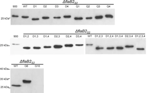
Fig 2. Western blot analysis of whole cell lysates of theΔflaB2S2strain complemented withflaB2S2 with mutations at variousN-glycosylation sites.Mutant FlaB2S2proteins missing single to quadrupleN
-glycosylation sites showed smaller apparent molecular mass than that of wildtype FlaB2S2. The G6 which
has extra glycosylation sequons migrated slower than wildtype FlaB2S2. The expression of G10 was not
detectable on this blot with normal exposure time. 900: wildtypeM. maripaludisS2Δhpt. WT:ΔflaB2S2 complemented with wildtypeflaB2S2.
possible exceptions of D4 and Q4 which ran at very close to wildtype size. The greater the num-ber ofN-glycosylation sites eliminated in a particular FlaB2S2mutant, the faster the mutant
proteins migrated, i.e., single-site mutants had the largest apparent molecular mass, and the quadruple-site mutant D1,2,3,4 had the smallest. However, the 8 single-site mutants did not migrate as proteins of the same apparent molecular mass. Of the 8 mutants, D1 and Q1, both of which had the 1stN-glycosylation site eliminated, had the smallest apparent molecular mass, while D4 and Q4 had the largest. Similar results were observed from the double-site and triple-site mutations. In the 6 double-triple-site mutations, FlaB2S2with D1,2 and D1,3 sites eliminated had
the smallest apparent molecular mass, followed by FlaB2S2with D1,4 and D2,3 sites eliminated,
while the archaellin having the D2,4 and D3,4 sites eliminated migrated with the largest appar-ent molecular mass. In the triple-site mutants, FlaB2S2with any of the 1stN-glycosylation site
eliminated (D1,2,3, D1,2,4 and D1,3,4) migrated at the same apparent molecular mass while FlaB2S2with the other triple combination of sites eliminated (D2,3,4) migrated more slowly.
One possible explanation for the observed different electrophoretic mobilities is that mutant FlaB2S2proteins lacking the same number ofN-glycosylation sites have the same number of
N-glycans attached but the attachment ofN-glycan on some sequons might have effects on the local protein structure so that the glycoprotein is not able to be totally denatured by SDS, thus resulting in an unusual migration pattern. This unusual electrophoretic mobility has been ob-served in other similar studies and been the suggested explanation. Human erythropoietin (Epo) has 3N-glycosylation sites, and the 3 single-site mutants generated by N to Q SDM showed uneven migrations on western blot, although all of the 3 mutants had the same theoret-ical molecular mass but differed only in the position of theN-glycans [52]. Similar uneven mi-gration was also observed in the 4 single-site mutations of hepatitis C virus envelope protein E1 each missing oneN-glycosylation site [53].
While local folding effects might explain the altered electrophoretic mobility, another possi-ble explanation for this unusual western blot result is that elimination of the 1stN-glycosylation site might interfere with the cell’s ability toN-glycosylate the remaining sites, resulting in FlaB2S2where not all the remaining sequons are actually occupied (and so run as smaller
mo-lecular mass proteins). On the other hand, it may be that elimination of one or more glycosyla-tion sites results in the attachment of glycan to the sequon26NTS28that is not glycosylated under our usual growth conditions. This could explain why, for example, the FlaB2S2mutants
that are missing the 4thN-glycosylation site (D4 and Q4) showed a larger apparent molecular mass than the other single mutants and close to wildtype size. If D4 and Q4, missing the 4th gly-cosylation site, now hadN-glycan attached at the normally unused sequon, these mutant pro-teins would have an identical mass as the wildtype. There is precedent for glycosylation at one sequon influencing what happens at distant sites. For example, it has been reported for rabies virus glycoprotein thatN-glycosylation at one sequon can influence processing of the
N-glycans at a different site on the protein [54]. InM. voltaePS, the archaella are composed of 4 archaellins FlaAMv, FlaB1Mv, FlaB2Mvand FlaB3Mv, which share high sequence similarity in
the N-terminal conserved region with FlaB1S2and FlaB2S2[41,55]. Interestingly, the first 40
amino acids in the mature FlaB1Mvand FlaB2Mvincluding the26NTS28sequon, are identical to
those of FlaB2S2, but in the case of theM. voltaePS archaellins, the26NTS28sequon was found
to be occupied withN-linked glycan [12]. Clearly, this region of the archaellin can be glycosy-lated and possibly the26NTS28sequon in FlaB2S2might be able to beN-glycosylated inM.
mar-ipaludisS2 under different conditions.
The G6 mutant FlaB2S2protein (identical to the FlaB2ΔRC) with additional glycosylation
sequons compared to wildtype FlaB2S2had a larger apparent molecular mass (~32 kDa) than
wildtype FlaB2S2(~27 kDa) in western blots. Since G6 has 6N-glycosylation sites (excluding
possibly all, of the extra sequons are, in fact, occupied since the mass of the tetrasaccharide is only 1036 Da [11]. Although the expression level of G10 was extremely low, on over-exposed western blots, the apparent molecular mass (37kDa) of this“artificially designed”glycoprotein was even larger than G6, indicating that AglB can recognize and transfer glycan to at least some of the newly introduced sequons in the hypervariable region.
Electron microscopy of the
Δ
flaB2
s2strain complemented with
flaB2
S2derivatives containing mutations at
N
-glycosylation sites
Complemented cells carrying FlaB2S2proteins having mutations atN-glycosylation sites were
examined by transmission electron microscopy for archaellation, and the results are listed in
Table 4. The majority of the complementations either restored archaellation to essentially all cells or were unable to restore archaellation to any cells. In only a couple of cases did the comple-mentation lead to a population which contained roughly equal number of archaellated and non-archaellated cells (D1,2 and D3,4).Fig. 3shows electron microscopy pictures of a number of se-lected complements (Q2, Q4, D2, D3, D4, D1,3, D2,4, D1,2,4, D2,3,4, D1,2,3,4, G6 and G10).
The archaellation state of the 4 control strains was as expected. Wildtype Mm900 cells (900) were archaellated while theΔflaB2S2mutant strain was non-archaellated. Archaella were
Table 4. Archaellation and swarming ability of complements bearing FlaB2S2mutants atN-glycosylation sites.
Complements Archaellationa Motility
Controls WT ++ (100%) ++
Blank - (0%)
-NQ single Q1 - (3%)b
-Q2 ++ (100%) ++
Q3 - (3%)b
-Q4 - (0%)
-ND single D1 - (3%)b
-D2 ++ (100%) ++
D3 - (0%)
-D4 ++ (93%)
-ND double D1,2 + (47%) +
D1,3 - (0%)
-D1,4 ++ (97%) ++
D2,3 ++ (100%) ++
D2,4 ++ (100%) +
D3,4 + (60%) +
ND triple D1,2,3 - (0%)
-D1,2,4 - (0%)
-D1,3,4 - (0%)
-D2,3,4 ++ (100%) ++
ND quadruple D1,2,3,4 - (0%)
-Additional sequons G6 ++ (100%) +++
G10 - (0%)
-aFor each strain a minimum of 30 random cells were assessed for the presence or absence of archaella. Values in parentheses describe the percentage
of cells with observable archaella.
bOnly a rare cell was observed with archaella, typically very few in number and abnormally short.
observed when theΔflaB2S2strain was complemented with the wildtype version offlaB2S2, but
not when theΔflaB2S2strain was complemented with the empty vector pWLG40.
In the 8 single-site mutation complements, 3 mutant versions of FlaB2S2(Q2, D2 and D4),
could restore archaellation. In the 3 archaellated complements, Q2 and D2 had different amino acid substitutions at the same 2ndN-glycosylation site. The N to D amino acid change at the 2ndsequon generated in this study replicates the spontaneous mutation inflaB2S2that we
pre-viously reported [43]. Both that spontaneous mutant and the complemented cells carrying the mutantD2gene generated in this study showed no impairment in archaellation or swarming motility [43]. These results indicate that missing the 2ndN-glycosylation site alone does not sig-nificantly interfere with archaellation. In contrast, complementation of theΔflaB2S2strain with
flaB2S2lacking the 4thN-glycosylation site differed markedly depending on what amino acid
the original Asn was changed to; cells complemented with the D4 version had archaella under electron microscopy while cells complemented with the Q4 version did not (Fig. 3). None of the other single-site mutation complements (Q1, Q3, Q4, D1 and D3 (Fig. 3)) were considered archaellated, although in each of the Q1, Q3 and D1 complementations a rare cell with short archaella was observed (Table 4).
Among the 6 double-site mutation complements, all but the D1,3 version could assemble archaella (electron micrographs of D1,3 and D2,4 complemented cells are shown inFig. 3). This was surprising since many of these double-site mutants contained eliminated sites which if deleted alone resulted in non-archaellated cells. In the 4 triple-site mutation complements, 3 complements D1,2,3, D1,2,4 and D1,3,4 were non-archaellated, while archaella could be assem-bled in D2,3,4 (Fig. 3). However, when all glycosylation sites were eliminated in the comple-menting version offlaB2S2(D1,2,3,4), theΔflaB2S2cells were not able to assemble archaella.
These results indicate that inM. maripaludisS2, archaella can be assembled using FlaB2S2
lack-ing as many as 3 out of the 4 glycosylation sites, as long as the first site remained intact (D2,3,4) but not when the archaellin is entirely non-glycosylated (D1,2,3,4).
In the two complementations where new sequons were introduced into FlaB2S2, different
re-sults were observed. In the G6 complemented cells, theΔflaB2S2strain were now archaellated,
suggesting that the FlaB2S2protein with extraN-glycan modifications in the hypervariable
re-gion could be incorporated into the archaellar filament by the archaella assembly apparatus in M. maripaludisS2. This was not unexpected since this version of FlaB2 already exists naturally in the archaellatedM. maripaludisstrainΔRC. In contrast, no archaella were observed on the
ΔflaB2S2strain complemented with the G10 version. The G10 version had extra glycosylation
sequons added to the internal hypervariable region of the protein. While this protein appeared to be modified at, at least, some of these additional sequons with glycan, judging from its higher apparent molecular mass in western blots, it was very poorly expressed in the cells under our normal growth conditions and this low expression may explain the lack of archaella observed by electron microscopy (Fig. 3).
Swarming assays of complements with mutant
flaB2
S2derivatives
containing mutations at
N
-glycosylation sites
In addition to the restoration of archaellation, the complemented cells were also examined for possible restoration of motility using semi-solid agar plates (Fig. 4A). Motility assay results are summarized inTable 4, which also incorporates the archaellation status of the complemented strains for comparison.
also non-motile on swarm plates, even after an extra 2-day incubation (Fig. 4B). The unusual exception was the complementation with the D4 version of FlaB2S2which was archaellated but
non-motile (Fig. 3,Fig. 4A). However, among the motile complemented cells, the swarming di-ameter was not always returned to the wildtype level. Cells complemented with theflaB2S2
genes carrying D2, Q2, D1,4, D2,3 and D2,3,4, mutations swarmed out to a similar distance on semi-solid agar (swarming diameter of D2/ WT = 1.01±0.11, Q2/WT = 1.10±0.09, D1,4/WT =
Fig 3. Transmission electron micrographs of selectΔflaB2S2strain complemented withflaB2S2with mutations at variousN-glycosylation sites.
Wildtype Mm900 cell (900) was archaellated, while theΔflaB2S2mutant was not. Wildtype FlaB2S2protein expressed inΔflaB2S2restored archaellation (WT), but the empty vector could not. Archaella were observed on surface of Q2, D2, D4, D2,4, D2,3,4 and G6 complemented cells. Cells complemented with Q4, D3, D1,3, D1,2,4, D1,2,3,4 and G10 were non-archaellated. Archaella are indicated by arrows. Bar equals 500 nm.
0.93±0.14, D2,3/WT = 0.96±0.20, D2,3,4/WT = 1.08±0.22), while complements with the flaB2S2genes carrying D1,2, D2,4 and D3,4 had a smaller swarming diameter (swarming
diam-eter of D1,2/WT = 0.51±0.05, D2,4/WT = 0.68±0.13, D3,4/WT = 0.52±0.07). For the D1,2 and D3,4 complementations, the smaller swarming distance may be explained by the lower per-centage of cells that were observed by electron microscopy to be archaellated, but in the case of the D2,4 all cells examined were archaellated. Interestingly, G6 appeared hyper-motile as it consistently swarmed further than cells complemented with the wildtype version offlaB2S2
(swarming diameter of G6/WT = 1.24±0.10).
InHfx. volcaniiH53, none of the 3 FlgA single-site mutation complements showed motility [18]. However, in this study,M. maripaludisS2 cells were still as motile as wildtype cells when theΔflaB2S2strains was complemented withflaB2S2with the D2,3,4 changes, in which
archae-lla were assembled using FlaB2S2lacking 3 out of the 4N-glycosylation sites. The structural
protein (flagellin) of the functionally analogous bacterial swimming organelle, the flagellum, can also be modified with glycan, especially in Gram-negative bacteria, although the linkage is O-glycosidic rather thanN-glycosidic [56,57]. TheO-glycan modification in bacterial flagellin can be critical for flagella assembly, stabilization, motility, and even virulence in pathogens [56,58–60]. InPseudomonas syringaepv.tabaci, flagellin FliC has 6O-glycosylation sites, and single-site mutations in any of these sites resulted in various impairments in motility, while a mutant carrying mutations to eliminate all 6O-glycosylation sites in FliC was non-motile [59]. The structural protein (pilin) from bacterial type-IV pili, structures which share several signifi-cant similarities with archaella [10,34], can also beO-glycosylated [61,62]. Elimination ofO -glycosylation of type IV pilin resulted in reduced twitching motility inPseudomonas aerugi-nosa1244 andP. syringaepv.tabacibut did not interfere with pili assembly [62,63]. However, inP. aeruginosa5196 in which a differentO-glycan was attached to the type IV pilin PilA, O-glycosylation played critical roles in both type IV pili assembly and twitching motility [2,64].
Fig 4. Swarming plates showing the motility of theΔflaB2S2strain complemented withflaB2S2with mutations at variousN-glycosylation sites.A.
Plates were incubated at 37°C for 4 days. B. Complemented cells that did not show motility or showed poor motility after 4 days incubation were incubated a further 2 days.
The results obtained with the G6 complemented cells indicate that an increase in glycosyla-tion can lead to hyper-motility. The western blots results indicate that the G6 version of FlaB2 is hyper-glycosylated compared to the wildtype FlaB2S2version. Of all the complemented
strains, only the G6 complement consistently demonstrated an increased zone of swarming compared to the wildtype. Interestingly, similar results were also observed in regards the O-glycosylation of flagellin inHelicobacter pylori[65] whereO-glycosylation of the flagellins FlaA and FlaB with pseudaminic acid is essential for flagella assembly and cell motility [66,67]. AH. pylorimutant defective in deglycosylation of flagellins showed both hyper-O -glycosyla-tion (3 fold more pseudominic acid) of FlaA as well as hyper-motility [65]. However, there is a limit to how many extra sequons can be added to archaellins since archaellin synthesis was very poor in the G10 complemented cells, even though the small amount of the G10 version detected was apparently modified at, at least, some of the extra sites.
The data obtained from the glycosylation site elimination mutants indicates that while no particular single site of glycosylation on FlaB2S2is essential, nonetheless glycosylation at some
site is necessary for archaella formation. In the case of the 4 triple-site mutants it is clear that glycosylation of only site 1 is sufficient for archaellation and motility. However, removal of the 1stsite did not always lead to non-archaellated cells as witnessed by the archaellated and motile cells observed in the D1,4 complementation, suggesting that glycosylation of FlaB2S2at several
different combinations of sites could be sufficient for incorporation of the subunits into func-tional archaella. In some ways, this is reminiscent of a situation in Wzc, a tyrosine autokinase essential for capsule formation inE. coli. Phosphorylation of tyrosine residues in the C-termi-nus of Wzc are necessary for its function but no single tyrosine is essential for phosphorylation and it was suggested that the overall level of phosphorylation rather than a precise combination of tyrosine residues accessible to phosphorylation is what is important for Wzc activity [68].
Western blot analysis of the
Δ
flaB2
s2strain complemented with
flaB2
S2scanning deletions
To determine which regions of the FlaB2S2protein are critical for archaella formation, a series
of FlaB2S2scanning deletion mutants that sequentially lacked 10 amino acids were generated
in the complementation vector pWLG40 and transformed into theΔflaB2S2mutant. The
scan-ning deletions inflaB2S2were identified since they migrated slightly faster than the wildtype
version offlaB2S2in 0.8% agarose gels due to the 30 bp deletion (Fig. 5shows an example for
screening ofΔ31–40). For the first 10 amino acids, two versions were generated. The first was deleted for amino acids 2–10 (named asΔ2–10), leaving the +1 amino acid which we thought might be important for successful cleavage of the 12 amino acid signal peptide. We also gener-ated a 4–10 amino acid deletion (named asΔ4–10) since the +3 glycine of the mature protein is needed for signal peptide removal in archaellins of the related methanogenM. voltaePS [69].
Mutant FlaB2S2proteins from whole cell lysates of the various complemented cells were
de-tected using anti-FlaB2S2specific antibody on western blot analysis, as shown inFig. 6. In the
21 FlaB2S2scanning deletions, 19 mutant proteins (all, exceptΔ4–10 andΔ11–20) were readily
detected on western blot by anti-FlaB2S2specific antibody, although the expression level ofΔ2–
10 andΔ21–31 was relatively lower compared to that of the other mutants. Evidence of some possible protein degradation was observed in theΔ2–10 andΔ161–170 FlaB2S2as multiple
lower molecular mass bands were detected in these two lanes. These results indicate that some of the mutant proteins were either not expressed or were unstable and degraded. Fourteen mu-tant proteins, includingΔ21–30,Δ31–40,Δ41–50,Δ51–60,Δ71–80,Δ81–90,Δ91–100,Δ131–
Fig 5. PCR screening of theΔ31–40scanning deletion.Following the deletion procedure, theflaB2S2 gene was amplified usingflaB2S2complementation primers and the PCR product analyzed by agarose gel electrophoresis along with the amplification product obtained with the wildtypeflaB2S2gene using the same primers. The scanning deletion is readily distinguished from wildtypeflaB2S2by the faster migration of its 30 bp smaller PCR product M: 100 bp DNA ladder;flaB2S2: PCR products using pKJ902 as template;Δ31–40: PCR products using plasmid isolated from one colony of theΔ31–40transformants as template.
doi:10.1371/journal.pone.0116402.g005
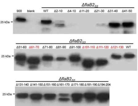
Fig 6. Western blot analysis of theΔflaB2S2strain complemented withflaB2S2scanning deletions.
Except forΔ4–10 andΔ11–20, all the FlaB2S2scanning deletion proteins were expressed, although the
expression level ofΔ2–10 andΔ21–30 was relatively low. FlaB2S2scanning deletion proteinsΔ61–70, Δ101–110,Δ111–120 whose 10-amino acid deletion contains anN-glycosylation site (shown in red) had smaller apparent molecular masses due to the loss ofN-glycan usually attached at this site.Δ121–130 missing the 4thN-glycosylation site (shown in red) had unusual bigger apparent molecular mass than the
or from theΔflaB2S2strain complemented with the wildtype version offlaB2S2. This is
consis-tent with the fact that all these mutation proteins are 10 amino acids shorter than wildtype FlaB2S2. The upper band in the mutantΔ2–10 lane appears slightly larger than the neighboring
WT lane. This might be due to the lack of processing of the archaellin signal peptide in this de-letion since inM. voltaePS the +3 glycine position was essential for cleavage of the signal pep-tide by the pre-archaellin peptidase FlaK and this is missing in theΔ2–10 version of FlaB2S2
[69]. The presence of the signal peptide does not prevent the attachment ofN-glycans [37] so this version of FlaB2S2would be expected to have a full complement of attachedN-glycans as
well as the extra amino acids of the signal peptide contributing to make it run slightly larger in the western blots than the processed wildtype version of FlaB2S2.
The FlaB2S2mutantsΔ61–70,Δ101–110 andΔ111–120 had even smaller apparent
molecu-lar masses compared to the FlaB2S2carrying other scanning deletions. This can be attributed to
the fact that these three deletions result in the loss of oneN-glycosylation sequon. Surprisingly, the scanning deletion that contains the remaining, 4th, glycosylation sequon, namelyΔ121–
131, does not follow this pattern. In this lone case, FlaB2S2migration in western blots is slower
than the other three deletion mutants missingN-glycosylation sites. This observation is consis-tent with the results of theN-glycosylation sequon mutation complementations. In the case of both single 4thsequon mutants (D4 and Q4), FlaB2S2had a larger apparent molecular mass
than the other single-site mutations (Fig. 2), suggesting either that the lack of glycosylation at this site has unusual effects on the migration of FlaB2S2in western blots or that when the 4th
glycosylation site is not available, the normally unoccupied26NTS28is now decorated with gly-can, adding to the molecular mass. Further studies are necessary to confirm the glycosylation status of this26NTS28sequon in FlaB2S2in these mutant proteins.
Electron microscopy of the
Δ
flaB2
s2strain complemented with FlaB2
S2scanning deletions
To examine ifΔflaB2S2cells could assemble archaella after being complemented with any of
the FlaB2S2scanning deletions, cells from each complementation were observed by
transmis-sion electron microscopy for the presence of archaella. All of the 21 10-amino acid scanning deletion complements were found to remain non-archaellated (data not shown) even though most of them produced FlaB2S2detected by western blot. These results suggested that either all
regions of the molecule were essential for archaella formation or that a certain critical archae-llin length is important for the archaella filament to be assembled.
Since none of the FlaB2S2scanning deletion mutants could restore archaellation, we
sus-pected that the 10-amino acid deletion in the scanning deletions was too long for FlaB2S2to be
assembled into archaella. To address this, we created a shorter 3-amino acid deletion in the hy-pervariable region of FlaB2S2. This protein was detected by western blot at a similar apparent
molecular mass as the wildtype FlaB2S2(Fig. 7A). The examination ofΔflaB2S2cells
comple-mented with theΔ3AA version offlaB2by electron microscopy revealed that even with this short deletion, only approximately half of the complemented cells were archaellated (Fig. 7B). These results suggest that a 3-amino acid deletion might be the shortest deletion that FlaB2S2
could tolerate and still be assembled into archaella filaments.
We also tried to examine the possible length requirement of archaellins in a different way. In a FlaB2S2that was already deleted for91TLSDGTTKTV100, we inserted into this spot
IIVSGVSFDT (originally from161IIVSGVSFDT170), creating a FlaB2S2hybrid that had amino
acids 91–100 replaced with a second copy of amino acids 161–170 so that the resulting length of the FlaB2S2(dubbed a substitution; SUB) was wildtype. Both the donor and the acceptor
sequons to minimally reduce the effects from disruption of the conserved regions that might be involved in subunit-subunit interaction or in glycosylation. The SUB protein was expressed in the complement cells and showed similar apparent molecular mass as that of WT protein, as expected (Fig. 7A). However, the SUB protein complement could not restore archaellation ei-ther (Fig. 7B), suggesting that the particular 10-amino acid sequence91TLSDGTTKTV100is critical for archaella assembly, despite the fact it is located in a hypervariable region of the mol-ecule. We had anticipated that at least some of the scanning deletions covering the hypervari-able region may have been tolerated and allow for formation of archaellation while those located in the conserved N-terminus believed to critical for subunit-subunit interactions in the filament would not be tolerated [70,71]. It is known that in the case of bacterial flagellins large internal deletions can be accepted; for instance inE. coli, the 493 amino acid flagellin can be re-duced by internal deletions so that only the N-terminal 193 residues and the 117 C-terminal amino acids are required for filament formation [72]. In addition, sequences in the internal hy-pervariable region of bacterial flagellin can be replaced with completely unrelated sequences [73]. This is also true for archaellins inHalobacterium salinarumwhere both FLAG (8 amino acid peptide) and a gold-binding 12 amino acid peptide have been inserted into variable re-gions of different archaellins and these mutant proteins were still able to be assembled into archaella [74]. However, for type IV pilins, it has been shown in a number of studies that very small changes at key amino acids in the major pilins can result in instability of the pilins and ones that cannot assemble into pili [75–77].
Archaella are unique swimming organelles that are thought to be assembled like bacterial type IV pili but function like bacterial flagella by filament rotation [10,33,34,78]. So far little is known about details of the incorporation of individual archaellins into the archaella filament. N-glycosylation seems to be a common modification of archaellin, but the relationship between N-glycosylation and archaella assembly is unclear [1,40]. In this study, we investigated the ef-fects of eliminating potentialN-glycosylation sites as well as scanning deletions of the archae-llin FlaB2S2on archaella assembly and function inM. maripaludisS2. InM. maripaludisS2,
functional archaella can be assembled using FlaB2S2lacking as many as 3 out of 4 glycosylation
sites (D2,3,4), but not when the archaellin is entirely non-glycosylated (D1,2,3,4). A hyper-N -glycosylated version of FlaB2S2(G6) resulted in hyper-motileM. maripaludisS2 cells. Attempts
to define essential and nonessential domains of the archaellin by scanning deletion analysis re-vealed that no contiguous 10 amino acid stretch could be deleted and still have the archaellin complement aΔflaB2s2strain back to an archaellated phenotype.
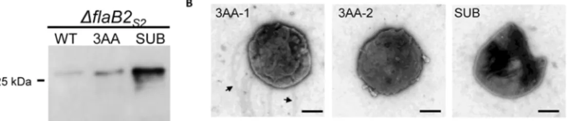
Fig 7. Analysis of theΔflaB2S2strain complemented withflaB2S2carrying the 3-amino acid deletion (3AA) or the substitution version offlaB2S2 (SUB).A. Western blot analysis of theΔflaB2S2strain complemented withflaB2S2carrying the 3-amino acid deletion (3AA) or the substitution version of
flaB2S2(SUB). 3AA and SUB had similar apparent molecular mass as wildtype FlaB2S2. B. Transmission electron micrographs of theΔflaB2S2strain complemented with the 3AA or SUB versions of FlaB2S2.In the case of the 3AA complemented cells, both archaellated cell (3AA-1) and non-archaellated cell
(3AA-2) are shown. Arrows show the archaella. Bar equals 500 nm.
Author Contributions
Conceived and designed the experiments: YD SIA JPJC KFJ. Performed the experiments: YD KU SIA KM AB CK KFJ. Analyzed the data: YD KU SIA KM AB CK KFJ. Contributed re-agents/materials/analysis tools: SIA CK JPJC KFJ. Wrote the paper: YD KFJ.
References
1. Jarrell KF, Ding Y, Meyer BH, Albers SV, Kaminski L, et al. (2014) N-linked glycosylation in archaea: A structural, functional, and genetic analysis. Microbiol Mol Biol Rev 78: 304–341. doi:10.1128/MMBR. 00052-13PMID:24847024
2. Nothaft H, Szymanski CM (2010) Protein glycosylation in bacteria: Sweeter than ever. Nature Rev Microbiol 8: 765–778. doi:10.1038/nrmicro2383PMID:20948550
3. Aebi M (2013) N-linked protein glycosylation in the ER. Biochim Biophys Acta 1833: 2430–2437. doi: 10.1016/j.bbamcr.2013.04.001PMID:23583305
4. Larkin A, Imperiali B (2011) The expanding horizons of asparagine-linked glycosylation. Biochemistry 50: 4411–4426. doi:10.1021/bi200346nPMID:21506607
5. Weerapana E, Imperiali B (2006) Asparagine-linked protein glycosylation: From eukaryotic to prokary-otic systems. Glycobiology 16: 91R–101R. PMID:16510493
6. Eichler J (2013) Extreme sweetness:Protein glycosylation in archaea. Nature Rev Microbiol 11: 151–156. doi:10.1038/nrmicro2957PMID:23353769
7. Burda P, Aebi M (1999) The dolichol pathway of N-linked glycosylation. Biochim Biophys Acta 1426: 239–257. PMID:9878760
8. Nothaft H, Scott NE, Vinogradov E, Liu X, Hu R, et al. (2012) Diversity in the protein N-glycosylation pathways within theCampylobactergenus. MolCell Proteomics 11: 1203–1219. doi:10.1101/pdb. ip071761PMID:23118356
9. Kaminski L, Lurie-Weinberger MN, Allers T, Gophna U, Eichler J (2013) Phylogenetic- and genome-derived insight into the evolution of N-glycosylation in archaea. Mol Phylogenet Evol 68: 327–339. doi: 10.1016/j.ympev.2013.03.024PMID:23567024
10. Jarrell KF, Albers SV (2012) The archaellum: An old motility structure with a new name. Trends Micro-biol 20: 307–312. doi:10.1016/j.tim.2012.04.007PMID:22613456
11. Kelly J, Logan SM, Jarrell KF, Vandyke DJ, Vinogradov E (2009) A novel N-linked flagellar glycan from
Methanococcus maripaludis. Carbohydr Res 344: 648–653. doi:10.1016/j.carres.2009.01.006PMID:
19203750
12. Voisin S, Houliston RS, Kelly J, Brisson JR, Watson D, et al. (2005) Identification and characterization of the unique N-linked glycan common to the flagellins and S-layer glycoprotein ofMethanococcus vol-tae. J Biol Chem 280: 16586–16593. PMID:15723834
13. Ng SYM, Wu J, Nair DB, Logan SM, Robotham A, et al. (2011) Genetic and mass spectrometry analy-sis of the unusual type IV-like pili of the archaeonMethanococcus maripaludis. J Bacteriol 193: 804–814. doi:10.1128/JB.00822-10PMID:21075925
14. Abu-Qarn M, Eichler J (2006) Protein N-glycosylation in archaea: DefiningHaloferax volcaniigenes in-volved in S-layer glycoprotein glycosylation. Mol Microbiol 61: 511–525. PMID:16762024
15. Kaminski L, Naparstek S, Kandiba L, Cohen-Rosenzweig C, Arbiv A, et al. (2013) Add salt, add sugar: N-glycosylation inHaloferax volcanii. Biochem Soc Trans 41: 432–435. doi:10.1042/BST20120142 PMID:23356324
16. Peyfoon E, Meyer B, Hitchen PG, Panico M, Morris HR, et al. (2010) The S-layer glycoprotein of the crenarchaeoteSulfolobus acidocaldariusis glycosylated at multiple sites with the chitobiose-linked N-glycans. Archaea doi:10.1155/2010/754101PMID:20936123
17. Wieland F, Paul G, Sumper M (1985) Halobacterial flagellins are sulfated glycoproteins. J Biol Chem 260: 15180–15185. PMID:3934156
18. Tripepi M, You J, Temel S, Önder Ö, Brisson D, et al. (2012) N-glycosylation ofHaloferax volcanii flagel-lins requires knownaglproteins and is essential for biosynthesis of stable flagella. J Bacteriol 194: 4876–4887. doi:10.1128/JB.00731-12PMID:22730124
20. Leigh JA, Albers SV, Atomi H, Allers T (2011) Model organisms for genetics in the domain archaea: Methanogens, halophiles, Thermococcales and Sulfolobales. FEMS Microbiol Rev 35: 577–608. doi:
10.1111/j.1574-6976.2011.00265.xPMID:21265868
21. Meyer BH, Albers SV (2013) Hot and sweet: Protein glycosylation in crenarchaeota. Biochem Soc Trans 41: 384–392. doi:10.1042/BST20120296PMID:23356316
22. Vandyke DJ, Wu J, Logan SM, Kelly JF, Mizuno S, et al. (2009) Identification of genes involved in the assembly and attachment of a novel flagellin N-linked tetrasaccharide important for motility in the archaeonMethanococcus maripaludis. Mol Microbiol 72: 633–644. doi:10.1111/j.1365-2958.2009.
06671.xPMID:19400781
23. Meyer BH, Albers SV (2014) AglB, catalyzing the oligosaccharyl transferase step of the archaeal N-glycosylation process, is essential in the thermoacidophilic crenarchaeonSulfolobus acidocaldarius. Microbiology Open 3: 531–543. doi:10.1002/mbo3.185PMID:24916761
24. Abu-Qarn M, Yurist-Doutsch S, Giordano A, Trauner A, Morris HR, et al. (2007)Haloferax volcaniiAglB and AglD are involved in N-glycosylation of the S-layer glycoprotein and proper assembly of the surface layer. J Mol Biol 374: 1224–1236. PMID:17996897
25. Kaminski L, Abu-Qarn M, Guan Z, Naparstek S, Ventura VV, et al. (2010) AglJ adds the first sugar of the N-linked pentasaccharide decorating theHaloferax volcaniiS-layer glycoprotein. J Bacteriol 192: 5572–5579. doi:10.1128/JB.00705-10PMID:20802039
26. Yurist-Doutsch S, Abu-Qarn M, Battaglia F, Morris HR, Hitchen PG, et al. (2008) AglF, aglG and aglI, novel members of a gene island involved in the N-glycosylation of theHaloferax volcaniiS-layer glyco-protein. Mol Microbiol 69: 1234–1245. doi:10.1111/j.1365-2958.2008.06352.xPMID:18631242
27. Yurist-Doutsch S, Magidovich H, Ventura VV, Hitchen PG, Dell A, et al. (2010) N-glycosylation in ar-chaea: On the coordinated actions ofHaloferax volcaniiAglF and AglM. Mol Microbiol 75: 1047–1058.
doi:10.1111/j.1365-2958.2009.07045.xPMID:20487296
28. Meyer BH, Peyfoon E, Dietrich C, Hitchen P, Dell A, et al. (2013) Agl16, a thermophilic glycosyltransfer-ase, mediating the last step of the N-glycan biosynthesis in the thermoacidophilic crenarchaeon Sulfo-lobus acidocaldarius. J Bacteriol 195: 2177–2186. doi:10.1128/JB.00035-13PMID:23475978
29. Meyer BH, Zolghadr B, Peyfoon E, Pabst M, Panico M, et al. (2011) Sulfoquinovose synthase—an im-portant enzyme in the N-glycosylation pathway of Ssulfolobus acidocaldarius. Mol Microbiol 82: 1150–
1163. doi:10.1111/j.1365-2958.2011.07875.xPMID:22059775
30. Guan Z, Naparstek S, Calo D, Eichler J (2012) Protein glycosylation as an adaptive response in ar-chaea: Growth at different salt concentrations leads to alterations inHaloferax volcaniiS-layer glycopro-tein N-glycosylation. Environ Microbiol 14: 743–753. doi:10.1111/j.1462-2920.2011.02625.xPMID: 22029420
31. Jarrell KF, Ding Y, Nair DB, Siu S (2013) Surface appendages of archaea: Structure, function, genetics and assembly. Life 3: 86–117. doi:10.3390/life3010086PMID:25371333
32. Lassak K, Ghosh A, Albers SV (2012) Diversity, assembly and regulation of archaeal type IV pili-like and non-type-IV pili-like surface structures. Res Microbiol 163: 630–644. doi:10.1016/j.resmic.2012.
10.024PMID:23146836
33. Shahapure R, Driessen RP, Haurat MF, Albers SV, Dame RT (2014) The archaellum: A rotating type IV pilus. Mol Microbiol 91: 716–723. doi:10.1111/mmi.12486PMID:24330313
34. Pohlschroder M, Ghosh A, Tripepi M, Albers SV (2011) Archaeal type IV pilus-like structures-evolution-arily conserved prokaryotic surface organelles. Curr Opin Microbiol 14: 1–7. doi:10.1016/j.mib.2011. 01.001PMID:21239215
35. Jarrell KF, VanDyke DJ, Wu J (2009) Archaeal flagella and pili. In: Jarrell KF, editor. Pili and Flagella: Current Research and Future Trends. Norfolk, U.K.: Caister Academic Press. pp. 215–234.
36. Bardy SL, Jarrell KF (2002) FlaK of the archaeonMethanococcus maripaludispossesses preflagellin peptidase activity. FEMS Microbiol Lett 208: 53–59. PMID:11934494
37. Bardy SL, Jarrell KF (2003) Cleavage of preflagellins by an aspartic acid signal peptidase is essential for flagellation in the archaeonMethanococcus voltae. Mol Microbiol 50: 1339–1347. PMID:14622420
38. Albers SV, Szabo Z, Driessen AJM (2003) Archaeal homolog of bacterial type IV prepilin signal pepti-dases with broad substrate specificity. J Bacteriol 185: 3918–3925. PMID:12813086
39. Tripepi M, Imam S, Pohlschroder M (2010)Haloferax volcaniiflagella are required for motility but are not involved in PibD-dependent surface adhesion. J Bacteriol 192: 3093–3102. doi:10.1128/JB. 00133-10PMID:20363933
41. Chaban B, Ng SY, Kanbe M, Saltzman I, Nimmo G, et al. (2007) Systematic deletion analyses of thefla
genes in the flagella operon identify several genes essential for proper assembly and function of flagella in the archaeon,Methanococcus maripaludis. Mol Microbiol 66: 596–609. PMID:17887963
42. Ding Y, Jones GM, Uchida K, Aizawa SI, Robotham A, et al. (2013) Identification of genes involved in the biosynthesis of the third and fourth sugars of theMethanococcus maripaludisarchaellin N-linked tetrasaccharide. J Bacteriol 195: 4094–4104. doi:10.1128/JB.00668-13PMID:23836872
43. Jones GM, Wu J, Ding Y, Uchida K, Aizawa S, et al. (2012) Identification of genes involved in the aceta-midino group modification of the flagellin N-linked glycan ofMethanococcus maripaludis. J Bacteriol 194: 2693–2702. doi:10.1128/JB.06686-11PMID:22408155
44. Moore BC, Leigh JA (2005) Markerless mutagenesis inMethanococcus maripaludisdemonstrates roles for alanine dehydrogenase, alanine racemase, and alanine permease. J Bacteriol 187: 972–979.
PMID:15659675
45. Keswani J, Orkand S, Premachandran U, Mandelco L, Franklin MJ, et al. (1996) Phylogeny and taxono-my of mesophilicmethanococcusspp. and comparison of rRNA, DNA hybridization, and phenotypic methods. Int J Syst Bacteriol 46: 727–735. PMID:8782682
46. Corder RE, Hook LA, Larkin JM, Frea JI (1983) Isolation and characterization of two new methane-producing cocci:Methanogenium olentangyi, sp. nov., andMethanococcus deltae, sp. nov. Arch Microbiol 134: 28–32.
47. Balch WE, Fox GE, Magrum LJ, Woese CR, Wolfe RS (1979) Methanogens: Reevaluation of a unique biological group. Microbiol Rev 43: 260–296. PMID:390357
48. Gardner WL, Whitman WB (1999) Expression vectors forMethanococcus maripaludis: Overexpression of acetohydroxyacid synthase and beta-galactosidase. Genetics 152: 1439–1447. PMID:10430574
49. Papworth C, Bauer JC, Braman J, Wright DA (1996) Site-directed mutagenesis in one day with>80%
efficiency. Strategies 9: 3–4. PMID:8740480
50. Tumbula DL, Makula RA, Whitman WB (1994) Transformation ofMethanococcus maripaludisand identification of apstI-like restriction system. FEMS Microbiol Lett 121: 309–314.
51. Towbin H, Staehelin T, Gordon J (1979) Electrophoretic transfer of proteins from polyacrylamide gels to nitrocellulose sheets: Procedure and some applications. Proc Natl Acad Sci U S A 76: 4350–4354.
PMID:388439
52. Yamaguchi K, Akai K, Kawanishi G, Ueda M, Masuda S, et al. (1991) Effects of site-directed removal of N-glycosylation sites in human erythropoietin on its production and biological properties. J Biol Chem 266: 20434–20439. PMID:1657925
53. Dubuisson J, Duvet S, Meunier JC, Op De Beeck A, Cacan R, et al. (2000) Glycosylation of the hepati-tis C virus envelope protein E1 is dependent on the presence of a downstream sequence on the viral polyprotein. J Biol Chem 275: 30605–30609. PMID:10882734
54. Wojczyk BS, Takahashi N, Levy MT, Andrews DW, Abrams WR, et al. (2005) N-glycosylation at one ra-bies virus glycoprotein sequon influences N-glycan processing at a distant sequon on the same mole-cule. Glycobiology 15: 655–666. PMID:15677380
55. Kalmokoff ML, Jarrell KF (1991) Cloning and sequencing of a multigene family encoding the flagellins ofMethanococcus voltae. J Bacteriol 173: 7113–7125. PMID:1718944
56. Logan SM (2006) Flagellar glycosylation—a new component of the motility repertoire? Microbiology 152: 1249–1262. PMID:16622043
57. Merino S, Tomás JM (2014) Gram-negative flagella glycosylation. Int J Mol Sci 15: 2840–2857. doi:10. 3390/ijms15022840PMID:24557579
58. Taguchi F, Shibata S, Suzuki T, Ogawa Y, Aizawa S, et al. (2008) Effects of glycosylation on swimming ability and flagellar polymorphic transformation inPseudomonas syringaepv.tabaci6605. J Bacteriol 190: 764–768. PMID:18024523
59. Taguchi F, Takeuchi K, Katoh E, Murata K, Suzuki T, et al. (2006) Identification of glycosylation genes and glycosylated amino acids of flagellin inPseudomonas syringaepv.tabaci. Cell Microbiol 8: 923–938. PMID:16681835
60. Ewing CP, Andreishcheva E, Guerry P (2009) Functional characterization of flagellin glycosylation in
Campylobacter jejuni81–176. J Bacteriol 191: 7086–7093. doi:10.1128/JB.00378-09PMID: 19749047
61. Castric P (1995)pilO, a gene required for glycosylation ofPseudomonas aeruginosa1244 pilin. Micro-biology 141: 1247–1254. PMID:7773418
62. Nguyen LC, Taguchi F, Tran QM, Naito K, Yamamoto M, et al. (2012) Type IV pilin is glycosylated in
63. Smedley JG 3rd, Jewell E, Roguskie J, Horzempa J, Syboldt A, et al. (2005) Influence of pilin glycosyla-tion onPseudomonas aeruginosa1244 pilus function. Infect Immun 73: 7922–7931. PMID:16299283 64. Harvey H, Kus JV, Tessier L, Kelly J, Burrows LL (2011)Pseudomonas aeruginosaD-arabinofuranose biosynthetic pathway and its role in type IV pilus assembly. J Biol Chem 286: 28128–28137. doi:10. 1074/jbc.M111.255794PMID:21676874
65. Asakura H, Churin Y, Bauer B, Boettcher JP, Bartfeld S, et al. (2010)Helicobacter pyloriHP0518 af-fects flagellin glycosylation to alter bacterial motility. Mol Microbiol 78: 1130–1144. doi: 10.1111/j.1365-2958.2010.07393.xPMID:21091500
66. Josenhans C, Vossebein L, Friedrich S, Suerbaum S (2002) TheneuA/flmDgene cluster of Helicobac-ter pyloriis involved in flagellar biosynthesis and flagellin glycosylation. FEMS Microbiol Lett 210: 165–
172. PMID:12044670
67. Schirm M, Soo EC, Aubry AJ, Austin J, Thibault P, et al. (2003) Structural, genetic and functional char-acterization of the flagellin glycosylation process inHelicobacter pylori. Mol Microbiol 48: 1579–1592. PMID:12791140
68. Paiment A, Hocking J, Whitfield C (2002) Impact of phosphorylation of specific residues in the tyrosine autokinase, wzc, on its activity in assembly of group 1 capsules inEscherichia coli. J Bacteriol 184: 6437–6447. PMID:12426330
69. Thomas NA, Chao ED, Jarrell KF (2001) Identification of amino acids in the leader peptide of Methano-coccus voltaepreflagellin that are important in posttranslational processing. Arch Microbiol 175: 263–
269. PMID:11382222
70. Trachtenberg S, Galkin VE, Egelman EH (2005) Refining the structure of theHalobacterium salinarum
flagellar filament using the iterative helical real space reconstruction method: Insights into polymor-phism. J Mol Biol 346: 665–676. PMID:15713454
71. Cohen-Krausz S, Trachtenberg S (2002) The structure of the archeabacterial flagellar filament of the extreme halophileHalobacterium salinarumR1M1 and its relation to eubacterial flagellar filaments and type IV pili. J Mol Biol 321: 383–395. PMID:12162953
72. Kuwajima G (1988) Construction of a minimum-size functional flagellin ofEscherichia coli. J Bacteriol 170: 3305–3309. PMID:3290204
73. Stocker BA, Newton SM (1994) Immune responses to epitopes inserted in Ssalmonellaflagellin. Int Rev Immunol 11: 167–178. PMID:7519231
74. Beznosov SN, Pyatibratov MG, Veluri PS, Mitra S, Fedorov OV (2013) A way to identify archaellins in
Halobacterium salinarumarchaella by FLAG-tagging. Cent Eur J Biol 8: 828–834.
75. Giltner CL, Nguyen Y, Burrows LL (2012) Type IV pilin proteins: Versatile molecular modules. Microbiol Mol Biol Rev 76: 740–772. doi:10.1128/MMBR.00035-12PMID:23204365
76. Yang Z, Hu W, Chen K, Wang J, Lux R, et al. (2011) Alanine 32 in PilA is important for PilA stability and type IV pili function inMyxococcus xanthus. Microbiology 157: 1920–1928. doi:10.1099/mic.0. 049684-0PMID:21493683
77. Kirn TJ, Lafferty MJ, Sandoe CM, Taylor RK (2000) Delineation of pilin domains required for bacterial association into microcolonies and intestinal colonization byVibrio cholerae. Mol Microbiol 35: 896–
910. PMID:10692166