Halogen light versus LED for bracket bonding:
Shear bond strength
Paulo Eduardo Guedes Carvalho1, Valdemir Muzulon dos Santos2, Hassan Isber2, Flávio Augusto Cotrim-Ferreira1
How to cite this article: Carvalho PEG, Santos VM, Isber H, Cotrim-Ferreira FA. Halogen light versus LED for bracket bonding: shear bond strength. Dental Press J Orthod. 2013 Jan-Feb;18(1):31.e1-6.
Submitted: August 28, 2008 - Revised and accepted: November 25, 2008
» The authors report no commercial, proprietary or inancial interest in the prod-ucts or companies described in this article.
Contact address: Paulo Eduardo Guedes Carvalho
Rua Cesário Galeno, 483, Tatuapé – CEP: 03.071-000 – São Paulo / SP – Brazil E-mail: [email protected]
1 Professor of the MSc course in Orthodontics, University of São Paulo City (UNICID).
2 MSc in Orthodontics, University of São Paulo City (UNICID).
Introduction: LED light-curing devices seek to provide a cold light activator which allows protocols of material polymerization with shorter duration. Objective: The present study aimed to evaluate the shear bond strength of bracket bonding using three types of light-curing devices: One with halogen light (Optilight Plus - Gnatus) and two with LEDs (Optilight CL - Gnatus and Elipar Freelight - 3M/ESPE). Results: Comparing the results by analysis of variance, the Gnatus LED device showed an inferior statistical behavior in relation to other light sources, when acti-vated by a short time. But, when it was used for 40 seconds, the polymerization results were consistent with the other evaluated sources. The device with the best average performance was the halogen light, followed by the 3M/ESPE LED. Conclusion: It was concluded that the LEDs may be indicated in orthodontic practice, as long as a protocol is used for the application of light with the activation time of 40 seconds.
Keywords: Orthodontics. Shear bond strength. Orthodontic brackets. Dental bonding.
Introdução:os aparelhos de fotopolimerização por LED buscam proporcionar uma luz ativadora fria, que possibilite pro-tocolos de polimerização do material com menor tempo de duração. Objetivo: avaliar a resistência à tração da colagem de braquetes, utilizando três tipos de aparelhos fotoativadores: um de luz halógena (Optilight Plus – Gnatus) e outros dois de LED (Optilight CL – Gnatus; e Elipar Freelight – 3M/Espe). Resultados: comparando os resultados por meio da análise de variância, o aparelho de LED Gnatus apresentou comportamento estatístico inferior em relação às outras fontes de luz, quando ativado por tempo reduzido. Já quando foi utilizado o tempo de 40 segundos, os resultados de polimerização foram compatíveis com as demais fontes avaliadas. O aparelho que apresentou melhor desempenho médio foi o de luz halógena, seguido pelo LED 3M/Espe. Conclusão: concluiu-se que os LEDs podem ser indicados na prática ortodôntica, uma vez que seja utilizado um protocolo de aplicação da luz com tempo de ativação de 40 segundos.
INTRODUCTION
In order to facilitate and improve the process of bracket bonding, researches are oten conducted with the halogen light, which is the one most used by profes-sionals so far. The device with halogen light source uses a broad spectrum of wavelengths, which has between 400 and 500 nm, and also a light ilter that disregards the waves, which do not sensitize the activator commonly used, camphorquinone. Thus, this makes the band of
light narrower usable for the resin polymerization.14
It is known that the quality of the resin polymer-ization is represented by the number of double carbon bonds of the methacrylate groups, which have reacted
during the resin polymerization.12,15 The best time of
application of the halogen light is 40 seconds, shown by
several studies with this equipment.5,10,17,18
Atmadja and Bryant2 carried out a study
evaluat-ing the microhardness of four resins (P 30, Prisma-Fil, Heliomolar and Durail). Test specimens were fabri-cated in cylindrical matrices with 4 mm in diameter and thickness ranging from 1 to 6 mm. The polymer-izing device used was Heliomat and it was activated in three diferent exposure times (20, 40 and 60 seconds). They concluded that: a) for each type of resin evalu-ated, the microhardness values decreased with the in-creasing depth of polymerization, b) the most efective exposure time was 40 seconds, c) reducing the thick-ness is the most correct way to achieve proper curing and it is better than to increase the exposure time, d) the light-cured resins continue their polymerization reaction ater the light source is removed.
Light intensity is another important factor for pres-ervation of the margins of the resin applied and
polym-erized.10,18 The resins have superior physical properties
and better marginal integrity when polymerized by the gradual technique when compared with polymerization
initiated with maximum intensity.11
Although there are few studies in the literature, it is believed that the LED device has real conditions of
replacing halogen devices in oices1,6 because it has a
cost similar to the halogen lamp and it is much lower when compared to argon laser and xenon plasma arc curing light. Either ways, it is known that LEDs have cold light emitted by a semiconductor, allowing an un-limited period of its use, with a low light spectrum
be-tween 460 and 480 nm.7 Since camphorquinone, the
photo-initiator most used in resin, works absorbing
visible light raging 350 to 550 nm with a peak absorp-tion around 470 nm, the values emitted by the LED seem to be favorable. The ideal light length for com-posite polymerization should range around 450 nm
and 470 nm.9,16 These data create the expectation of
an optimal polymerization reaction, compensating the lower intensity of light produced by these units,
be-sides generating a reduced heat emission.7
The light-curing devices with halogen lamps have a vast literature on the subject, unlike LEDs, which are currently being investigated more in-tensely. With this in mind, this work aimed to study the shear bond strength of orthodontic brackets, bonded to human teeth, using different resin light-curing protocols, according to the variation of the light type and light-activation time.
MATERIAL AND METHODS
This experiment was submitted to the Ethics Com-mittee in Research of the University where it was held, having been fully approved.
For this study the following equipment were compared:
» Light source based on halogen lamp with 600 mW/cm² - Optilight Plus device (Gnatus Medical Dental Equipment Ltda, Ribeirão Pre-to - SP, Brazil).
» Light source based on LED with 700 mW/cm² - LED device Optilight CL (Gnatus Medical Den-tal Equipment Ltda, Ribeirão Preto - SP, Brazil). » Light source based on LED with 1000 mW/cm²
- Elipar Freelight LED device (3M/ESPE Dental Products, Monrovia - CA, USA).
Fity premolar teeth that were previously selected were used, teeth with fractures, cracks, issures, or which had not a perfect facial surface were refused. This sample was divided randomly into ive groups with ten teeth each, always kept in saline solution.
The samples were prepared by a dentist with clinical experience in orthodontics and in bracket bonding, not needing help from other operators in the experiment, in order to minimize the possibilities of intoperator er-rors. The buccal surfaces of the teeth were irst cleaned and polished with rubber cup and pumice for 20 sec-onds each, at low speed and then washed with water.
washed with water for 20 seconds and dried with compressed air. For the bonding procedure, a specific caliper for bracket bonding and a light-cured
orth-odontic adhesive system were used – TransbondTM
XT (3M/Unitek, Dental Products, Monrovia - CA, USA). This adhesive was applied on the base of each of the fifty Unitek “full-size” brackets used in this study (3M/Unitek, Dental Products, Monrovia - CA, USA). With the aid of a microbrush, liquid adhesive was applied, from the same adhesive system, onto the etched surface of the tooth. Then, the set brack-et / resin was positioned in the center of the clinical crown of the tooth, performing a light pressure with the explorer probe in search of excess material. This material was removed with the tip of this probe in order to prevent the resin covering the bracket base, thereby increasing its retention and, consequently, influencing the results.
From this moment, the polymerization of the speci-mens was initiated, according to the time set for each of the groups, divided as follows:
» Group 1: Application of halogen light for 40 sec-onds on each bracket (control group).
» Group 2: Application of LED light with the 3M/ESPE device for 40 seconds on each bracket. » Group 3: Application of LED light with the
3M/ESPE device for 10 seconds on each bracket. » Group 4: Application of LED light with the
Gna-tus device for 40 seconds on each bracket. » Group 5: Application of LED light with the
Gna-tus device for 10 seconds on each bracket.
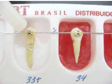
After carrying out the light-curing of all speci-mens, as each studied protocol, the set tooth / bracket was placed in plastic matrices that received acrylic resin, in order to create the structure of the speci-mens (Fig 1).
It was decided to bond the brackets irst, followed by the stabilization of the set in the plastic matrix and the inclusion of the resin. This avoided the instability in positioning the tooth during the insertion of the resin. Care was taken so the specimens were perpendicular to the hook that was used for traction, which accurately it the bracket wings, reducing the opportunities for lever-age and unequal traction movements during the test.
The samples were conditioned in moist environ-ment (saline) at a temperature of 37 °C in the dark, for 24 hours. Then they were taken to the Plastics and
Rubbers Laboratory, in the Chemistry Division at the Institute of Technological Research of São Paulo - IPT, where the tests were performed.
The specimens were subjected to tensile testing by a universal testing machine (EMIC 10,000), with a speed of 1.0 mm/min and with a load cell 50 kgf. To perform the tests an extractor clutch was used made at the Met-al Laboratory in the Engineering Division of the IPT. This clutch itted the wing of the brackets perpendicu-lar to the specimen, and subsequently ixed on top of the traction machine EMIC (Fig 2).
Each specimen underwent the tensile test and for each moment of bracket bonding rupture a certain val-ue was recorded. These valval-ues referred to the load sup-ported by the bracket at the exact moment of rupture. The tests were performed in all 50 teeth in the same day. Statistical tests were used to validate the results and comparison of the groups, the analysis of variance (ANOVA) was used, with signiicance level of 5%, fol-lowed by Tukey test when diferences were found.
RESULTS
It was intended to arrive to some conclusions from the resin resistance measurements that were made, considering five types of light curing protocols. To study the comparisons between the groups, since the measured data followed approximately normal dis-tributions, the analysis of variance was chosen when comparing the statistical similarity of the five groups.
DISCUSSION
The purpose of researching the LEDs is related to its recent availability in the market, and there is a large number of scientiic studies proving its real eiciency. Since the band of light emitted by the LEDs matches the absorption peak of camphorquinone, it creates a great expectation of the polymerization reaction, com-pensating the reduced intensity of light produced by these units, besides generating a low heat emission.
Once the braces are bonded in the intraoral environ-ment, it receives force applications due to mastication and also by biomechanic forces itself. Various types of force will be acting at this time such as traction, shear and torsion forces, which can act in isolated or com-bined ways. This work uses tensile forces because it is a force also used in Orthodontics and less studied.
For this study, first the means and standard deviations were calculated for the measurements studied, and from these results, the analysis of variance test was applied. Since a significant difference in the analysis of variance was found for the total sample, the Tukey test was applied, when comparing the groups, indi-cating sufficient differences to represent statistical significance between which protocols.
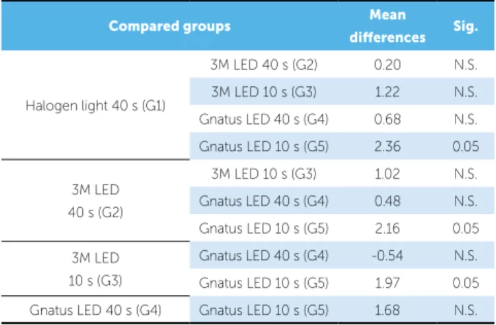
Table 1 presents the mean values and standard devia-tions for the many light-curing protocols studied. The values obtained on analysis of variance and Tukey’s test for comparisons of various measurements, are shown in Table 2.
To facilitate the comparison of data here obtained with the ones in literature, Table 3 presents the mean values of each sample also converted to values of pres-sure in mega Pascal (MPa), veriied from base meapres-sure- measure-ments of the brackets used. The “full-size” brackets have their base with 0.399 x 0.335 cm, generating a base
area of 0.133665 cm2. The values in MPa are of great
importance when interpreting the data on other works, because they eliminate the variation in the size of the diferent brackets used.
Figure 2 - EMIC 10,000 traction machine. Table 1 - Mean values and standard deviations of the ive groups studied, in kgf.
SD = Standard Deviation; s = seconds. Light-curing
protocol
Halogen 3M LED Gnatus LED
40 s 40 s 10 s 40 s 10 s
Group 1 2 3 4 5
Mean 6.21 6.01 4.99 5.53 3.85
SD 1.74 1.34 1.00 2.28 1.05
Compared groups Mean
diferences Sig.
Halogen light 40 s (G1)
3M LED 40 s (G2) 0.20 N.S.
3M LED 10 s (G3) 1.22 N.S.
Gnatus LED 40 s (G4) 0.68 N.S.
Gnatus LED 10 s (G5) 2.36 0.05
3M LED 40 s (G2)
3M LED 10 s (G3) 1.02 N.S.
Gnatus LED 40 s (G4) 0.48 N.S.
Gnatus LED 10 s (G5) 2.16 0.05
3M LED
10 s (G3)
Gnatus LED 40 s (G4) -0.54 N.S.
Gnatus LED 10 s (G5) 1.97 0.05
Gnatus LED 40 s (G4) Gnatus LED 10 s (G5) 1.68 N.S.
Table 2 - Statistical tests for analysis of variance and of Tukey.
Minimum Signiicant Diference (MSD) = 1.9136; NS = non-signiicant.
Table 3 - Mean values from the ive assessed groups, in MPa. Light-curing
protocol
Halogen 3M LED Gnatus LED
40 s 40 s 10 s 40 s 10 s
Group 1 2 3 4 5
In this study values in MPa and kgf were obtained for the activation time of 40 seconds, which show
agree-ment with the indings of Silva,15 who used types of
de-vices compatible with this work, however only evalu-ating longer light-curing protocols (40 to 60 seconds). It is worth noting that few works are available in the literature studying shear bond strength of brackets, es-pecially with proposals for shorter curing time (less than 20 seconds) and that it should always be taken into ac-count the methodology, forms and types of brackets ex-tractors, controllable and uncontrollable variations, and the number of people involved.
CONCLUSIONS
Based on the methodology applied in this study, and according to the results obtained and applied to the statistical analysis, it was considered reasonable to conclude that:
» The halogen lamp provided the highest mean shear bond strength of brackets, but without sta-tistical signiicance in relation to three other pro-tocols performed with LED devices.
» The 3M/ESPE LED device had shear bond strength of brackets similar to that obtained by halogen source, even with the protocol with 10 seconds of activation.
» The Gnatus LED device showed shear bond strength of brackets similar to the one obtained by halogen source, only with the activation proto-col of 40 seconds, being signiicantly lower when used for 10 seconds.
However, it must be emphasized that at the time of the laboratory force application by the stress machine, there is no possibility of obtaining a pure tensile force, but a dominant force with tensile stress and shear forces, secondarily present. Thus the test may provide more complex rupture patterns, involving several types of
forces on a single moment.13 This factor may be
pres-ent in most of the studies prespres-ented in the literature and be a variant of inluence in the absolute comparison of results between the various others displayed.
The objective of this study was to permeate ive po-lymerization time protocols in orthodontics without worrying about the protocols recommended in works of the dentistry area, recommending as viable times
of 40 s and 60 s to use LEDs.8 Diferent measurement
units were used (MPa and kgf) only in order to provide a comparative parameter with previous works.
In analyzing the results obtained by analysis of vari-ance, four groups were statistically similar to each other, where only the group 5 (LED Gnatus 10 s) was signii-cantly diferent. This work showed higher values for the halogen lamp when compared with the other four groups of LEDs. Analyzing the groups, which had the highest light incidence values, it is observed that they possess the best results. These groups, halogen (6.21 kgf), 3M LED 40 s and 20 s (6.01 kgf and 4.99 kgf) and Gnatus LED 40 s and 20 s (5.53 kgf and 3.85 kgf) tend to conclude that the greater the exposure time, the greater the en-hancement in retention of the bracket onto the tooth. As comparison to this shear bonding brackets, similar mean
1. Asmussen E, Peutzfeldt A. Light emitting diode curing: inluence on selected properties of resin composites. Quintessence Int. 2003;34(1):71-5. 2. Atmadja G, Bryant RW. Some factors inluencing the depth of cure of
visible light-activated composite resin. Aust Dent J. 1990;35(3):213-8. 3. Bengtson NG, Bengtson AL, Carvalho DS, Rossetto SM. Estudo
comparativo da força adesiva de quatro materiais para colagem de braquetes. Rev Dental Press Ortod Ortop Facial. 2003;8(3):43-7. 4. Bishara SE, Ajlouni R, Oonsombat C. Evaluation of a new curing light
on the shear bond strength of orthodontic brackets. Angle Orthod. 2003;73(4):431-5.
5. Carillo VEB. Estudo comparativo in vitro da capacidade adesiva da resina fotoativada pela luz halógena e por laser de argônio, utilizando-se bracketes metálicos em pré-molares humanos [tese]. São Paulo (SP): Universidade de São Paulo; 2004.
6. Fujibayashi K, Norihiko M, Masaaki O, Hirokazu I, Atsushi K. Newly developed curing unit using blue light emitting diodes. Dent Japan. 1998;34(1):49-53.
7. Kurachi C, Eduardo CP, Magalhães DV, Bagnato VS. Human teeth exposed to argon laser irradiation: determination of power-time-temperature working conditions. J Clin Laser Med Surg. 1999;17(6):255-9. 8. Kurachi C. Estudo comparativo do laser, do led azul e da lâmpada
convencional no processo de polimerização da resina composta dental [dissertação]. São Carlos (SP): Universidade de São Paulo; 2000. 9. Leonard DL, Charlton DG, Roberts HW, Cohen ME. Polymerization
eiciency of led curing lights. J Esthet Restor Dent. 2002;14(5):286-95.
REFERENCES
10. Mehl A, Hickel R, Kunzelmann KH. Physical properties and gap formation of light-cured composites with and without soft start-polymerization. J Dent. 1997;25(3-4):321-30.
11. Mills RW, Jandt KD, Ashworth SH. Dental composite depth of cure with halogen and blue light emitting diode technology. Brit Dent. 1999;186(8):388-91.
12. Peutzfeldt A. Correlation between recordings obtained with a light-intensity tester and degree of conversion of curing resin. Scand J Dent Rest. 1994;102(1):73-5.
13. Phillips RW. Materiais dentários de Skinner. 8a ed. Rio de Janeiro: Interamericana; 1984.
14. Santos LA, Turbino ML, Youssef MN, Matson E. Microdureza de resinas compostas: efeito de aparelhos e tempos de polimerização em diferentes profundidades. Pesq Odontol Bras. 2000;14:65-70.
15. Silva PCG. Estudo in vitro da colagem de braquetes sobre o esmalte dental humano: utilizando leds como fonte de luz - análise comparativa com a luz halógena [tese]. Araraquara (SP): Universidade Estadual Paulista; 2003. 16. Teshima W, Nomura Y, Tanaka N, Urabe H, Okazaki M, Nahara Y. ESR
study of camphorquinone/amine photoinitiator systems using blue light-emitting diodes. Biomaterials. 2003 May;24(12):2097-103.
17. Turbino ML, Vinha D, Centola AL, Campos GM. Photopolymerized resins: surface hardness variation in relation to time of polymerization and setting. Braz Dent J. 1993;3(2):87-94.