Shear bond strength evaluation of metallic
brackets using self-etching system
Camilo Aquino Melgaço*, Graciele Guerra de Andrade**, Mônica Tirre de Souza Araújo***, Lincoln Issamu Nojima****
Objective: To evaluate the shear bond strength of metallic brackets using the self-etching system after its activation and storage for 2, 5 and 9 day periods. Methods: A total of 64 bovine teeth were divided in four groups and prepared to receive the brackets. Initially (T1), 7 self-etching primer blisters were activated and used to bond the brackets of group I. The blisters were stored at a constant temperature of 4ºC for 2 (T2), 5 (T3) and 9 (T4) days and used to bond the brackets of groups II, III and IV, respectively. Results: No statistic difference was found in shear bond strength comparing groups I, II and III. However, a significant difference was found in group IV. Conclu-sion: The shear bond strength seems not to be affected by activation and storage of the self-etching primer at a mean temperature of 4°C for a 5 day period. More studies are necessary to evaluate other characteristics of the material after its activation and storage for long periods of time. Abstract
Keywords: Shear bond strength. Self-etching system. Bovine enamel. Bond.
* PhD Student, Federal University of Rio de Janeiro. Professor of the Specialization Course in Orthodontics, FO-UFMG. ** DDS, FO-UFRJ. Specialist in Pediatric Dentistry.
*** Master and PhD in Orthodontics, FO-UFRJ. Associate Professor, FO-UFRJ. **** Master and PhD in Orthodontics, FO-UFRJ. Associate Professor, FO-UFRJ.
intROduCtiOn
Since Buonocore3 in 1955 proposed the acid
etching technique, the concept of bonding to enamel allowed the development and applica-tion of various techniques and new materials in all dentistry fields.19 The use of phosphoric acid on
dental surface results in microporosity into which composite can flow and be polymerized to form a mechanical adhesion to enamel. Therefore, several adhesive materials used in orthodontics have been
developed, making necessary new studies to prove its efficacy.12,17,18
The use of direct bonding techniques al-lowed great benefits to orthodontists: esthetics improvement, less risk of enamel damage, easy control of dental plaque by the patient, minor gingival irritation and reducing patient chair time. These important gains were possible since direct bonding is easier than the adaptation of bands on all teeth.20
» The authors report no commercial, proprietary, or inancial interest in the
products or companies described in this article. How to cite this article: Melgaço CA, Andrade GG, Araújo MTS, Nojima LI.
Among the huge variety of new materials, self-etching system (acid already incorporated into the primer) uses chemical and different etching technology than traditional products.14
It was first available for use in restorative den-tistry8 and recently has been used for
orthodon-tic acessory bonding.15
The adhesive used in this study is the Trans-bond Plus Self Etching Primer-3M. This adhesive has esters of phosphoric methacrylate acid (hy-droxyethyl-metacrylate), forming a bifunctional molecule. When rubbed on enamel surface, the acid component exposes the enamel prisms while the primer components simultaneously penetrate on these exposed prisms. Phosphoric acid removes calcium of the hydroxyapatite. The phosphate group forms a composite with calcium which is incorporated to the product during polymerization, since there is no wash-ing step in this system.5 The self-etching
adhe-sive pH is around 1, promoting a good enamel penetration. The lower the pH of the substance, the greater its capacity of penetration in dental tissues. This product solvent is water, enabling the ionization of acid monomers and contrib-uting for enamel demineralization.14 After
ap-plying the adhesive on dental surface, the tooth must be dried with oil and moisture-free com-pressed air, in order to permit solvent evapo-ration. If drying is not performed properly, the adhesion forces can be compromised.10
Some studies show no statistical significant differences between conventional and self-etching systems on shearing bond strength of metallic brackets.7,18
Some authors defend the use of bovine teeth instead of human teeth in dental re-searches. The use of bovine teeth became well-documented and preferred due to its enamel composition (similar to human’s enamel) and its easy acquisition.11,13,15,18
The objective of the present study is to eval-uate shearing bond strength of metallic brackets
on bovine teeth using self-etching system im-mediately after its activation and with intervals of time of 2, 5 and 9 days.
MAtERiAL And MEtHOdS
Sixty-four bovine incisors were used. The teeth were selected based on the same criteria adopted by Sponchiado et al18 and included: Intact enamel
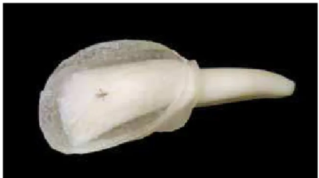
surface, absence of prior application of chemical substances such as thymol, hydrogen peroxide, alcohol or formalin. The teeth were cleaned and preserved in distilled water at an average tempera-ture of 4ºC. A landmark was printed on the most labial projection of the vestibular surface of each tooth for later identification of the region to re-ceive the brackets. The crowns were involved in acrylic resin (manipulated according to the manu-facturer’s instructions), and then pressed against a glass plate until the landmark became visible (Fig 1). In order to prevent the formation of porosities, polymerization occurred under hydrostatic pres-sure, inside a specific container.
After complete polymerization, the acrylic res-in was worn usres-ing sandpapers (numbers 150, 400 and 600) until it was exposed the enamel surface with sufficient area for placement of an orthodontic bracket (Edgewise standard - dimensions of 3.3 mm by 3.6 mm and 0.022x0.028-in slot). The radicular
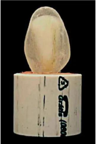
portions of the teeth were included in PVC rings, filled with type IV gypsum (stone) manipulated according to the manufacturer’s specifications. The teeth were positioned with their vestibular surfaces perpendicular to the base of the ring. A square template was specially constructed for this purpose (Figs 2 and 3). After gypsum crystalliza-tion, all teeth were immersed in distilled water and stored in an oven at a temperature of 37±1ºC. All test samples were equally and randomly di-vided into 4 groups.
Before bracket bonding, the teeth received prophylaxis with non-fluoridated pumice and rubber prophylactic cups at a low speed for 15 seconds. Then a water/air spray washed the sur-face for over 15 seconds. Enamel drying occurred using oil and moisture-free compressed air. The activation of the self-etching system proceeded according to the manufacturer’s instructions in sufficient quantity (10 blisters) to bond 64 brack-ets. At the time of activation, 16 brackets of group I were bonded as follows:
- Adhesive was rubbed on the surface of each tooth for three seconds and dried with a hair dryer for the same period.
- Transbond XT was applied on bracket base following the manufacturer’s recommendations. The accessories were positioned and pressed against the enamel surface. The excess of compos-ite was removed. The polymerization occurred for 10 seconds on each side of the bracket perimeter with visible halogen light appliance (Photopo-lymerizer 3M - USA - 5560AF model), with in-tensity of 488mW/cm2 (measured by radiometer
Demetron, model 100).
- The bracket slot was positioned perpendicu-lar to the PVC ring (Fig 4). A template for this purpose was used.
The test samples of Group I were stored in dis-tilled water at 37±1°C for 7 days. The activated blisters (containing the self-etching system) were stored inside a refrigerator at a mean temperature of 4ºC for future use in groups II, III and IV.
FIGURE 2 - Teeth were included in PVC rings.
Following the same methodology described before, 2 days after the activation of the self-etch-ing system, 16 brackets of groups II were bonded. The 16 brackets of groups III and IV were bonded 5 and 9 days after initial activation of the blisters, respectively. All test samples of groups II, III and IV were also stored for 7 days in distilled water at a temperature of 37±1°C.
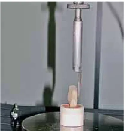
After the storage period, the shear tests were performed in an EMIC DL-10,000 test-ing machine of the Instituto Militar de Engen-haria - RJ, Brazil, using MTest program version 1.01 (Figs 5 and 6). Non-paired t tests were used for statistical analysis.
RESuLtS And diSCuSSiOn
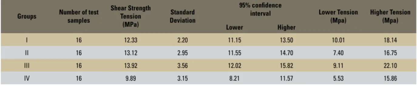
The results of shearing bond strength in Mega-pascal (MPa) can be visualized in Table 1, for all groups. The groups I and III presented, respective-ly, the smallest and the highest values of standard deviation: 2.2 and 3.56, respectively.
No statistical significant differences were ob-served when comparing the results of shearing
bond strength between groups I, II and III. However, a statistical significant difference can be observed when comparing these groups with group IV (Table 2).
Some studies9,16 suggested that bracket
adhe-sion force should vary between 6 and 8 MPa, since these values would be sufficient to resist chewing and orthodontic forces. Other studies using this same kind of material found shearing bond strength with values ranging from 7.5 to 10.57 MPa.7,18 All
results found in this study, including group IV, are near these values (Fig 7). The results indicate that the self-etching system could be used maintaining its characteristic of optimal shearing strength until 9 days after its activation. However, to obtain the best results using this material, the operator shall follow carefully all manufacturer’s recommenda-tions. Sponchiado et al18 stated that variations on
the technique that uses the self-etching system can adversely affect shear resistance. Other factors that may affect the shear strength and should be con-sidered are the bracket size, enamel chemical com-position,1 dental prophylaxis2 and the conditions
30
20
10
0
N 16 16 16 16
15
in which the procedure was conducted.6 In
excep-tion of enamel chemical composiexcep-tion, all other fac-tors can be controlled by the orthodontist.
Another aspect that must be considered is that bovine enamel offers different levels of
adhesion, when compared to human enamel.11
Besides, the manipulation, preparation and sur-face treatment during the experiment is crucial for reliable results.4 However, the use of bovine
teeth in shearing bond strength studies became preferred due to its easy acquisition and histo-logical and histochemical characteristics similar to human enamel.11,13,15,18
COnCLuSiOnS
Based on the results obtained in this study we can conclude that:
» The shearing bond strength levels found in all groups were higher than the levels recom-mended by the literature.
» The storage of the self-etching system for 5 days after activation at an mean temperature of 4ºC seems not to affect shearing bond strength levels.
» Compared to other groups, shearing bond strength levels presented significant statisti-cal difference in group IV (9 days after adhe-sive activation).
» New materials and technologies are daily presented. Self-etching system technique simplifies bracket bonding process with-out compromising its adhesion. However, it is very important to follow manufactur-er’s instructions to obtain best results. » New studies are necessary to evaluate the
properties of the material when it is stored for a long period after being activated. Groups Number of test
samples
Shear Strength Tension
(MPa)
Standard Deviation
95% conidence
interval Lower Tension (Mpa)
Higher Tension (Mpa)
Lower Higher
I 16 12.33 2.20 11.15 13.50 10.01 18.14
II 16 13.12 2.95 11.55 14.70 7.40 16.75
III 16 13.92 3.56 12.02 15.82 9.11 22.10
IV 16 9.89 3.15 8.21 11.57 5.53 15.86
TABLE 1 - Means, standard deviations, higher and lower values for shearing bond strength in groups I, II, III and IV.
TABLE 2 - Comparison of shearing bond strength forces in groups I, II, III and IV. Significance p ≤ 0.05.
FIGURE 7 - Shear bond strength distribution in all groups. Group comparison Signiicance p ≤ 0.05
G I x G II 0.3958
G I x G III 0.1404
G I x G IV 0.0168
G II x G III 0.4989
G II x G IV 0.0055
G III x G IV 0.0020
REfEREnCES
Contact address
Camilo Aquino Melgaþo
Rua Espírito Santo, n. 1111/1401 – Centro CEP: 30.160-031 – Belo Horizonte / MG, Brazil E-mail: [email protected]
Submitted: August 7, 2008 Revised and accepted: August 8, 2008
1. Bishara SE, VonWald L, Olsen ME, Laffoon JF. Effect of time on the shear bond strength of glass ionomer and composite orthodontic adhesives. Am J Orthod Dentofacial Orthop. 1999;116(6):616-20.
2. Bryant S, Retief DH, Russell CM, Denys FR. Tensile bond strengths of orthodontic bonding resins and attachments to etched enamel. Am J Orthod Dentofacial Orthop. 1987;92(3):225-31.
3. Buonocore MG. A simple method of increasing the adhesion
of acrylic illing materials to enamel surfaces. J Dent Res.
1955;34(6):849-53.
4. Cacciafesta V, Jost-Brinkmann PG, Subenberger U, Miethke RR. Effects of saliva and water contamination on the enamel shear bond strength of a light-cured glass ionomer cement. Am J Orthod Dentofacial Orthop. 1998;113(4):402-7.
5. Eversoll DK, Moore RN. Bonding orthodontic acrylic resin to enamel. Am J Orthod Dentofacial Orthop. 1988;93(4):477-85. 6. Grandhi RK, Combe EC, Speidel TM. Shear bond strength
of stainless steel orthodontic brackets with a moisture-insensitive primer. Am J Orthod Dentofacial Orthop. 2001;119(3):251-5.
7. Grubisa HIS, Heo G, Raboud D, Glover KE, Major PW. An evaluation and comparison of orthodontic bracket bond strengths achieved with self-etching primer. Am J Orthod Dentofacial Orthop. 2004;126(5):213-9.
8. Kanemura N, Sano H, Tagami J. Tensile bond strength to and SEM evaluation of ground and intact enamel surfaces. J Dent Res. 1999;27(3):523-30.
9. Lopes GC, Baratieri LN, Andrada MA, Vieira LC. Dental adhesion: present state of the art and future perspective. Quintessence Int. 2002;33(3):213-24.
10. Miyazaky M, Hirohata N, Takagaki K, Onose H, Moore BK.
Inluence of self-etching primer drying time on enamel bond
strength of resin composites. J Dent Res. 1999;27(5):203-7. 11. Nakamichi I, Iwaku M, Fusayama T. Bovine teeth as
possible substitutes in the adhesion test. J Dent Res. 1983;62(10):1076-81.
12. Newman GV. Epoxy adhesives for orthodontic attachments: progress report. Am J Orthod. 1965;51(12):901-12. 13. Oesterle LJ, Shellhart WC, Belanger GK. The use of bovine
enamel in bonding studies. Am J Orthod Dentofacial Orthop. 1998;114(5):514-9.
14. Pashley DH, Tay FR. Aggressiveness of contemporary self-etching adhesives: Part II: self-etching effects on unground enamel. Dent Mater. 2001;17(5):430-44.
15. Paskosky TN. Shear bond strength of a self-etching primer in the binding of orthodontics brackets. Am J Orthod Dentofacial Orthop. 2003;123(1):101-8.
16. Reynolds IR. A review of direct orthodontic bonding. Br J Orthod. 1975;2(3):171-8.
17. Retief DH. The mechanical bond. Int Dent J. 1978;28(1):18-27. 18. Sponchiado AR, Wunderlich AE Jr, Galletta OS, Rosa M.
Avaliação do uso do Self etching primer na colagem de braquetes ortodônticos metálicos. Rev Dental Press Ortod Ortop Facial. 2005;10(3):66-74.
19. Surmont P, Dermont L, Martens L, Moors M. Comparison in
shear bond strength of orthodontic brackets between ive
bonding systems related to different etching times: an in vitro study. Am J Orthod Dentofacial Orthop. 1992;101(5):414-9. 20. Zachrisson BU. Bonding in orthodontics. In: Graber TM,