Use of maggot therapy for treating a diabetic foot ulcer colonized by
multidrug resistant bacteria in Brazil
Marilia A.R.Q. Pinheiro3, Julianny B. Ferraz2, Miguel A.A. Junior1, Andrew D. Moura4, Maria E.S.M. da Costa1,
Fagner J.M.D. Costa1, Valter F.A. Neto4, Renato M. Neto1 & Renata A. Gama3
1Laboratory of Mycobacteria, Department of Microbiology and Parasitology, Biosciences Center, Federal
University of Rio Grande do Norte. 2Surgical Clinic Department, Committee of the Curative Inirmary University, Hospital Onofre Lopes, Federal University of Rio Grande do Norte, 3Laboratory of insect & Vectors, Department of Microbiology & Parasitology, Biosciences Center, Federal University of Rio Grande do Norte, & 4Laboratory of Malaria and Toxoplasmosis, Department of Microbiology & Parasitology, Biosciences Center, Federal University of Rio Grande do Norte, Natal/RN, Brazil
Received October 11, 2013
This study reports the eficacy of maggot therapy in the treatment of diabetic foot ulcer infected with multidrug resistant microorganisms. A 74 year old female patient with diabetes for over 30 years, was treated with maggot therapy using larvae of Chrysomya megacephala.The microbiological samples were collected to evaluate aetiology of the infection. The therapy done for 43 days resulted in a reduction of necrosis and the ulcer’s retraction of 0.7 cm2 in area. Analysis of the bacteriological swabs revealed the
presence of Escherichia coli,Klebsiella pneumoniae and Pseudomonas aeruginosa. Further studies need
to be done to conirm the role of maggot therapy in wound healing using a large sample and a proper study design.
Key words Chrysomya megacephala - diabetic foot ulcer - Klebsiella pneumoniae - Pseudomonas aeruginosa Indian J Med Res 141, March 2015, pp 340-342
340
Maggot therapy is known to be used in chronic wounds to remove necrotic tissue, stimulate granulation tissue formation and kill bacteria1,2. In
diabetic foot ulcers with the problem of bacterial resistance, this therapy has been used as an alternative treatment of these ulcers. We report here a case study in Brazil using maggot therapy for treating chronic ulcers infected with multidrug-resistant bacteria in a diabetic patient.
A 74 year old female patient having diabetes for over 30 years, reported to the Surgical Clinic Department, University Hospital Onofre Lopes (HUOL)- University Federal do Rio Grande do Norte, Brazil, with a foot ulcer in August 2012. The second instar larvae of the Chrysomya megacephala (Diptera: Calliphoridae) were used in this study for maggot therapy and the maintenance and eggs disinfection processes were performed using procedures recommended
by Marcondes3. The larvae were obtained from
established colonies in the insectory of Laboratory of Insect and Vectors, Department of Microbiology and Parasitology, Biosciences Center, Federal University of Rio Grande do Norte. The larvae were removed from the medium, washed with sterile water and transferred to a sterile container. The technique of maggot therapy consisted of an initial wash with saturated sodium chloride followed by collection of clinical specimens for microbiological analysis and application of the free-range larvae (5 per cm2 of tissue compromised)
directly at the ulcer. The clinical specimen was sent in Stuart transport medium (Difco Laboratories, USA) to Mycobacteria Laboratory, department of Microbiology and Parasitology, Federal University of Rio Grande do Norte. The processing of clinical specimens was performed using procedures and culture media recommended by Murray et al4. Biochemical tests were
performed for bacteria identiication at species level. To perform the antimicrobial sensibility test (AST), the Kirby-Bauer’s agar disk diffusion method was used5. The bacterial strains that showed the multidrug
resistance pattern were subjected to genotypic analysis to conirm the presence of genes encoding TEM, SHV, and CTX-M6. Strains of Escherichia coli (ATCC
25922) and Klebsiella pneumoniae (ATCC 700603) were included as controls.
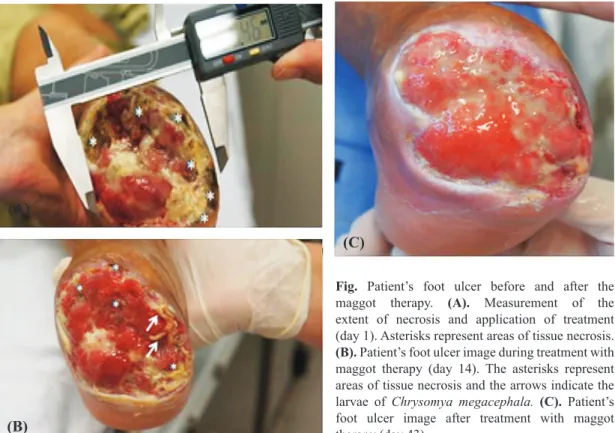
The study was approved by the ethics committee of the Federal University of Rio Grande do Norte, Brazil. Six biological dressings were done with an interval of every 48 h. Before therapy, an extension of tissue impairment of 8.4 cm2 was obtained (Fig. A) and after
two weeks of treatment (14 days) a retraction of the ulcer 0.7 cm2 was observed (Fig. B). At the end of treatment
(43 days) the ulcer’s surface area was occupied by granulation tissue (Fig. C). The maggot therapy was found to be an effective and inexpensive method of debridement, providing a rapid acceleration in the process of wound healing7,8. Importantly, the effect of
using the debridement maggot therapy occurs not only by the physical activity of larvae (by their mandibles) but also by secretions and excretions such as trypsin, collagenase, and chymotrypsin, showing that the lytic activity is able to promote the dissolution of necrotic tissue. This therapy also eliminated multidrug resistant microorganisms (K. pneumoniae, E. coli, Pseudomonas aeruginosa) present in the wound, as reported earler9.
The SHV and CTX-M genotypes were observed only in the K. pneumoniae isolates. These results indicated the
Fig. Patient’s foot ulcer before and after the maggot therapy. (A). Measurement of the extent of necrosis and application of treatment (day 1). Asterisks represent areas of tissue necrosis.
(B). Patient’s foot ulcer image during treatment with maggot therapy (day 14). The asterisks represent areas of tissue necrosis and the arrows indicate the larvae of Chrysomya megacephala. (C). Patient’s foot ulcer image after treatment with maggot therapy (day 43).
PINHEIRO et al: ALTERNATIVE TREATMENT FOR DIABETIC FOOT ULCERS 341
presence of ESBL (extended-spectrum beta-lactamase)
in bacteria. Bexield et al10demonstrated the eficacy of
these compounds against strains of methicillin-resistant Staphylocccus aureus and other bacteria. Several peptides produced in whole body extract of maggots with high antimicrobial activity as well as compounds present in their haemolymph have been shown to have activity against P. aeruginosa11.
In conclusion, considering the role of maggot therapy in wound healing, particularly in diabetic ulcer and its low cost compared with the other usual treatments, there is a need to evaluate its safety and
eficacy on a large number of patients.
references
Martini RK, Sherman RA. Maggot debridement therapy. 1.
J Bras Med 2003; 85 : 82-5.
Sherman RA, Hall MJR, Thomas S. Medicinal Maggots: an 2.
ancient remedy for some contemporary aflictions. Annu Rev Entomol2000; 45 : 55-81.
Marcondes CB.
3. Use of maggot therapy for treating a diabetic foot ulcer colonized by multidrug resistant bacteria in Brazil. 1st ed, Florianópolis: Editora da UFSC; 2006.
Murray PR, Baron EJ, Pfaller MA, Tenover FC, Yolken RH. 4.
Manual of clinical microbiology. 7th ed. Washington DC: ASM Press; 1999.
Clini
5. cal and Laboratory Standards Institute (CLSI).
Performance standards for antimicrobial susceptibility testing; 23rd informational supplement. Wayne, PA, USA: CLSI; 2013.
de Oliveira CF, Salla A, Lara VM, Rieger A, Horta JA, 6.
Alves SH. Prevalence of extended spectrum beta-lactamases producing microorganisms in noscomial patients and molecular characterization of the shv type isolates. Braj J Microbiol 2010; 41 : 278-82.
Tanyuksel M, Araz E, Dundar K, Uzun G, Gumus T, Alten B, 7.
et al. Maggot debridement therapy in the treatment of chronic wounds in a military hospital setup in Turkey. Dermatology
2005; 210 : 115-8.
Chambers L, Woodrow S, Brown AP, Harris PD, Philips D, 8.
Hall M, et al. Degradation extracellular matrix components
by deined proteinases from the green bottle larva Lucillia sericata used for the clinical debridement of non-healing wounds. Br J Dermatol 2003; 148 : 14-23.
Mclnnes W, Ruzehaji N, Wright N, Cowin AJ, Fitridge R. 9.
Venous ulceration contaminated by multi-resistant organisms: larval therapy & debridement. J Wound Care 2013; 22 (Suppl 10): s27-30.
Bexield A, Nigam Y, Thomas S, Ratcliffe NA. Detection and
10.
partial characterization of two antibacterial factors from the excretions/secretions of the medicinal maggot Lucilia sericata
and their activity against methicillin-resistant Staphylococcus aureus (MRSA). Microbes Infect2004; 6 : 1297-304.
Hubernan L, Gollop N, Mumcuoglu KY, Block C, Galun 11.
R. Antibacterial properties of whole body extracts and haemolymph of Lucilia sericata maggots. J Wound Care
2007; 16 : 123-7.
Reprint requests: Dr Renato Motta Neto, Department of Microbiology & Parasitology, Biosciences Center, Federal University of Rio Grande Do Norte, Av. Senador Salgado Filho, 3000,
Candelaria, Natal, RN, Brasil e-mail: [email protected]
342 INDIAN J MED RES, MARCH 2015