Original Article
Artigo Original
Maria Elaine Trevisan1 José Humberto Bellinaso2 Andrielle de Bitencourt Pacheco3 Luciana Barros Augé4 Ana Maria Toniolo da Silva5 Eliane Castilhos Rodrigues Corrêa1
Keywords
Palate, Hard Measures Nasal obstruction Mouth breathing Respiratory system
Descritores
Palato Duro Medidas Obstrução Nasal Respiração Bucal Trato Respiratório
Correspondence address:
Maria Elaine Trevisan
Rua Silva Jardim, 2.141, Apto. 701, Santa Maria (RS), Brasil, CEP: 97010-493. E-mail: [email protected]
Received: 10/01/2014
Accepted: 01/07/2015
Study carried out at the Graduate Program in Human Communication Disorders, Universidade Federal de Santa Maria – UFSM – Santa Maria (RS), Brazil
(1) Physical Therapy and Rehabilitation Department, Graduate Program in Human Communication Disorders, Universidade Federal de Santa Maria – UFSM – Santa Maria (RS), Brazil.
(2) Graduate Program in Implantodonty, Sobresp Faculdade de Ciências da Saúde – Santa Maria (RS), Brazil. (3) Graduate Program in Human Communication Disorders, Universidade Federal de Santa Maria –UFSM – Santa Maria (RS), Brazil.
(4) Hospital de Guarnição de Santa Maria (RS), Brazil.
(5) Speech Language Pathology and Audiology Department, Graduate Program in Human Communication Disorders, Universidade Federal de Santa Maria – UFSM – Santa Maria (RS), Brazil.
Conlict of interests: nothing to declare.
Respiratory mode, nasal patency
and palatine dimensions
Modo respiratório, patência nasal
e dimensões palatinas
ABSTRACT
Purpose: To investigate the inluence of breathing mode and nasal patency in the dimensions of the hard palate by comparing mouth breathing (MB) and nasal breathing (NB) adults. Methods: Seventy-seven individuals, distributed into the MB group (n=38) and the NB group (n=39), of both genders and aged between 18 and 30 years old, took part in the study. The respiratory mode diagnosis was based on anamnesis, physical characteristics, and otorhinolaryngological examination. The volunteers were evaluated in terms of nasal patency, with a peak nasal inspiratory low (PNIF) meter, and obstruction symptoms, by a Nasal Obstruction Symptom Evaluation (NOSE) scale, and had their transversal and vertical hard palate dimensions measured with a digital caliper in plaster models. Results: Comparing both groups, the MB group presented signiicantly higher values in the NOSE scale, lower values in the PNIF, lower values in the transversal distance of the palate in the intercanine region, and signiicantly higher values in the vertical distance in the regions of the irst and second premolars and molars. There was a negative correlation between PNIF and NOSE, and a positive correlation between PNIF and transversal distance of the palate in the region of the irst premolars.
Conclusion: MB adults presented reduced nasal patency and a higher degree of nasal obstruction symptoms. The hard palate was morphologically narrower and deeper in adults with the MB mode compared to the NB mode. Moreover, it was concluded that the smaller the nasal patency, the greater the obstruction symptoms and the narrower the hard palate.
RESUMO
INTRODUCTION
Nasal breathing (NB), which is inherent to the human being and has the important role of preparing the air to reach the greater structures of the respiratory system, stimulates the adequate growth of the craniofacial complex, with a direct inluence on the development of the jawbones, the posture of the jaw, the positioning of the tongue, and the maintenance of the rhinopharyngeal space(1-3).
Any factor that promotes obstruction of the upper airways leads to the partial or complete substitution of NB for mouth breathing (MB), and the persistence of this is responsible for major muscle imbalances, with repercussions on morphofunc-tional craniofacial features and the stomatognathic system(3,4).
The reduction in nasal patency, a phenomenon that persists during the growth period, is an important etiologic factor for maxillary atresia in the MB adult(5) and is a result of the fact
that the adequate jaw growth is stimulated both by the passage of air through the nostrils and the proper positioning of the tongue on the hard palate(6).
Alterations in the vertical and transversal dimensions of the hard palate in MB(7-10) adults induce other adaptations of form
and function, such as malocclusion, high arched, narrow, and deep palate, lack of lip closure, tongue lowered on the mouth loor or with interdental placement, laccid orofacial muscles, and atypical swallowing(11). These alterations contribute to the
increase in the resistance to nasal airlow and to the reduction in the peak nasal inspiratory low (PNIF)(12,13).
The PNIF meter is a viable, noninvasive, easy handling, and cost-effective alternative for the evaluation of nasal patency. It is an indirect measure of nasal obstruction, which considers that the increase in nasal resistance modiies the nasal airlow and, consequently, the PNIF(12,13). Previous studies(14,15)
evalu-ated and considered the measuring features of this instrument appropriate. It is a commonly used method for evaluating the effectiveness of drugs or treatments, as well as for home moni-toring the variations in circadian rhythms or the environmental effects on nasal patency(12).
Another possibility to assess the effect of reduced nasal patency is the Nasal Obstruction Symptom Evaluation (NOSE) scale, which measures how much the obstruction interferes in the life of participants with increased nasal resistance(16).
There are few studies about nasal patency and palatine coniguration with adult participants. It is believed that the investigation of these aspects in adults may provide more spe-ciic information for the understanding of the impact of this syndrome over time, which contributes to a global diagnostic approach and a more complete therapeutic intervention.
On the basis of the points explained earlier, the objective of this study was to investigate the inluence of the respiratory mode and nasal patency on the dimensions of the hard palate in view to comparing MB and NB adults.
METHODS
Methods involved an exploratory, cross-sectional, and controlled study, approved by the Ethics Committee
at Universidade Federal de Santa Maria (Process No. 04039912.7.0000.5346), with the informed consent signed by all participants. The participants in the study were adults of both genders, aged between 18 and 30 years old, with mouth breathing mode (MB group) and nasal breathing mode (NB group), normoponderal, with no evidence of lung or neuromuscular diseases and chest deformities, no history of smoking and/or exposure to a risk environment, and without previous maxillary expansion. Individuals who used topical or systemic corticosteroids, nasal vasoconstrictors, muscle relaxants, and/or barbiturates, who had flu for the previous 3 weeks or an allergic crisis on the evaluation day, or who had prior thoracic, abdominal and/or rhinosinusal surgery were excluded from the study. The diagnosis of MB was based on anamnesis, signs and symptoms, physical characteristics associated to MB, and otorhinolaryngologi-cal examination(17).
The individuals referred to or who voluntarily came forward for the research underwent an initial interview with collection of demographic and anthropometric data that was relevant to the research. Then, the anamnesis was done by a speech language pathologist based on the orofacial myofunctional evaluation protocol (MBGR)(18).
The tests were carried out with the individuals in a sitting position, upright torso, relaxed arms, hands on the thighs, eyes open oriented by the Frankfurt plane, and feet lat on the loor.
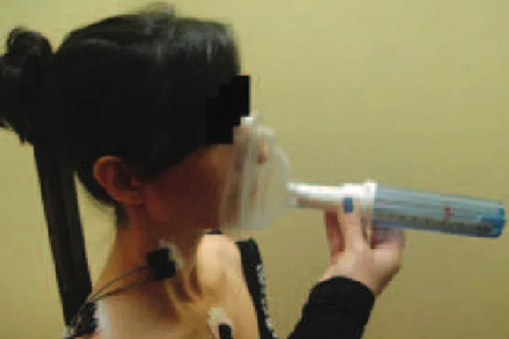
The PNIF was evaluated by the main researcher using the In-Check Inspiratory Flow Meter (Clement Clarke International, the United Kingdom) with the method of residual volume (RV), that is, a full exhale followed by a nasal inhale as fast and strong as possible, with the mouth closed and the mask well-adapted to the face(12,19). When they achieved RV, participants signaled
with the right thumb so that, simultaneously, the examiner would adapt the mask to the face for a strong and fast nasal inhale, with the following verbal stimulus “Release the air, close your mouth and draw (the air) strongly..” (Figure 1). The test was repeated at least three times or until three technically satisfactory measures could be obtained, with less than 10% variation between them, with the highest value being the one registered(14,19,20). The time interval between each measure was
30 seconds(20). Before the test, the demonstration was given and
a brief training was provided.
The nasal obstruction was assessed subjectively by the main researcher using the NOSE scale. The scale consists of ive questions that address how nasal obstruction represented a problem in the past month:
1. Nasal congestion or feeling of suffocation; 2. Nasal blockage or obstruction;
3. Dificulty of breathing through the nose; 4. Dificulty during sleep, and
5. Incapacity to inhale suficiently through the nose during exercise or exertion.
These questions can have scores from zero to four (0=not a problem, 1=very light problem, 2=moderate problem, 3=very bad problem, 4=severe problem). These values are added and multiplied by ive. The obtained score can range from 0, mean-ing that there is no problem, to 100, showmean-ing the worst possible problems when it comes to the nasal obstruction(16).
The measures of the hard palate were obtained from molding the jaw using alginate and the subsequent prepara-tion of plaster models, both performed by a dentist. In these models, points of reference were marked using a mechanical pencil with 0.05 mm graphite, and the transversal (width) (Figure 2) and vertical (height) (Figure 3) distances between these points were calculated using a 150 mm digital caliper (Western®, DC-60 Model, the United States), with resolution
of 0.01 mm and accuracy of ±0.03 mm. In the regions of the canines and of the irst and second premolars, the points were marked in the more apical gingival margin. For the irst molars, the marking was made in the junction of the gingival margin with the palatine sulcus of these teeth, bilaterally(8).
For the vertical measurements, a stainless steel wire 1 mm thick and with the length corresponding to the transversal distance was ixed with hot wax between the previously marked points in each of the considered teeth. This wire served as a support for sliding the caliper rod and obtaining the vertical measure of the palate, which corresponded to the perpendicular distance between the median palatine line and
the steel wire, minus 1 mm (the value that corresponds to the thickness of the wire)(8). Two dentists who collaborated in the
research, previously trained, performed two measures each, starting with the canines and continuing with the irst and second premolars and the irst molars. Then, it was veriied the intra- and interexaminer agreement.
Through the palatine height index (PHI = palatine height x 100/palatine width) in the region of the irst molars, the height of the palate was assessed. The centesimal relationship between the palatine height and width helps in the classiication of this structure in chamestaphyline (low palate) – values ≤27.9 mm, orthostaphyline (intermediate palate) – values between 28.0 and 39.9 mm, or hypsistaphyline (high palate) – values >40.0 mm(21,22). The palatine weight index (PWI) was not
de-termined in this study because the plaster models do not allow the visualization of the staphyline anthropometric point, which is necessary to determine the length of the hard palate and the subsequent calculation of this index(22).
The sample size calculation was estimated to achieve a signiicance level (alpha) of 5% (p<0.05) and power level (1-beta) of 80% (WinPepi software, version 1.5). For the calculation, the results of the vertical distance of the palate in the second premolar region was considered because this was the variable that presented greater variability in the pilot study with 10 participants in each group. Thus, the estimate was a sample of 36 subjects in each group.
The data were analyzed using descriptive statistics, the normality was veriied through the Shapiro-Wilk test, and the groups were compared using the Student’s t-test and Mann-Whitney test. The relationship (r) between the variables as-sessed using Spearman’s correlation test was as follows: weak correlation (0<r<0.3), moderate correlation (0.3<r<0.7), and
strong correlation (r≥0.7)(23). The intraclass correlation
coef-icient (ICC) was used to verify the reliability of the palate measures. The ICC values were classiied in poor reliability (ICC≤0.4), moderate reliability (0.4<CHF<0.75), and excellent reliability (ICC≥0.75)(24).
The analyses were performed using the Statistical Package for the Social Sciences software (SPSS, version 17.0), and a signiicance level of 5% was assumed in all tests.
RESULTS
The sample consisted of 77 volunteers, 39 (28 women, 11 men) in the NB group and 38 (25 women, 13 men) in the MB group. According to the otorhinolaryngological examination, all the members of the MB group were classiied as functional MBs of nonorganic cause, primarily allergic problems, and parafunctional habits.
The anthropometric and nasal permeability characteristics presented in Table 1 show that the nasal permeability (PNIF) was signiicantly lower (147.69 versus 121.45), whereas the perception of nasal obstruction (NOSE) was signiicantly higher (6.15 versus 47.11) in the MB group.
For the reliability of palate measures, inter- and intraex-aminers presented an ICC with a level higher than 0.97 in all measures, assuming it excellent at >0.75(24). Thus, the measures
of the transversal and vertical distances of the palate in the MB and NB groups are a result of the arithmetic mean of intra- and interexaminers results.
There was a signiicant difference between the groups, with lower transversal distance in the intercanine region (Table 2) and higher vertical distance in the regions of the irst and second premolars and the molars in the MB group (Table 3).
The PHI in the NB group was 39.9±6.5, which corresponds to the intermediate palate classiication. In the MB group, it was 45.1±8.5, which corresponds to the high palate classiication.
The correlations between NOSE scale versus PNIF (r=-0.34) and PNIF versus transversal distance between the irst premolars (r=0.31) was moderate to weak (Figures 4 and 5).
NB Group (n=39) MB Group (n=38)
p-value
Mean±SD Mean±SD
Age (years) 22.56±2.89 22.71±3.50 0.99
Body mass (kg) 59.89±11.06 65.00±12.19 0.05
Height (m) 1.68±0.09 1.69±0.10 0.45
PNIF (L/min) 147.69±35.65 121.45±30.99 <0.01*
NOSE 6.15±13.15 47.11±19.85 <0.01*
Table 1. Anthropometric and nasal permeability characteristics in the nasal breathing and mouth breathing groups
*Mann-Whitney test.
Caption: NB = nasal breather; MB = mouth breather; SD = standard deviation; PNIF = peak nasal inspiratory flow; NOSE = nasal obstruction symptom evaluation scale
*Mann-Whitney test.
Caption: mm = millimeters; NB = nasal breather; MB = mouth breather
Table 2. Measure of the transversal distances of the palate in nasal breathing and mouth breathing groups
Transversal distance NB Group (n=39) MB Group (n=38) p-value Mean±SD Mean±SD
Intercanine (mm) 24.65±1.84 23.95±2 0.04* First premolar (mm) 27.38±2 26.5±2.47 0.09 Second premolar (mm) 32.05±2.44 31.11±2.88 0.12 First molar (mm) 34.28±2.74 34.35±3.39 0.93
*Student’s t-test for independent samples; **Mann-Whitney test. Caption: mm = millimeters; NB = nasal breather; MB = mouth breather
Table 3. Measure of the vertical distances of the palate in nasal breathing and mouth breathing groups
Vertical distance NB Group (n=39) MB Group (n=38) p-value Mean±SD Mean±SD
Intercanine (mm) 5.76±1.80 6.13±1.92 0.20 First premolar (mm) 10.33±1.80 11.26±1.81 0.03* Second premolar (mm) 13.51±1.90 14.86±2.15 0.03* First molar (mm) 13.68±2.23 15.45±3.16 <0.01**
r=-0.34; p<0.01
NOSE
PNIF (L/min) 100
90 80 70 60 50 40 30 20 10 0
0 50 100 150 200 250
Caption: PNIF = peak nasal inspiratory flow; NOSE = nasal obstruction symptom evaluation scale
Figure 4. Correlation between the nasal obstruction symptom evaluation scale and the peak nasal inspiratory flow
r=0.31; p<0.01
Dist
ance - 1
st pr
emolar (mm)
PNIF (L/min) 35
34
32
30
28
26
24
22
20
0 100 150 200 250
Caption: PNIF = peak nasal inspiratory flow
DISCUSSION
In the objective assessment of the variation of nasal patency between the groups, it was observed that PNIF showed lower values in the MB group compared to the NB group. Similar results were found in a study of adults with and without nasal obstruction by rhinitis, in which the group with rhinitis had lower PNIF values (114.0 versus 154.3 L/min), with the same pattern being observed in the comparison between adults with and without nasal obstruction symptoms (123.6 l/min versus 151.4 L/min)(25). These indings were expected as the MB mode
is established when there is a reduction in nasal patency. The subjective evaluation of nasal obstruction (NOSE scale) found a moderate impact on the quality of life in the MB group, with a higher score than the one found for the NB group (47.1 versus 6.2), showing the perception and the interference of obstructive symptoms, also during sleep and exercise. High scores were also observed in studies(26,27) of individuals with
nasal obstruction by septal deviation (75 and 60.2), who had lower scores (10 and 11.3) after septoplasty.
In this study, a negative correlation between the NOSE scale scores and the PNIF values was observed, which showed that the more reduced the nasal patency, the worse is the perception of the obstruction symptoms. These same correla-tions between objective and subjective evaluation methods of nasal obstruction were also found in other studies(25,27).
Kahveci et al.(27) showed that the evaluation using the NOSE
scale had good correlation with the indings of nasal obstruc-tion obtained by computed tomography (CT) scan, without correlation with acoustic rhinometry. The assessment of nasal obstruction through a visual analog scale had a negative cor-relation with the PNIF measure(25). These indings show that
the objective and subjective evaluations add different informa-tion that is complementary and can be taken into account in clinical practice(28).
The inluence of the MB mode on the morphology of the hard palate of MBs, which was investigated in this study, re-vealed that the MBs had a palate with lower transversal distance (width) and higher vertical distance (height) compared to the NBs, except in the transversal measure for the molar region. Using the ratio of the means of vertical and transversal distances (PHI) in the region of the irst molars, it was observed that the MBs presented a palate that could be classiied as high. It was found that the morphological appearance of the adult palate was similar to that identiied in MB children(7-10), who also had
lower values for the transversal measures and higher values for the vertical measures, compared to NB children. These indings were compared with results obtained in children, as studies with similar methodology, comparing the palatine dimensions linked to the breathing mode in adults, were not found.
In this study, a lower transversal measure was found in the canines region, a result similar to that found in MB children(29),
in which the dental and skeletal alterations, including the nar-rowing of the upper arch, were more frequent in the region of the canines. However, they were different from the ind-ings of other studies(7-9), in which this measure was lower in
the molar region, suggesting that in MB children there is a
tendency of narrowing palate in the posterior region without interference in the anterior region.
The palate height in the adult MB group was higher in the region of the irst and second premolars and the irst molars. Similar results were observed in MB children, with greater height in the regions of the second premolars(7,8) and the second molars(9).
The discovery of greater height in the posterior regions as a whole and of palate narrowing only in the region of the canines in adults that maintained the MB mode since childhood seems to indicate a trend that the impact of oral breathing becomes more evident in the vertical palatine dimension over the years. The indings of this study, supported by previous studies(7-10,29,30),
conirm that breathing through the mouth inluences the hard palate morphology of mouth breathers.
According to the literature(5,6), the palatine alterations
evi-denced in the MB group may be due to the loss of negative pressure in the nasal cavity, with consequent reduction in the stimulus for the lowering of the palate and of the positioning of the tongue in the loor of the mouth, preventing its expander stimulus on the jaw. In addition, the decrease in tension of orofacial muscles, often seen in the MB group(3,11), causes the
soft tissues to exert less force on the bone tissues that constitute the face, which could alter the growth and development of the craniofacial bone structure, including the jaw and the hard pal-ate(8). However, one cannot ignore the inluence of genetic
fac-tors on the craniofacial growth and of the dolichofacial growth pattern, which can also determine morphological alterations in the palate, regardless of breathing mode(30).
Considering that PNIF had a weak correlation with the width of the palate, only in the region of the irst premolars, and that a relation to the vertical measures has not been found, the hypothesis that nasal patency affects the height of the hard palate requires more studies for conirmation.
The evaluation of the palatine measures and nasal patency in MBs could provide objective parameters to rate the intensity/ severity of this respiratory mode. However, because of the ab-sence of normative parameters for these variables, especially in adults, this classiication is not yet possible. The assessment by the NOSE scale showed a difference between the NB and MB groups, with a moderate level of nasal obstruction symptoms in the MB group. Nevertheless, it is important to consider that the scale is a perceptual evaluation.
The dificulty to control variables, such as installation time, duration of oral breathing, and parafunctional habits, can be considered a limitation of this study.
It is believed that the magnitude of MB is associated with the impact on orofacial morphology. Therefore, this study owes its contribution for the clinical practice to the use of PNIF for evaluating and quantifying the nasal patency through an objec-tive and of accessible use measure. In addition, it allows one to control the homeostasis and nasal congestion relections, as well as the environmental effects on nasal patency.
includes, in addition to orthodontic aspects, those of ventilatory, postural, and orofacial motricity nature. Because it is a com-mon condition, one recognizes the need for the awareness of parents, teachers, patients, and professionals about the systemic impacts of this breathing mode and the compromised quality of life of these patients.
CONCLUSION
The adults with MB mode showed lower nasal patency, lower transversal distance (width), and higher vertical distance (height) of the hard palate when compared to NB adults.
*MET was responsible for the elaboration of the project, data collection and tabulation, analysis, interpretation, and preparation of the manuscript; JHB contributed with the data collection and tabulation; ABP contributed with the data collection and tabulation; LBA contributed with the data collection; AMTS contributed with guidance in the stages of implementation and review of the manuscript; ECRC was responsible for the overall orientation of the project and for the steps of implementation and preparation of the manuscript.
REFERENCES
1. Pacheco AB, Silva AMT, Mezzomo CL, Berwig LC, Neu AP. Relação da respiração oral e hábitos de sucção não-nutritiva com alterações do sistema estomatognático. Rev CEFAC. 2012:14(2):281-9.
2. Lessa FCR, Enoki C, Feres MFN, Valera FCP, Lima WTA, Matsumoto MAN. Inluência do padrão respiratório na morfologia craniofacial. Rev Bras Otorrinolaringol. 2005;71(2):156-60.
3. Lemos CM, Wilhelmsen NSW, Mion OG, Mello Júnior JF. Alterações funcionais do sistema estomatognático em pacientes com rinite alérgica: estudo caso-controle. Braz J Otorhinolaryngol. 2009;75(2):268-74.
4. Armijo Olivo S, Magee DJ, Paritt M, Major P, Thie NM. The association between the cervical spine, the stomatognathic system, and craniofacial pain: a critical review. J Orofac Pain. 2006;20(4):271-87.
5. Cappellette Jr. M, Carlini D, Pignatari SS, Cruz OLM, Weckx LLM. Rinometria acústica em crianças submetidas à disjunção maxilar. Rev Dent Press Ortodon Ortop Facial. 2006;11(2):84-92.
6. DiFrancesco RC, Bregola EGP, Pereira LS, Lima RS. A obstrução nasal e o diagnostico ortodôntico. Rev Dent Press Ortodon Ortop Facial. 2006;11(1):107-13.
7. Berwig LC, Silva AMT. Análise quantitativa do palato duro em respiradores orais: revisão de literatura. Rev Soc Bras Fonoaudiol. 2011;16(4):483-7.
8. Berwig LC, Silva AMT, Corrêa ECR, Moraes AB, Montenegro MM, Ritzel RA. Quantitative analysis of the hard palate in different facial typologies in nasal and mouth breathers. Rev CEFAC. 2012;14(4):616-25.
9. Feres MFN, Enoki C, Sobreira CR, Matsumoto MAN. Dimensões do palato e características oclusais de crianças respiradoras nasais e bucais. Pesqui Bras Odontopediatria Clín Integr. 2009;9(1):25-9.
10. Lione R, Buongiorno M, Franchi L, Cozza P. Evaluation of maxillary arch dimensions and palatine morphology in mouth-breathing children by using digital dental casts. Int J Pediatr Otorhinolaryngol. 2014;78(1):91-5. 11. Cattoni DM, Fernandes FD, Di Francesco RC, Latorre M do R.
Characteristics of the stomatognathic system of mouth breathing children: anthroposcopic approach. Pro Fono. 2007;19(4):347-51. 12. Kjærgaard T, Cvancarova M, Steinsvåg SK. Does nasal obstruction mean
that the nose is obstructed? Laryngoscope. 2008;118(8):1476-81. 13. Wheeler SM, Corey JP. Evaluation of upper airway obstruction-an ENT
perspective. Pulm Pharmacol Ther. 2008;21(3):433-41.
14. Ottaviano G, Scadding GK, Coles S, Lund VJ. Peak nasal inspiratory low: normal range in adult population. Rhinology. 2006;44(1):32-5. 15. Dufour X, Gohler C, Delagranda A, Fontanel JP, Klossek JM. Peak
Nasal Inspiratory Flow: apprentissage de la méthode de mesure et reproductibilité. Ann Otolaryngol Chir Cervico-faciale. 2007;124(3):115-9. 16. Stewart MG, Witsell DL, Smith TL, Weaver EM, Yueh B, Hannley MT.
Development and validation of the nasal obstruction symptom evaluation (NOSE) scale. Otolaryngol Head Neck Surg. 2004;130(2):157-63. 17. Milanesi JM, Weber P, Berwig LC, Ritzel RA, Silva AMT, Corrêa ECR.
Childhood mouth-breathing consequences at adult age: ventilatory function and quality of life. Fisioter Mov. 2014;27(2):211-8.
18. Genaro KF, Berretin-Felix G, Rehder MIBC, Marchesan I. Avaliação miofuncional orofacial – Protocolo MBGR. Rev CEFAC. 2009;11(2):237-55.
19. Ottaviano G, Lund VJ, Coles S, Stafieri A, Scadding GK. Does peak nasal inspiratory flow relate to peak expiratory flow? Rhinology. 2008;46(3):200-3.
20. Lee DK, Haggart K, Lipworth BJ. Reproducibility of response to nasal lysine-aspirin challenge in patients with aspirin-induced asthma. Ann Allergy Asthma Immunol. 2004;93(2):185-8.
21. Oliveira MO, Vieira MM. Influência da respiração bucal sobre a profundidade do palato. Pró-Fono. 1999;11(1):13-20.
22. Sicher H, Dubrul EL. Anatomia Oral. 8ª edição. São Paulo: Artes Médicas; 1991.
23. Chan YH. Biostatistics 104: correlational analysis. Singapore Med J. 2003;44(12):614-9.
24. Fleiss JL, Levin B, Cho Paik M. The measurement of interrater agreement. In: Fleiss JL, Levin B, Cho Paik M, editors. New York: John Wiley & Sons; 2000.
25. Teixeira RU, Zappelini CE, Alves FS, da Costa EA. Peak Nasal Inspiratory Flow Evaluation as an objective method of measuring nasal airlow. Braz J Otorhinolaryngol. 2011;77(4):473-80.
26. Bezerra TF, Stewart MG, Fornazieri MA, Pilan RR, Pinna F de R, Padua FG, et al. Quality of life assessment septoplasty in patients with nasal obstruction. Braz J Otorhinolaryngol. 2012;78(3):57-62.
27. Kahveci OK, Miman MC, Yucel A, Yucedag F, Okur E, Altuntas A. The eficiency of nose obstruction symptom evaluation (NOSE) scale on patients with nasal septal deviation. Auris Nasus Larynx. 2012;39(3):275-9. 28. Braun T, Rich M, Kramer MF. Correlation of three variables describing nasal patency (HD, MCA, NOSE score) in healthy subjects. Braz J Otorhinolaryngol. 2013;79(3):354-8.
29. Harari D, Redlich M, Miri S, Hamud T, Gross M. The effect of mouth breathing versus nasal breathing on dentofacial and craniofacial development in orthodontic patients. Laryngoscope. 2010;120(10):2089-93.