CASE REPORT
Contact-lens-related corneal ulcer caused by
klebsiella pneumoniae
Tongabay Cumurcu,IPembegul Firat,IErcan O¨ zsoy,IMufide Cavdar,IYusuf YakupogullariII
IInonu University School of Medicine, Department of Ophthalmology, Malatya/Turkey. IIInonu University School of Medicine, Department of Microbiology, Malatya/Turkey.
Email: [email protected] Tel.: 90-422-342 06 60/4003
INTRODUCTION
The use of contact lenses (CLs) has been widely associated with corneal alterations that range in symptomatology and severity. Lesions may vary from small peripheral sterile infiltrates to infectious central ulcers with sight-threatening potential.1Contact lens wear has been described as the most important predisposing factor to microbial keratitis world-wide.2
Infections are more frequent in soft-lens users, and the risk with gas-permeable rigid lenses is estimated to be one-third of that with daily-wear soft lenses. Extended wear is also associated with a higher risk of microbial keratitis.3
Bacterial keratitis is most commonly caused by aggressive gram-negativePseudomonas spp.; Klebsiella spp.is rarely the causative agent of keratitis due to contact lens wear.4
We present a case of contact-lens-related bacterial keratitis caused byKlebsiella pneumoniae.
Case Presentation
A 19-year-old man presented to our service complaining of pain and hyperemia of two days’ duration in the right eye. He had been a regular contact lens wearer for two years. The patient used multipurpose solutions for cleaning, disinfect-ing, and storing daily-wear soft contact lenses. Recently, the lenses had been worn for extended periods of time.
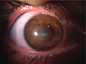
His visual acuity was at the level of finger counting. Ophthalmologic examination revealed central corneal ker-atitis in his right eye (Figure 1)
Samples from conjunctival swabbing and corneal scraping, the contact lens, the cleaning solution, and the case were all submitted for microbiological analysis. Gram staining revealed gram-negative microorganisms in the corneal scrap-ing and in the contact lens cleanscrap-ing solution. Cultures on appropriate media were positive forKlebsiella pneumoniae in these specimens. The organism was susceptible to aminogly-cosides and to fourth-generation fluoroquinolones. The patient was treated topically with moxifloxacin (0.5%) and tobramycin (15 mg/ml) drops every hour and cyclopentolate 2% three times daily in the right eye. N-acetylcysteine eye drops were administered four times per day.
After clinical improvement (four days later), topical corticosteroid drops were prescribed. The infection resolved, but residual central stromal opacification remained (Figure 2). The patient achieved a final visual acuity of 20/100 in the right eye.
DISCUSSION
Contact-lens–related corneal infections continue to pre-sent a major challenge to ophthalmologists. With the continuous improvement in lens materials and design, as well as in disinfecting and storing solutions, contact lens use is on the rise. Currently, patients can use a single solution to rinse, disinfect, and store their lenses without the need for rubbing or enzymatic cleaning.5
The patient described herein used multipurpose solution for cleaning, disinfecting, and storing daily-wear soft contact lenses. The extended wear reported may have caused this case of microbial keratitis.3Gram-negative bacteria include opportunistic enterobac-teria (such as Serratia spp. and Klebsiella spp.) that can survive in contact lens fluid and on plastic surfaces, which explains their increased numbers in contact-lens-induced corneal infections.6
Contact-lens-related keratitis may vary from small per-ipheral sterile infiltrates to infectious central ulcers with sight-threatening potential, as in our case.7
Overnight wear and, to a lesser extent, improper lens care are known to be major risk factors for corneal infection.8
Copyrightß2011CLINICS– This is an Open Access article distributed under
the terms of the Creative Commons Attribution Non-Commercial License (http:// creativecommons.org/licenses/by-nc/3.0/) which permits unrestricted non-commercial use, distribution, and reproduction in any medium, provided the
original work is properly cited. Figure 1 -Central corneal keratitis in the right eye.
CLINICS 2011;66(8):1509-1510 DOI:10.1590/S1807-59322011000800036
Lam et al. reported a five-fold increase in the risk of microbial keratitis among patients who wear their contact lenses overnight.3
As reported by several authors, contact-lens-induced keratitis is mainly caused by gram-negative micro-organ-isms.4,8However,Klebsiella spp.are not a common cause of bacterial keratitis.
One study that investigated the causes of microbial keratitis showed that 3.2% of microbial keratitis cases were due to contact lens use. The same study showed that in 2.2% of these keratitis patients,Enterobacter spp.was the causative agent.9 In another study, 33 patients with contact-lens-related keratitis were followed.Klebsiella spp.were observed in the cultures of only two patients.10
N-acetylcysteine is a potential candidate for use as an inhibitor ofKlebsiellabiofilm formation. Therefore, we added N-acetylcysteine eye drops to the patient’s treatment regimen.11
The majority of keratitis was caused by soft contact lens wear. One-fifth of these comparatively young patients displayed corneal scars.12This case highlights the need for clinicians to be aware thatKlebsiella spp.can induce contact-lens-related corneal ulcers.
REFERENCES
1. Moriyama AS, Hofling-Lima AL. Contact lens-associated microbial keratitis. Arq Bras Oftalmol. 2008;71:32-6, doi: 10.1590/S0004-27492-008000700007.
2. Bourcier T, Thomas F, Borderie V, Chaumeil C, Laroche L. Bacterial keratitis: predisposing factors, clinical and microbiological review of 300 cases. Br J Ophthalmol. 2003;87:834-8.
3. Lam DS, Houang E, Fan DS, Lyon D, Seal D, Wong E, et al. I˙ncidence and Risk Factors for Microbial Keratitis in Hong Kong: comparison with Europe and North America. Eye. 2002;16:608-18, doi: 10.1038/sj.eye. 6700151.
4. Stapleton F, Keay LJ, Sanfilippo PG, Katiyar S, Edwards KP, Naduvilath T. Relationship between climate, disease severity, and causative organism for contact lens-associated microbial keratitis in Australia. Am J Ophthalmol. 2007;144:690-8.
5. Najjar DM, Aktan SG, Rapuano CJ, Laibson PR, Cohen EJ. Contact lens-related corneal ulcers in compliant patients. Am J Ophthalmol. 2004;137:170-2.
6. Holden BA, La Hood D, Grant T, Newton-Howes J, Baleriola-Lucas C, Willcox MD, et al. Gram-negative bacteria can induce contact lens related acute red eye (CLARE) responses. CLAO J. 1996;22:47–52.
7. Efron N, Morgan PB. Can subtypes of contact lens-associated corneal infiltrative events be clinically differentiated? Cornea. 2006;25:540-4, doi: 10.1097/01.ico.0000214219.67872.3c.
8. Cheng KH, Leung SL, Hoekman HW, Beekhuis WH, Mulder PG, Geerards AJ, et al. Incidence of contact-lens-associated microbial keratitis and its related morbidity. Lancet. 354:181–5, doi: 10.1016/S0140-6736(98)09385-4.
9. Yilmaz S, Ozturk I, Maden A. Microbial keratitis in West Anatolia, Turkey: aretrospective review. Int Ophthalmol. 2007;27:261-8, doi: 10. 1007/s10792-007-9069-2.
10. Palamar M, Masarogulları M, Egrilmez S, Aydemir S, Yagcı A. Our Microbiological Analysis Results in Microbial Contact Lens Keratitis. Turk J Ophthalmol. 2010;40:349-53.
11. Pinna A, Sechi LA, Zanetti S, Carta F. Detection of Virulance Factors in a corneal Isolate of Klebsiella pneumoniae. Ophthalmology. 2005;112:883-7, doi: 10.1016/j.ophtha.2004.12.024.
12. Neumaier-Ammerer B, Stolba U, Feichtinger H, Bindler S. Contact lens related corneal infiltrates and ulcers – a retrospective study of 134 eyes. Spektrum Augenheilkd. 2008;22:297–300, doi: 10.1007/s00717-008-0284-7.
Figure 2 -The infection resolved, but residual central stromal opacification remained in the right eye.
Klebsiella pneumoniaekeratitis
Cumurcu T et al. CLINICS 2011;66(8):1509-1510