Experimental implantation of an arterial substitute
made of silicone reinforced with polyester fabric in rabbits
Laila Massad Ribas,* Inez Ohashi Torres, Fernanda Appolonio, Karina Paula Domingos Rosa, Fabio Rodrigues Ferreira do Espı´rito-Santo, Nelson De Luccia
Departamento de Cirurgia, Faculdade de Medicina FMUSP, Universidade de Sao Paulo, Sao Paulo, SP, BR.
OBJECTIVES:The aim of this study was to analyze silicone tubes with an internal diameter of 4 mm as a possible material for vascular prostheses.
METHODS:Grafts were implanted into the infrarenal aortas of 33 rabbits. Fluoroscopic examinations were performed within 150 days after surgical implantation. Sample grafts were analyzed via electron microscopy to evaluate the eventual endothelialization of the prostheses.
RESULTS:The patency rates of the prostheses were 87% (±6.7%) after 30 days, 73% (±9.3%) after 60 days and 48% (±12%) after 120 days. The material presented characteristics that support surgical implantation: good tolerance promoted by polyester tear reinforcement, ease of postoperative removal and a lack of pseudo-aneurysms. However, intimal hyperplasia was a limiting factor for the patency rate.
CONCLUSIONS:We concluded that polydimethylsiloxane has limited potential as an alternative material for small vascular prostheses.
KEYWORDS: Vascular Grafting; Polydimethylsiloxane; Polyesters; Silicon; Aorta; Blood Vessel Prosthesis.
Ribas LM, Torres IO, Appolonio F, Rosa KP, Espirito Santo FR, De Luccia N. Experimental implantation of an arterial substitute made of silicone reinforced with polyester fabric in rabbits. Clinics. 2017;72(12):780-784
Received for publication onMay 15, 2017;First review completed onJuly 6, 2017;Accepted for publication onAugust 4, 2017 *Corresponding author. E-mail: [email protected]
’ INTRODUCTION
Graft replacement of large vessels, such as the aorta and the iliac and femoral arteries, is reasonably achieved using currently available synthetic tubes, such as those composed of polytetrafluoroethylene (PTFE) and Dacron (1-5). However, in small vessels, none of these materials have proved to be superior or equal to the saphenous vein (6-10), which is regarded as the conduit of choice for peripheral revascularization (11-14).
A challenge that can arise is the lack of an available vein for arterial replacement. A substantial amount of research has been performed in this field, and there are several options for solving this problem. Cryopreserved donor veins (15,16) and biosynthetic materials are examples of alternatives described in vascular surgery studies (17,18). However, the search for an effective alternative for the replacement of small vessels remains open (19,20).
Polydimethylsiloxane (PDMS), or silicone, has been used in medicine since the 1960s (21,22). Due to its characteristics, silicone has become one of the most frequently used materials for prosthetic replacement in various contexts, such as breast
reconstruction (23). The use of PDMS in the form of various types of catheters for the intravenous administration of sub-stances is also widespread and universally accepted (24,25).
The characteristics that make this material attractive for these uses are its excellent thermal stability, good wetting properties, physiological inertness, excellent long-term biost-ability and low thrombogenicity (26-28).
Given that the search for an ideal vascular prosthesis con-tinues and that silicone is a material that promotes few tissue reactions, this study aims to propose an experimental model and test the hypothesis that PDMS is a suitable substitute for small vessels.
’ MATERIALS AND METHODS
All surgical procedures were performed in domestic rabbits (Oryctolagus cuniculus)in the Department of Surgery in the Faculty of Medicine of the University of São Paulo. The animals were provided by that institution’s animal house and were cared for in accordance with principles established by the Animal Welfare Act and the NIH Guide for Care and Use of Laboratory Animals.
This study was conducted with the approval of the ethics committee of the Faculty of Medicine of the University of São Paulo.
A tubular prosthesis made ??of PDMS (silicone) reinforced with polyester fabric was implanted into the infrarenal aorta of each animal.
DOI:10.6061/clinics/2017(12)10
Copyright&2017CLINICS–This is an Open Access article distributed under the terms of the Creative Commons License (http://creativecommons.org/licenses/by/ 4.0/) which permits unrestricted use, distribution, and reproduction in any medium or format, provided the original work is properly cited.
Synthetic prosthesis
The prostheses were prepared in accordance with a patent-registered design (29). Medical-grade silicone in liquid form was mixed with a curing agent and applied over a metal mandrel covered with a polyester fabric to increase the silicone’s tear resistance. To achieve uniform curing, rota-tional movement of the mandrel was maintained, initially at room temperature and then in an oven to achieve a post-cure temperature of 110o
C for 30 minutes. The PDMS prosthesis exhibited a tubular wall of 0.4 mm and an internal diameter of 4 mm (Figure 1). Despite attempts to create a porous tube, the final product was nonporous, and the internal and external surfaces were composed of silicone. The fabric material remained inside the wall simply to provide reinforcement.
Anesthetic procedures
Animals were anesthetized with a mixture of ketamine hydrochloride (35 mg/kg i.m. Ketalar 10%; Cristalia, São Paulo, Brazil) and xylazine hydrochloride (5 mg/kg i.m. Rompum 2%; Bayer AG, Leverkusen, Germany). During the surgical procedures, the animals received saline solution (0.9% sodium chloride) through a 22G catheter cannulated in the marginal ear vein.
Surgical technique
An anterior laparotomy was performed, and a transper-itoneal approach allowed for the dissection of 3 to 4 cm of the infrarenal aorta. A specifically developed self-static retractor was used to avoid evisceration during this exposure. The lumbar arteries were carefully preserved. Before the aorta was clamped with microsurgical clamps, sodium heparin (200 U/kg i.v.; Hepamax, Blausiegel, São Paulo, Brazil) was administered.
Anastomoses of the PDMS in the aorta were completed using an end-to-side technique and a continuous 7-0 poly-propylene suture. At the end of the proximal anastomosis, blood flow was released for a few minutes for ischemic conditioning.
At the end of the vascular anastomosis, pulses were checked, and the aorta was ligated with 4-0 cotton and cut between the anastomotic sites.
The animals were evaluated for up to 150 days. All surviv-ing animals were subjected to fluoroscopic examination via retrograde femoral contrast injection.
Prosthesis evaluations
The prostheses were evaluated using aortic fluoroscopy. Images were acquired on a Diasonics OECs
9000 fluoro-scopy scanner (Salt Lake City, Utah, USA). Dissection of the femoral artery was performed via unilateral inguinal incision to perfuse the contrast agent diatrizoate meglumine (Reliev 60%; BerliMed S.A., São Paulo, Brazil).
At the end of the experimental procedures, the animals were euthanized while they remained anesthetized with 19.1% potassium chloride (Isofarma, Eusébio, CE, Brazil), and their bodies were disposed of in accordance with the surgical department’s routine procedures.
The prostheses were removed after euthanasia, and samples were sent for scanning electron microscopy; images were obtained using a Philips XL30 system (FEI, Hillsboro, Oregon, USA).
Statistical analysis
The Kaplan-Meier method was used to analyze risk of occlusion and patent condition. Statistical analyses were performed using SPSS 18.0 software.
’ RESULTS
Surgical procedures were performed in 64 animals. Thirty animals (46.9%) survived until late evaluations. Early mor-tality was observed in 23 animals (35.9%). Eleven animals (17.2%) developed paraplegia in the immediate postopera-tive period, and these animals were not considered for long-term follow-up.
All 30 surviving animals were submitted to angiographic control at the end of the observational period. In three para-plegic animals that were not considered for late follow-up, angiography was performed to diagnose the status of the graft. Another three animals were excluded from late follow-up because early occlusion (at less than 7 days) of the graft was detected. The remaining 27 animals were analyzed using Kaplan-Meier survival/patency curves.
Surgical characteristics
The walls of the prostheses had characteristics of flexibility and hardness that allowed for easy passage of the needle and retention and containment of the suture lines. Thus, satis-factory hemostasis was achieved at the end of the experi-ments, and the presence of a pulse distal to the anastomoses attested to the immediate patency of the prostheses in all animals.
Prosthesis evaluations
The patency rates of the prosthesis were 87% (±6.7%) after
thirty days, 73% (±9.3%) after sixty days, 57% (±11%) after
ninety days and 48% (±12%) after one hundred twenty days
(Figure 2). The risk of occlusion is indicated in Figure 3.
Macroscopic analysis
Peri-implant fibrous tissue was easy to identify, and a cleavage plane between the periprosthetic tissue reaction and the tube was observed. Neither aneurysmal dilatation of the implant nor pseudoaneurysm formation in the suture lines was observed. Inside the occluded grafts, whitish thrombi
Figure 1 -PDMS prosthesis with a tubular wall of 0.4 mm and an internal diameter of 4 mm.
CLINICS 2017;72(12):780-784 Implantation of an arterial substitute in rabbits
vwere observed that were likely caused by intimal hyper-plasia, which was subsequently confirmed via scanning electron microscopy.
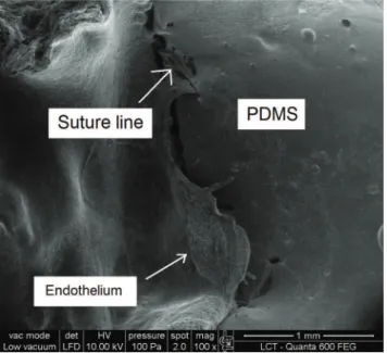
Scanning electron microscopy
Electron microscopy was used to provide additional infor-mation in this study. It was possible to observe endothelial growth from the native vessel toward the PDMS graft, a finding consistent with intimal hyperplasia (Figure 4).
’ DISCUSSION
Vascular prostheses have been used for several decades to restore blood flow at many sites. However, although the functions of such prostheses have been well established for
large vessels, the use of these prostheses is greatly limited in vessels with a diameter of less than four millimeters (9).
This study demonstrated that the patency rate for PDMS prostheses after ninety days was 57% (±11%). Nordestgaard
et al. (30) performed a similar study but used ePTFE pros-theses with a diameter of three millimeters that were also implanted into rabbit aortas. Those authors reported a patency rate of 82% after ninety days. In 2012, Zheng et al. (31) tested polycaprolactone prostheses coated with arginine-glycine-aspartic acid in the carotid arteries of ten rabbits, and the patency rate after four weeks was 100%. However, the authors discussed their short observation period and acknowl-edged that one month is not sufficient for vascular regenera-tion. If we considered only thirty days of follow-up, then we would have observed 87% patency (±6.7%) in our
experiments.
Intimal hyperplasia is the result of natural cicatrization after vascular injuries, especially in anastomotic regions, and is a major cause of vascular stenosis and occlusions (32). Anastomotic intimal hyperplasia is more likely to develop for synthetic prostheses than for vein grafts. Given that the endothelium is responsible for preservation of the intravas-cular structure (33,34), it is natural to search for graft endothelialization. One method for achieving endothelial growth relates to graft porosity. The porosity concept for vascular grafts and the growth of endothelial cells that could pave the inner surface of such grafts remain controversial in medical science.
Theoretically, pores are scaffolds for the passage and habitation of vascular cells through the graft lumen. Thus, it was thought that larger spaces between graft nodes would correspond to greater cell housing (35). However, intermedi-ate porosity is considered to be more suitable for preventing intimal hyperplasia (36). In contrast, Lumsden et al. (37) achieved satisfactory results from coating PTFE prostheses with non-porous silicone to promote a smooth and uniform surface and limit internal tissue growth beyond the trans-anastomotic region. Based on that study, we developed an
Figure 4 -Electron microscopy image showing endothelial growth
from the native vessel toward the PDMS graft, which is consistent with intimal hyperplasia.
Figure 2 - Proportion of "survivors" in the patent condition.
The patency rates of the prosthesis were 87% (±6.7%) after thirty days, 73% (±9.3%) after sixty days, 57% (±11%) after ninety days and 48% (±12%) after one hundred twenty days.
experimental model to test the hypothesis that non-porous silicone could be a suitable prosthesis material for small vessels. We believe that our patency results are consistent with the development of intimal hyperplasia in the anasto-motic region. Electron microscopy generated images compa-tible with endothelial growth from the anastomotic region. Further studies with porous silicone could demonstrate the difference between porous and non-porous grafts and elucidate the cause of intimal cell growth in synthetic prostheses.
In evaluations of prostheses, patency has always been demonstrated via contrast injection into the aorta (using arteriography performed via retrograde catheterization) and long-term monitoring. Occlusions that occur in less than thirty days are related to surgical failures, such as technical failures with respect to anastomosis, prosthesis placement, prosthesis folding or insufficient blood flow (38). In our study, we performed prosthesis evaluation for up to 150 days. Within this period, we aimed to perform arteriography as late as possible.
The use of polyester as reinforcement for the PDMS was intended to prevent tears during prosthesis suturing because PDMS has a low tear tolerance. This coating appeared to be useful because these events did not occur during any surgical procedure. Moreover, pseudoaneurysms were not observed. Although the silicone graft did not show satisfactory results with respect to long-term patency, the use of this material in other forms, such as sheets, has already been tested. An experimental study that used the same animal model produced extremely good results for patches in the aortas of rabbits (39).
The tested material exhibited characteristics that support surgical implantation: good tolerance promoted by polyester tear reinforcement, ease of postoperative removal and lack of pseudoaneurysms. However, intimal hyperplasia was a limiting factor for the patency rate. We concluded that PDMS has limited potential as an alternative small vascular prosthesis material.
’ ACKNOWLEDGMENTS
We would like to thank Professor Eduardo Massad (LIM 01) and the team in the Department of Surgery in the Faculty of Medicine at the University of São Paulo for supporting this study and treating the study animals respectfully.
’ AUTHOR CONTRIBUTIONS
All of the co-authors participated in the surgical procedures. De Luccia N also contributed to the statistical analysis and supervised the entire study.
’ REFERENCES
1. Moore WS. Vascular and endovascular surgery: a comprehensive review expert consult. 8th ed. Philadelphia: Elsevier Saunders. 2013;1020 p. 2. Sarkar S, Salacinski HJ, Hamilton G, Seifalian AM. The mechanical
properties of infrainguinal vascular bypass grafts: their role in influencing patency. Eur J Vasc Endovasc Surg. 2006;31(6):627-36, http://dx.doi.org/ 10.1016/j.ejvs.2006.01.006.
3. Moll FL, Powell JT, Fraedrich G, Verzini F, Haulon S, Waltham M, et al. Management of abdominal aortic aneurysms clinical practice guidelines of the European society for vascular surgery. Eur J Vasc Endovasc Surg. 2011;41 Suppl 1:S1-S58, http://dx.doi.org/10.1016/j.ejvs.2010.09.011. 4. Stollwerck PL, Kozlowski B, Sandmann W, Grabitz K, Pfeiffer T.
Long-term dilatation of polyester and expanded polytetrafluoroethylene tube grafts after open repair of infrarenal abdominal aortic aneurysms. J Vasc Surg. 2011;53(6):1506-13, http://dx.doi.org/10.1016/j.jvs.2011.02.028. 5. Yamamoto H, Yamamoto F, Ishibashi K, Liu KX, Yamaura G, Chida Y,
et al. Long-term outcomes of open surgical repair for ruptured iliac artery
aneurysms. Ann Vasc Surg. 2011;25(6):740-7, http://dx.doi.org/10.1016/ j.avsg.2010.11.011.
6. Johnson WC, Lee KK. A comparative evaluation of polytetrafluoro-ethylene, umbilical vein, and saphenous vein bypass grafts for femoral-popliteal above-knee revascularization: a prospective randomized Department of Veterans Affairs cooperative study. J Vasc Surg. 2000;32(2):268-77, http://dx.doi.org/10.1067/mva.2000.106944.
7. Curi MA, Skelly CL, Meyerson SL, Woo DH, Desai TR, McKinsey JF, et al. Conduit choice for above-knee femoropopliteal bypass grafting in patients with limb-threatening ischemia. Ann Vasc Surg. 2002;16(1):95-101, http://dx. doi.org/10.1007/s10016-001-0134-4.
8. SolakovićE, TotićD, SolakovićS. Femoro-popliteal bypass above knee with saphenous vein vs synthetic graft. Bosn J Basic Med Sci. 2008;8(4):367-72. 9. Cavallaro A, Sterpetti AV, DiMarzo L, Sapienza P. Worsening of pre-operative foot ischemia after occlusion of polytetrafluoroethylene femor-otibial grafts: a comparison with saphenous vein grafts. Ann Vasc Surg. 2013;27(5):634-7, http://dx.doi.org/10.1016/j.avsg.2012.05.030. 10. Loh SA, Howell BS, Rockman CB, Cayne NS, Adelman MA, Gulkarov I,
et al. Mid- and long-term results of the treatment of infrainguinal arterial occlusive disease with precuffed expanded polytetrafluoroethylene grafts compared with vein grafts. Ann Vasc Surg. 2013;27(2):208-17, http://dx. doi.org/10.1016/j.avsg.2012.04.018.
11. Davis MG, Dalen H, Austerheim AM, Gulbrandsen TF, Svendsen E, Hagen PO. Suppression of intimal hyperplasia in experimental vein grafts by oral L-arginine supplementation and single ex vivo immersion in deferoxamine manganese. J Vasc Surg. 1996;23(3):410-20, http://dx.doi. org/10.1016/S0741-5214(96)80005-X.
12. Green RM, Abbott WM, Matsumoto T, Wheeler JR, Miller N, Veith FJ, et al. Prosthetic above-knee femoropopliteal bypass grafting: five-year results of a randomized trial. J Vasc Surg. 2000;31(3):417-25, http://dx. doi.org/10.1067/mva.2000.103238.
13. Ku DN, Allen RC. Vascular Grafts. In: Bronzino JD. The biomedical engineering handbook. 2nd ed. Boca Raton: CRC Press LLC. 2000; p.128. 14. Fulton GJ, Davies MG, Barber L, Gray JL, Svendsen E, Hagen PO. Local effects of nitric oxide supplementation and suppression in the development of intimal hyperplasia in experimental vein grafts. Eur J Vasc Endovasc Surg. 1998;15(4):279-89, http://dx.doi.org/10.1016/S1078-5884(98)80030-0.
15. Martin RS, Edwards WH, Mulherin JL Jr, Edwards WH Jr, Jenkins JM, Hoff SJ. Cryopreserved saphenous vein allografts for below-knee lower extremity revascularization. Ann Surg. 1994;219(6):664-70, http://dx.doi. org/10.1097/00000658-199406000-00009.
16. Barshes NR, Ozaki CK, Kougias P, Belkin M. A cost-effectiveness analysis of infrainguinal bypass in the absence of great saphenous vein conduit. J Vasc Surg. 2013;57(6):1466-70, http://dx.doi.org/10.1016/j.jvs.2012.11.115. 17. Alcántara EM, Marshall LM, Rodrigues VV, Rosado CF. Mechanics of biomaterials: vascular graft prosthesis. Appli Engin Mech Med. 2005; 5:A1-25.
18. Cleary MA, Geiger E, Grady C, Best C, Naito Y, Breuer C. Vascular tissue engineering: the next generation. Trends Mol Med. 2012;18(7):394-404, http://dx.doi.org/10.1016/j.molmed.2012.04.013.
19. Conte MS. The ideal small arterial substitute: a search for the Holy Grail? FASEB J. 1998;12(1):43-5, http://dx.doi.org/10.1096/fj.1530-6860. 20. Kakisis JD, Liapis CD, Breuer C, Sumpio BE. Artificial blood vessel: the
Holy Grail of peripheral vascular surgery. J Vasc Surg. 2005;41(2):349-54, http://dx.doi.org/10.1016/j.jvs.2004.12.026.
21. Sanislow CA Jr, Zuidema GD. The use of silicone t-tubes in reconstructive biliary surgery in dogs. J Surg Res. 1963;3:497-502, http://dx.doi.org/ 10.1016/S0022-4804(63)80029-3.
22. Leininger RI, Mirkovitch V, Peters A, Hawks WA. Change in properties of plastics during implantation. Trans Am Soc Artif Intern Organs. 1964; 10:320-2.
23. Gladilin E, Gabrielova B, Montemurro P, Hedén P. Customized plann-ing of augmentation mammaplasty with silicon implants usplann-ing three-dimensional optical body scans and biomechanical modeling of soft tissue outcome. Aesthetic Plast Surg. 2011;35(4):494-501, http://dx.doi.org/ 10.1007/s00266-010-9642-3.
24. Oliver DW, Walker MS, Walters AE, Chatrath P, Lamberty BG. Anti-silicone antibodies and Anti-silicone containing breast implants. Brit J Plast Surg. 2000;53(5);410-4, http://dx.doi.org/10.1054/bjps.2000.3344. 25. Montague DK. Penile prosthesis implantation in the era of medical
treatment for erectile dysfunction. Urol Clin North Am. 2011;38(2):217-25, http://dx.doi.org/10.1016/j.ucl.2011.02.009.
26. Kuo ACM. Poly (dimethylsiloxane). In: Mark JE. Polymer data handbook. Oxford: Oxford University Press. 1999; p. 411-35.
27. Spiller D, Losi P, Briganti E, Sbrana S, Kull S, Martinelli I, et al. PDMS content affects in vitro hemocompatibility of synthetic vascular grafts. J Mater Sci Mater Med. 2007;18(6):1097-104, http://dx.doi.org/10.1007/ s10856-006-0067-0.
28. Simmons A, Padsalgikar AD, Ferris LM, Poole-Warren LA. Biostability and biological performance of a PDMS-based polyurethane for controlled drug release. Biomaterials. 2008;29(20):2987-95, http://dx.doi.org/10.1016/ j.biomaterials.2008.04.007.
CLINICS 2017;72(12):780-784 Implantation of an arterial substitute in rabbits
29. De Luccia N, De Luccia TPB. Prótese vascular feita em silicone. Patent PI 0704867-0, Instituto Nacional de Propriedade Industrial, BR, 2007. 30. Nordestgaard AG, Buckels JA, Wilson SE. Platelet antagonists eliminate
thromboembolic complications of small-diameter arterial prostheses. J Vasc Surg. 1987;5(1):110-7, http://dx.doi.org/10.1016/0741-5214(87) 90201-1.
31. Zheng W, Wang Z, Song L, Zhao Q, Zhang J, Li D, et al. Endotheliali-zation and patency of RGD-functionalized vascular grafts in a rabbit carotid artery model. Biomaterials. 2012;33(10):2880-91, http://dx.doi. org/10.1016/j.biomaterials.2011.12.047.
32. Fulton GJ, Davies MG, Barber L, Gray JL, Svendsen E, Hagen PO. Local effects of nitric oxide supplementation and suppression in the development of intimal hyperplasia in experimental vein grafts. Eur J Vasc Endovasc Surg. 1998;15(4):279-89, http://dx.doi.org/10.1016/S1078-5884(98)80030-0.
33. Rotmans JI, Heyligers JM, Verhagen HJ, Velema E, Nagtegaal MM, de Kleijn DP, et al. In vivo cell seeding with anti-CD34 antibodies successfully accelerates endothelialization but stimulates intimal hyperplasia in porcine arteriovenous expanded polytetrafluoroethylene grafts. Circulation. 2005;112(1): 12-8, http://dx.doi.org/10.1161/CIRCULATIONAHA.104.504407.
34. Jobst BJ, Riegger GA, Griese DP. Endothelial cell seeding fails to prevent intimal hyperplasia following arterial injury in the rat carotid model.
Cardiovasc Drugs Ther. 2009;23(5):343-53, http://dx.doi.org/10.1007/ s10557-009-6191-6.
35. Berger K, Sauvage LR, Rao AM, Wood SJ. Healing of arterial prostheses in man: its incompleteness. Ann Surg. 1972;175(1):118-27, http://dx.doi. org/10.1097/00000658-197201000-00018.
36. Golden MA, Hanson SR, Kirkman TR, Schneider PA, Clowes AW. Healing of polytetrafluoroethylene arterial grafts is influenced by graft porosity. J Vasc Surg. 1990;11(6):838-44, http://dx.doi.org/10.1016/0741-5214(90) 90082-L.
37. Lumsden AB, Chen C, Coyle KA, Ofenloch JC, Wang JH, Yasuda HK, et al. Nonporous silicone polymer coating of expanded polytetra-fluoroethylene grafts reduces graft neointimal hyperplasia in dog and baboon models. J Vasc Surg. 1996;24(5):825-33, http://dx.doi.org/ 10.1016/S0741-5214(96)70019-8.
38. Davies MG, Hagen PO. Pathophysiology of vein graft failure: a review. Eur J Vasc Endovasc Surg. 1995;9(1):7-18, http://dx.doi.org/10.1016/ S1078-5884(05)80218-7.
39. Sassaki Neto PI. Estudo experimental comparativo de remendos arteriais de polidimetilsiloxano com reforc¸o de tecido de poliéster (PDMSr) versus