An acid-sensing ion channel that
detects ischemic pain
1Departamento de Fisiologia e Biofísica, Instituto de Ciências Biológicas,
Universidade Federal de Minas Gerais, Belo Horizonte, MG, Brasil
2Vollum Institute, Oregon Health Science University, Portland, OR, USA
L.A. Naves1 and
E.W. McCleskey2
Abstract
Ischemic pain occurs when there is insufficient blood flow for the metabolic needs of an organ. The pain of a heart attack is the prototypical example. Multiple compounds released from ischemic muscle likely contribute to this pain by acting on sensory neurons that innervate muscle. One such compound is lactic acid. Here, we show that ASIC3 (acid-sensing ion channel #3) has the appropriate expres-sion pattern and physical properties to be the detector of this lactic acid. In rats, it is expressed only in sensory neurons and then only on a minority (~40%) of these. Nevertheless, it is expressed at extremely high levels on virtually all dorsal root ganglion sensory neurons that innervate the heart. It is extraordinarily sensitive to protons (Hill slope 4, half-activating pH 6.7), allowing it to readily respond to the small changes in extracellular pH (from 7.4 to 7.0) that occur during muscle ischemia. Moreover, both extracellular lactate and extracellular ATP increase the sensitivity of ASIC3 to protons. This final property makes ASIC3 a “coincidence detector” of three molecules that appear during ischemia, thereby allowing it to better detect acidosis caused by ischemia than other forms of systemic acidosis such as hypercapnia.
Correspondence
L.A. Naves
Departamento de Fisiologia e Biofísica, ICB-UFMG Avenida Antônio Carlos, 6627 31270-901 Belo Horizonte, MG Brasil
Fax: +55-31-3499-2924 E-mail [email protected]
Presented at the XIX Annual Meeting of the Federação de Sociedades de Biologia Experimental, Águas de Lindóia, SP, Brazil, August 25-29, 2004.
Received February 23, 2005 Accepted May 19, 2005
Key words
•Pain •Ischemia
•Dorsal root ganglia •Acid-sensing ion channels •Lactic acidosis
Ischemic pain
Three diseases are characterized by is-chemic pain: angina, intermittent claudica-tion, and sickle cell anemia. Angina is the chest pain caused by coronary vascular dis-ease and intermittent claudication the pain caused by peripheral vascular disease. Mis-shapen red blood cells can block blood flow in sickle cell anemia, causing severe, unpre-dictable bouts of pain. Ischemic pain feels distinctly different from other forms of pain such as tissue damage, burning, or inflam-mation. The sensation can be experienced simply by running or swimming at a sprint for about 40 or 50 s. The severity of the
muscle pain will stop you from proceeding further. It is this sensation that appears unex-pectedly in sickle cell or angina patients, or upon mild exercise, such as walking, in a patient with intermittent claudication. Middle distance athletes and thoroughbred racehorses train to maximize the time they can sprint before being stopped by muscle ischemia.
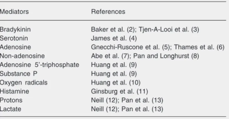
from working muscle that accumulate when there is insufficient blood flow and then act upon certain nociceptive sensory neurons that innervate the muscle. Much of the ensu-ing effort in the field has asked what the mediating compounds and their receptors on sensory neurons are. Table 1 lists the most widely considered compounds that are re-ported to cause pain when perfused into the hearts of intact animals (2-13).
The lactic acid paradoxes
Lactic acid is produced by anaerobic metabolism when muscle gets insufficient oxygen. This and the fact that acid is com-monly considered a painful stimulus suggest that lactic acid mediates ischemic pain. But two experimental facts argue otherwise. First, extracellular pH drops to about neutral dur-ing ischemic pain, but not much further (14,15). Neutral pH solutions surely do not cause pain when poured onto a skinned knee, so the field has been reluctant to believe that such a modest pH change could be the source of ischemic pain. The second fact is that similar pH changes can occur during meta-bolic problems involving the lungs or kid-ney. However, metabolic acidosis is not ac-companied by angina-like chest pain.
Nevertheless, a compelling experiment by Pan et al. (13) argues that pH must play a role. These investigators measured the elec-trical activity of sensory nerve fibers coming
from the heart and then blocked blood flow to a coronary artery. The resulting increase in nerve firing was greatly diminished if a high concentration of a pH buffer was pres-ent when the artery was blocked. Evidpres-ently, the buffer intercepted the protons before they reached their receptor on the sensory axon and thereby diminished the resulting pain signal. But, what receptor works to detect the small change in pH and how can it distinguish between a pH change that is caused by ischemia from one caused by systemic metabolic problems?
Acid-sensing ion channels
The results presented in this paper will demonstrate that acid-sensing ion channel number 3 (ASIC3) has the appropriate physi-cal properties and expression pattern to se-lectively sense the acid that accompanies ischemia. ASIC were discovered in the early 1980’s by Oleg Krishtal and his colleagues in Kiev (16-18). These investigators demon-strated that acid triggered the opening of ion channels in sensory neurons and showed that there were several kinetically distinct acid-sensing channels expressed on differ-ent sensory neurons. Krishtal demonstrated the basic properties of the channels: gating by acid, sodium-selective pores, and weak block by amiloride. Further work also showed the presence of ASIC on neurons of the central nervous system (19,20).
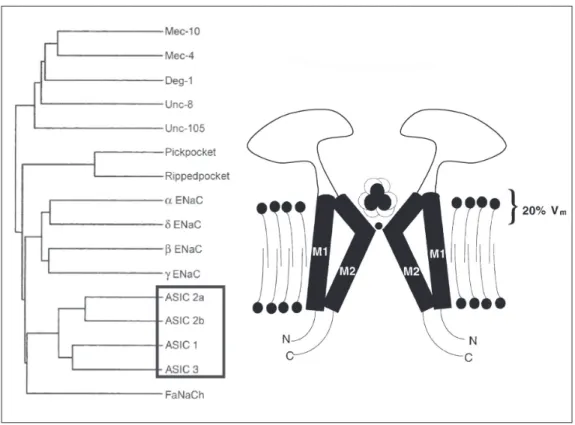
Largely dormant for a decade after Krishtal’s work (21), the field was resur-rected in 1997 when Michel Lazdunski’s laboratory cloned an acid-sensing channel and demonstrated that it is a member of a large family of channels that have been ex-tensively studied. The family is commonly called the degenerins/epithelial sodium chan-nels (ENaC) (18) and is characterized by amiloride sensitivity and sodium selectivity (Figure 1). These channels have no homol-ogy to voltage-gated sodium channels but, instead, have about 500 amino acids, two
Table 1. Putative mediators of ischemic pain.
Mediators References
Bradykinin Baker et al. (2); Tjen-A-Looi et al. (3)
Serotonin James et al. (4)
Adenosine Gnecchi-Ruscone et al. (5); Thames et al. (6) Non-adenosine Abe et al. (7); Pan and Longhurst (8) Adenosine 5'-triphosphate Huang et al. (9)
Substance P Huang et al. (9)
Oxygen radicals Huang et al. (10)
Histamine Ginsburg et al. (11)
Protons Neill (12); Pan et al. (13)
putative transmembrane domains, intracel-lular N and C terminals, and a substantial extracellular domain. At least three subunits must bind together to form a functional chan-nel. The most studied members of this fam-ily are the ENaCs. ENaCs are constitutively active, sodium selective channels that are expressed on epithelia and support selective sodium transport across them. Degenerin channels are expressed in C. elegans worms. A mutation that causes them to be overactive causes degeneration of mechanosensing neu-rons, this being one reason why they are considered to be mechanosensing channels in worms.
Extensive cloning work has found four distinct genes with appropriate homology to
be considered in the ASIC family (22). There are also two known splice variants of ASIC1 and two of ASIC2. ASIC4 forms no func-tional channel and it takes fairly extreme acid (pH 5 and below) to open ASIC2. But ASIC1 and ASIC3 are opened by pH changes that are clearly relevant physiologically. ASIC3 is expressed predominately in the sensory peripheral nervous system and is not seen in the central nervous system. ASIC1 is the predominant ASIC in the central nervous system and is also present in sensory neu-rons. ASIC2 is present both in the central and peripheral nervous systems and forms heteromultimers with ASIC1 and ASIC3, causing detectable changes in either kinetics or pharmacology.
A method to isolate ischemia-sensing neurons
Our contributions to the study of ischemic pain use the single-cell patch clamp method applied to dissociated sensory neurons. The most significant disadvantage of this ap-proach compared to the traditional whole animal preparations is that one cannot readily distinguish the sensory modality of indi-vidual neurons once they have been dissoci-ated from the animal. We addressed this problem with an in vivo retrograde labeling strategy (Figure 2).
The rationale of the method takes
advan-tage of the fact that the heart is a most unusual sensory organ. Pain is the only con-scious sensation that ever arises from the heart muscle and the only thing that can cause the pain is ischemia. Cutting or burn-ing heart muscle causes no pain, but block-ing a blood vessel does. There may also be subconscious sensory signals involved in cardiovascular reflexes. But it can be said with certainty that most, and perhaps all, sensory neurons that innervate heart muscle are specialized to detect ischemia. Thus, if we could label them, we would have labeled ischemia sensors.
We injected into the pericardial space a
lipid-soluble dye called 1,1'-dioctadecyl-3,3,3',3'-tetramethylindocarbocyanine per-chlorate (DiI). DiI intercalates into lipid membranes at the site of injection, including local nerve endings. The resulting fluores-cent membrane gets endocytosed and the intracellular fluorescent vesicles then use axoplasmic transport to travel to the cell body, located just outside the spinal cord at the level of the upper thoracic dorsal root ganglia. Therefore, when we dissect and dissociate dorsal root ganglia several weeks after injecting the dye, we find some fluores-cent neurons that must have innervated the heart muscle. We took these to be special-ized ischemia sensors (23).
Ischemia sensors express high levels of ASIC3
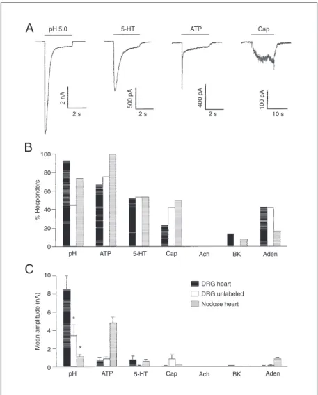
Do ischemia-sensing neurons have prop-erties that are different from those of other types of sensory neurons? We examined their array of ion channels using patch clamp methods. The most remarkable property is that they all have extraordinarily high cur-rents evoked by decreasing pH (Figure 3). These currents dwarf those caused by other pain-causing agents, they are present in vir-tually all of the labeled neurons, and they are relatively rare in unlabeled neurons (23). This implies that the acid-triggered ion chan-nel is important for detecting ischemia.
Which of the various ASICs mediate the current on the cardiac sensory neurons? To answer this, we expressed cDNA for each of the ASICs in COS7 cells and compared the functional properties of these known clones to the functional properties of the native channels in the cardiac sensory neurons (25). ASIC3 and the native channel were identical in all respects, whereas all other ASICs var-ied dramatically from the native channel. The most significant properties are: 1) the extremely steep activation curve (Hill slope = 4; half-activation pH 6.7); 2) pH at which half of the channels are desensitized (pH
7.2); 3) the rate of recovery from desensiti-zation (ASIC3 and the native current re-cover quickly (less than 5 s), whereas other ASICs take about 20 s); 4) calcium perme-ability and block. We conclude that the bulk of the acid-triggered current in cardiac sen-sory neurons is carried through ion channels that include ASIC3. We have not yet
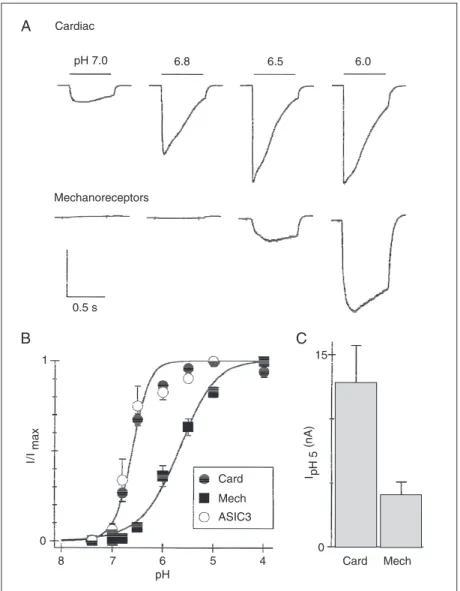
Figure 4. Labeled cardiac dorsal root ganglion neurons have highly sensitive acid-sensing ion channels (ASICs) which are similar to ASIC3 and distinct from those expressed by mechanoreceptors from the mesencephalic nucleus of the trigeminal nerve. A, Currents evoked from a cardiac afferent or a mechano-sensing afferent by pulses to the indicated pH from pH 8.0. Vertical scales: 8 nA (upper), 1 nA (lower). B, Mean (± SEM) fractional current vs pH for cardiac cells (Card), mechanoreceptors (Mech) and cells expressing ASIC3. C, Mean (± SEM) amplitude of currents evoked by pH 5. Reproduced, with permission, from Sutherland et al. (25), National Academy of Sciences of the United States.
mined if these channels are homomers of ASIC3 or heteromers formed between ASIC3 and other ASICs.
Paradox number 1 answered: extreme acid sensitivity of ASIC3
Recall the lactic acid paradoxes men-tioned above. The first was that the
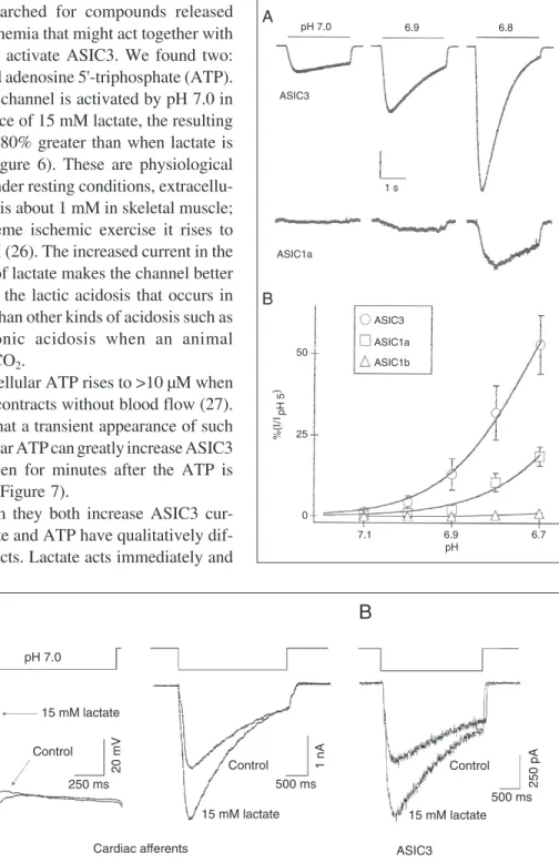
extracel-lular pH changes that occur during ischemia are so small (1/2 pH unit) that people have doubted that this could be sufficient to trig-ger pain. ASIC3 answers this issue because it has extraordinarily high pH sensitivity in the range of extracellular pH that occurs during a heart attack (25). Figure 4 shows three activation curves. The ones for ASIC3 and the channel native to cardiac sensory neurons exactly overlap; another for a dif-ferent kind of sensory neuron is far less sensitive to pH. Figure 5 shows a higher resolution view of the pH range, 7.1 to 6.7, that can occur during heart attack. ASIC3 reaches about 50% activation over this range, whereas other cloned ASICs are not so sen-sitive. The Hill coefficient for ASIC3 is about 4; this more or less means that ASIC3 is four times as sensitive as a pH meter over this critical range of pH. Thus, we appear to have an answer to paradox number 1: ische-mia-sensing neurons express high levels of ASIC3, which readily respond to the small pH changes that accompany a heart attack.
Paradox number 2 answered: coincident detection of lactate, ATP and acid
extracellular pH greatly diminishes axon fir-ing durfir-ing artery occlusion? The simple in-terpretation is that protons must be neces-sary to activate the sensory axons, but can-not by themselves be sufficient. In other words, something must act together with protons to activate the axons.
We searched for compounds released during ischemia that might act together with protons to activate ASIC3. We found two: lactate and adenosine 5'-triphosphate (ATP). When the channel is activated by pH 7.0 in the presence of 15 mM lactate, the resulting current is 80% greater than when lactate is absent (Figure 6). These are physiological values. Under resting conditions, extracellu-lar lactate is about 1 mM in skeletal muscle; after extreme ischemic exercise it rises to 15-30 mM (26). The increased current in the presence of lactate makes the channel better at sensing the lactic acidosis that occurs in ischemia than other kinds of acidosis such as the carbonic acidosis when an animal breathes CO2.
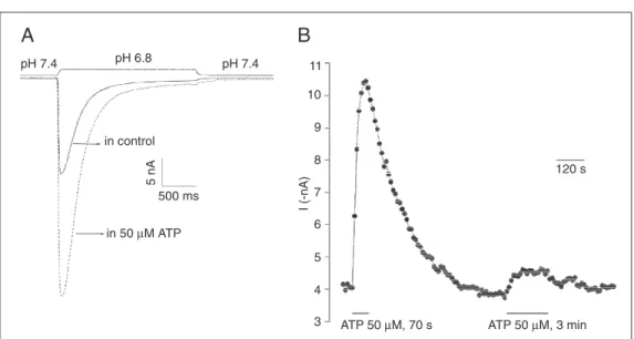
Extracellular ATP rises to >10 µM when a muscle contracts without blood flow (27). We find that a transient appearance of such extracellular ATP can greatly increase ASIC3 current even for minutes after the ATP is removed (Figure 7).
Though they both increase ASIC3 cur-rent, lactate and ATP have qualitatively dif-ferent effects. Lactate acts immediately and
Figure 5. Acid-sensing ion chan-nel 3 (ASIC3) opens at ischemic pH. A, Currents evoked from cells expressing either ASIC3 or ASIC1a by steps to pH 7.0, 6.9, and 6.8. The vertical scale bar represents 10% of the current evoked by pH 5 (2 nA ASIC3; 46 pA ASIC1a). B, Average cur-rents of the indicated clones nor-malized to the value at pH 5.0 and fitted with the Hill equation. Hill coefficients are 4.3 (ASIC3, pH0.5 6.7) and 3.9 (ASIC1a, pH0.5 6.4). Reproduced, with permission, from Sutherland et al. (25), National Academy of Sciences of the United States.
Figure 6. Lactate enhances the activity of acid-sensing ion chan-nels (ASICs). A, Representative voltage (left) and current (right) recordings from a cardiac affer-ent dorsal root ganglion neuron briefly exposed to pH 7.0 (rest-ing pH = 8.0) in the presence or absence of 15 mM lactate. B, Representative current record-ings from a cell expressing ASIC3 using the same protocol as in A. Reproduced, with per-mission, from Immke and Mc-Cleskey (24), Nature Publishing Group [www.nature.com/neuro/].
ASIC3 whereas ATP acts on some cells but not others. We find that lactate acts by alter-ing the basic gatalter-ing of the channel, which, surprisingly, involves binding of calcium in addition to protons (28). In contrast, the ATP binding site must not be the ASIC3
channel itself; there are a variety of puriner-gic receptors, some of which are ion chan-nels and some of which are G-protein-coupled receptors. We are presently asking if any of these known receptors might medi-ate ATP modulation of ASIC3.
Figure 7. Slow and persistent adenosine 5'-triphosphate (ATP) modulation of ASIC3-like cur-rents in dorsal root ganglion neu-rons. A, Representative current recordings with changing exter-nal pH from 7.4 to 6.8, before (control) and after 50 µM ATP application. B, Time course of the ATP effect in one cell. Cur-rent sizes before, during and af-ter ATP application for 70 s. A second ATP application did not enhance the acid current to the same extent. From Fierro LP, Naves LA, Spelta V and Mc-Cleskey EW (unpublished data).
References
1. Lewis T (1932). Pain in muscular ischemia. Archives of Internal Medicine, 49: 713-727.
2. Baker DG, Coleridge HM, Coleridge JC et al. (1980). Search for a cardiac nociceptor: stimulation by bradykinin of sympathetic afferent nerve endings in the heart of the cat. Journal of Physiology, 306: 519-536.
3. Tjen-A-Looi SC, Pan HL & Longhurst JC (1998). Endogenous brady-kinin activates ischaemically sensitive cardiac visceral afferents through kinin B2 receptors in cats. Journal of Physiology, 510: 633-641.
4. James TN, Rossi L & Hageman GR (1988). On the pathogenesis of angina pectoris and its silence. Transactions of the American Clini-cal and ClimatologiClini-cal Association, 100: 81-99.
5. Gnecchi-Ruscone T, Montano N, Contini M et al. (1995). Adenosine activates cardiac sympathetic afferent fibers and potentiates the excitation induced by coronary occlusion. Journal of the Autonomic Nervous System, 53: 175-184.
6. Thames MD, Kinugawa T & Dibner-Dunlap ME (1993). Reflex sympathoexcitation by cardiac sympathetic afferents during myo-cardial ischemia. Role of adenosine. Circulation, 87: 1698-1704. 7. Abe T, Morgan D, Sengupta JN et al. (1998). Attenuation of
ische-mia-induced activation of cardiac sympathetic afferents following brief myocardial ischemia in cats. Journal of the Autonomic Nervous System, 71: 28-36.
8. Pan HL & Longhurst JC (1995). Lack of a role of adenosine in
activation of ischemically sensitive cardiac sympathetic afferents. American Journal of Physiology, 269: H106-H113.
9. Huang MH, Horackova M, Negoescu RM et al. (1996). Polysensory response characteristics of dorsal root ganglion neurones that may serve sensory functions during myocardial ischaemia. Cardiovascu-lar Research, 32: 503-515.
10. Huang HS, Pan HL, Stahl GL et al. (1995). Ischemia and reperfu-sion-sensitive cardiac sympathetic afferents: influence of H2O2 and hydroxyl radicals. American Journal of Physiology, 269: H888-H901. 11. Ginsburg R, Bristow MR, Kantrowitz N et al. (1981). Histamine provocation of clinical coronary artery spasm: implications concern-ing pathogenesis of variant angina pectoris. American Heart Jour-nal, 102: 819-822.
12. Neill WA (1968). Myocardial hypoxia and anaerobic metabolism in coronary heart disease. American Journal of Cardiology, 22: 507-515.
13. Pan HL, Longhurst JC, Eisenach JC et al. (1999). Role of protons in activation of cardiac sympathetic C-fibre afferents during ischaemia in cats. Journal of Physiology, 518: 857-866.
14. Cobbe SM & Poole-Wilson PA (1980). The time of onset and sever-ity of acidosis in myocardial ischaemia. Journal of Molecular and Cellular Cardiology, 12: 745-760.
15. Reeh PW & Steen KH (1996). Tissue acidosis in nociception and pain. Progress in Brain Research, 113: 143-151.
nerve cell membrane. Neuroscience, 5: 2325-2327.
17. Krishtal OA & Pidoplichko VI (1981). A “receptor” for protons in small neurons of trigeminal ganglia: possible role in nociception. Neuro-science Letters, 24: 243-246.
18. Krishtal O (2003). The ASICs: signaling molecules? Modulators? Trends in Neurosciences, 26: 477-483.
19. Ueno S, Nakaye T & Akaike N (1992). Proton-induced sodium current in freshly dissociated hypothalamic neurones of the rat. Journal of Physiology, 447: 309-327.
20. Akaike N & Ueno S (1994). Proton-induced current in neuronal cells. Progress in Neurobiology, 43: 73-83.
21. Waldmann R, Champigny G, Bassilana F et al. (1997). A proton-gated cation channel involved in acid-sensing. Nature, 386: 173-177.
22. Waldmann R & Lazdunski M (1998). H(+)-gated cation channels: neuronal acid sensors in the NaC/DEG family of ion channels. Current Opinion in Neurobiology, 8: 418-424.
23. Benson CJ, Eckert SP & McCleskey EW (1999). Acid-evoked cur-rents in cardiac sensory neurons: A possible mediator of myocardial ischemic sensation. Circulation Research, 84: 921-928.
24. Immke DC & McCleskey EW (2001). Lactate enhances the acid-sensing Na+ channel on ischemia-sensing neurons. Nature Neuro-science, 4: 869-870.
25. Sutherland SP, Benson CJ, Adelman JP et al. (2001). Acid-sensing ion channel 3 matches the acid-gated current in cardiac ischemia-sensing neurons. Proceedings of the National Academy of Sci-ences, USA, 98: 711-716.
26. Cohen RD & Woods HF (1983). Lactic acidosis revisited. Diabetes, 32: 181-191.
27. Forrester T (1972). An estimate of adenosine triphosphate release into the venous effluent from exercising human forearm muscle. Journal of Physiology, 224: 611-628.