Update on hypertrophic scar treatment
Felipe Bettini Rabello,ICleyton Dias Souza,IIJayme Adriano Farina Ju´niorIII
IUniversidade de Sa˜o Paulo, Faculdade de Medicina de Ribeira˜o Preto, Ribeira˜o Preto/SP, Brazil.IIUniversidade de Sa˜o Paulo, Faculdade de Medicina de
Ribeira˜o Preto, Programa de Po´s-Graduac¸a˜o da Clinica Ciru´rgica, Ribeira˜o Preto/SP, Brazil.IIIUniversidade de Sa˜o Paulo, Hospital das Clı´nicas da Faculdade
de Medicina de Ribeira˜o Preto, Departamento de Cirurgia e Anatomia, Divisa˜o de Cirurgia Pla´stica, Ribeira˜o Preto/SP, Brazil.
Scar formation is a consequence of the wound healing process that occurs when body tissues are damaged by a physical injury. Hypertrophic scars and keloids are pathological scars resulting from abnormal responses to trauma and can be itchy and painful, causing serious functional and cosmetic disability. The current review will focus on the definition of hypertrophic scars, distinguishing them from keloids and on the various methods for treating hypertrophic scarring that have been described in the literature, including treatments with clearly proven efficiency and therapies with doubtful benefits. Numerous methods have been described for the treatment of abnormal scars, but to date, the optimal treatment method has not been established. This review will explore the differences between different types of nonsurgical management of hypertrophic scars, focusing on the indications, uses, mechanisms of action, associations and efficacies of the following therapies: silicone, pressure garments, onion extract, intralesional corticoid injections and bleomycin.
KEYWORDS: Hypertrophic Scar; Review; Pathological Scars; Update.
Rabello FB, Souza CD, Farina Jr JA. Update on hypertrophic scar treatment. Clinics. 2014;69(8):565-573.
Received for publication onSeptember 13, 2013;First review completed onDecember 2, 2013;Accepted for publication onFebruary 10, 2014 E-mail: [email protected]
Tel.: 55 16 3602-2593/2961
& INTRODUCTION
Hypertrophic scars (HTSs) are defined as visible and elevated scars that do not spread into surrounding tissues and that often regress spontaneously (1). These scars are characterized by proliferation of the dermal tissue, with excessive deposition of fibroblast-derived extracellular matrix (ECM) proteins and especially collagen, over long periods and by persistent inflammation and fibrosis (2).
Numerous methods have been described for the treat-ment of abnormal scars, but to date, the optimal treattreat-ment method has not been established. A wide variety of treatments have been advocated for HTSs. Among these treatments are surgical excision with or without grafting (1), pressure therapy (3), intralesional interferon (4), topical and intralesional corticosteroids (5), intralesional bleomycin (6), laser therapy (7), silicone gel sheeting (8), onion extract gel and other therapies directed at collagen synthesis (9).
Distinguishing hypertrophic scars from keloids
When faced with patients seeking treatment for patholo-gical scars, many physicians have difficulty in differentiating
HTSs from keloids; therefore, it is crucial to establish criteria to distinguish them.
HTSs are usually raised, although rarely elevated more than 4 mm above the skin; red or pink in color; hard; and pruritic. Additionally, these scars do not extend beyond the general geographic margins of the wound and tend to regress over time (10) (Figure 1). HTSs primarily contain type III collagen, oriented parallel to the epidermal surface and with abundant nodules and large extracellular collagen filaments (11).
In contrast, keloids continue to evolve over time, without a quiescent or regressive phase and do infiltrate the surrounding tissue (Figure 2). Keloids appear as firm, mildly tender, bosselated tumors with a shiny surface and occasional telangiectasia. The epithelium is thinned and there may be focal areas of ulceration. The color is pink to purple and may be accompanied by hyperpigmentation (12). The initial lesions are erythematous and become brownish red, followed by paling as they age. The lesions preferentially develop on the earlobes, shoulders and presternal skin; are void of hair follicles and other glands; and usually project above the level of the surrounding skin (13). Keloids are primarily composed of abnormally thick, irregularly branched and septal disorganized type I and III collagen bundles without nodules and with excess myofi-broblasts (11) and overproduction of multiple fibroblast proteins, indicating the persistence of wound healing or even a failure to downregulate wound-healing cells. In addition, keloids are not triggered to enter the final phase of wound healing, or the ‘‘remodeling’’ phase, whereas HTSs will eventually do so (14).
Copyrightß2014CLINICS– This is an Open Access article distributed under the terms of the Creative Commons Attribution Non-Commercial License (http:// creativecommons.org/licenses/by-nc/3.0/) which permits unrestricted non-commercial use, distribution, and reproduction in any medium, provided the original work is properly cited.
No potential conflict of interest was reported.
Distinguishing HTSs from keloids histopathologically is occasionally difficult because thickened hyalinized collagen (keloidal collagen), the hallmark of keloids, is not always detectable and becausea-smooth muscle actin (a-SMA), a differentiating marker of HTSs, is variably expressed in both types of scars (15).
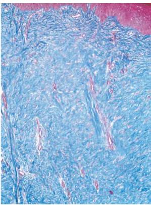
The histopathological findings most commonly observed in HTSs are flattening of the epidermis and replacement of the papillary and reticular dermis by scar tissue with prominent vertically oriented blood vessels (Figure 3). In keloids, there is no flattening of the overlying epidermis, no scarring of the papillary dermis, the presence of a significant amount of keloidal collagen, an absence of prominent vertically oriented blood vessels and the presence of a significant disarray of fibrocollagenous fascicles (Figure 4) (15).
Differential and exclusive diagnosis
The differential and exclusive diagnosis of diseases that are similar to keloids and HTSs is important because various types of malignant tumors resemble these scars (16–19). For example, malignant dermatofibrosarcoma pro-tuberans (DFSP) tumors have been mistaken for keloids or HTSs (16,17).
Nodular scleroderma and keloidal scleroderma are rare benign tumors with lesions that clinically resemble keloids (20). The skin lesions are characteristically nodules or plaques, hard in consistency, and nontender, with a predilection for the superior portions of the thorax and sparing of the face and hands (21).
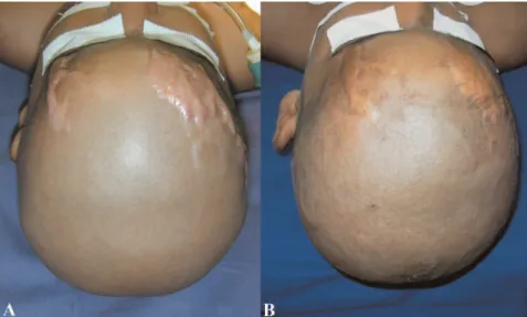
Gonzalez-Vela et al. (22) described keloids and HTSs as differential diagnoses of sclerotic neurofibroma. Sclerotic neurofibroma is differentiated from a keloid by an absence Figure 1 -Hypertrophic scar regression in a burned child after four years (A and B). Hypertrophic scars are usually raised, although rarely elevated more than 4 mm above the skin; red or pink in color; hard; and pruritic. Additionally, these scars do not extend beyond the general geographic margins of the wound and tend to regress over time.
of previous surgical excision and by the positivity of the sclerotic neurofibroma cells for the protein S100 (22).
Other rare, benign keloid and HTS-like diseases include keloidal granuloma (23), erythema elevatum diutinum (24), infantile digital fibromatosis (25), dermatofibroma,(26) penile edema (27), pseudoangiomatous hyperplasia (28) and lichen sclerosus (29).
Hair folliculitis occasionally leads to hypertrophic scar-ring (30) but more often leads to progressive folliculitis caused by bacterial or fungal infection. The nape is an area
of predilection for hair folliculitis. Treatment of any infection should be primary and steroid injection is contra-indicated for infection. Moreover, a fungus may cause a skin nodule that mimics a keloid (31). Thus, examination for fungal infection should be conducted in cases of nape hair folliculitis, even if the nodule appears to be a keloid or HTS. In conclusion, the following issues are considered important in the examination of a keloid or HTS: a biopsy should be conducted in anomalous cases because malignant disease may be the original or secondary problem, steroid injection should be performed only after careful considera-tion because malignancy or infecconsidera-tions may be present, making a careful differential diagnosis is particularly challenging in African-Americans because the skin and the tumor colors are often similar and the presence of bacterial or fungal infection should be investigated (32).
Demographics
The majority of individuals who develop HTSs and keloids are young, with ages ranging from 10 to 30 years old. The elderly rarely develop these lesions (33). This observation is partly attributed to the following facts: young individuals are more prone to trauma; their skin generally possesses more elastic fibers, resulting in greater tension; and the rate of collagen synthesis is greater in younger individuals (34). Keloids are more common in patients with darker skin, with an incidence of 4.5% to 16% in the black and Hispanic populations (34).
HTSs are a common complication of burn injury. In the developed world, approximately four million patients acquire scars due to burns each year and the incidence is even greater in developing countries (4). Previous studies have reported diverging incidences of hypertrophic scar-ring, with incidence rates varying from 40% to 94% following surgery and from 30% to 91% following burns (35,36).
Etiology
Wound healing is classically divided into four stages: hemostasis, inflammation, proliferation and tissue remodel-ing. In these four stages, there are complicated interactions within a complex network of profibrotic and antifibrotic molecules, such as growth factors, proteolytic enzymes and ECM proteins (37). Each molecule has its own function in the different phases of the wound healing process. As soon as an injury occurs, the process of hemostasis begins and the bleeding is controlled by the aggregation of platelets at the site of injury. The subsequent formation of a fibrin clot helps to stop the bleeding and provides a scaffold for the attachment and proliferation of cells. Growth factors and cytokines are mainly secreted by inflammatory cells and contribute to the initiation of the proliferative phase of wound healing. Later, angiogenesis and collagen synthesis, followed by tissue remodeling complete the stages of the wound healing process.
The delicate balance of the deposition and degradation of ECM proteins is disrupted when either excessive produc-tion of collagen, proteoglycans and fibronectin by fibro-blasts or deficient degradation and remodeling of the ECM occur (38). HTSs occur when the inflammatory response to injury is prolonged, leading to the pathological character-istics of HTSs, including increased vascularization, hyper-cellularity, excessive collagen deposition and a decrease in small leucine-rich proteoglycans (SLRPs) (39,40).
Figure 3 -The histology of hypertrophic scars is characterized by replacement of the papillary and reticular dermis by scar tissue with prominent vertically oriented blood vessels. The fibrous bundles are parallel and horizontal in the upper dermis. (Masson’s trichrome, 100X).
HTS tissue contains enhanced amounts of fibroblasts that exhibit an altered phenotype and higher expression of transforming growth factor beta-1 (TGF-b1) than normal fibroblasts do (41). An increase in or prolonged activity of TGF-b1 leads to overproduction and excess deposition of collagen by fibroblasts, often resulting in HTS formation (42). Fibroblasts in HTSs may differentiate into myofibro-blasts and account for increased ECM synthesis and contraction of tissue. These cells have a particular pheno-type that differs from that of fibroblasts based on their expression ofa-SMA (43). Myofibroblasts in HTSs are less sensitive to apoptotic signals, coupled with an ability to produce more collagen and play an important role in HTS formation (43).
Clinical manifestations
The clinical manifestations of HTSs are variable and correlate with a variety of causes that initiate HTS formation. HTSs can develop anywhere on the body. In contrast, keloids preferentially develop on the earlobes, shoulders and presternal skin; are void of hair follicles and other glands; and usually project above the level of the surrounding skin (13). Curiously, pathological scars are rare on the scalp. Farina Jr et al. recently described a casuistic study of 413 surgical procedures involving collection of thin skin grafts from the scalp, without development of HTSs or keloids among 295 cases over a period of ten years (44).
Hypertrophic scarring following surgical procedures, trauma and especially burns is a significant concern for patients and a challenging problem for clinicians because it can be painful, pruritic, erythematous, raised and cosmeti-cally unacceptable. A previous study reported that the most common and distressing complications in burn patients who developed HTSs were abnormal appearance (75.2%), pruritus (73.3%) and pain (67.6%) (45). The cause of pruritus in HTSs and keloid scars is not yet well characterized, but recent studies have indicated the probable involvement of direct activation of opioid receptors identified in the skin (46).
Prevention and non-surgical treatment
There is evidence suggesting that increased mechanical tension can initiate HTS formation (47). Based on this hypothesis, it makes sense to minimize mechanical forces after surgery. Surgical excision scars should be positioned along, rather than across, relaxed skin tension lines whenever possible. An appropriate strength, depth and number of sutures should ensure that the risk of dehiscence is minimized.
Inflammation is also known to contribute to hypertrophic scarring, (49) and every attempt to minimize the inflamma-tory response should be made by ascertaining clean surgery and good wound care to prevent infection thereafter. Using inert suture materials is also important in this context (48). In patient candidates for skin grafts, the donor site must be well chosen by the surgeon in consultation with the patient to try to hide or avoid HTSs or keloids. In burn patients, the corresponding author believes that using the scalp as a source of thin skin grafts can reduce the level of visible aesthetic deformities at donor sites in patients who have already suffered the immense trauma that being a burn victim entails.
Conceptually and practically, treatment and prevention regimens can be similar and the following section presents
the clinical data for both. Early diagnosis can considerably affect the outcome. There is evidence that the most successful non-surgical treatment of an HTS or keloid is achieved when the scar is immature and the overlying epithelium is intact, although further studies are necessary to confirm this concept (50).
Silicone
Silicone gel sheeting (SGS) has been widely used in clinical practice for the treatment of HTSs since the early 1980s. There is good evidence of the efficacy of the SGS, which has become standard practice among plastic sur-geons (51).
Although gel sheeting is effective for HTS treatment, patient compliance may not be satisfactory for the following reasons: skin reaction to the tape used for fixation; excessive sweating; difficulty in its application; and the visibility of the treatment in the case of scars located in visible areas, such as the face (52).
In contrast, silicone gel does not require fixation and is nearly invisible when dry, suggesting that it could be especially useful in visible areas (52). However, there are certain problems in its application. For example, silicone gel requires multiple applications in a day and one must wait long enough for drying because the dressing may be smudged. Friction by clothes may also contribute to early removal of the silicone film (51). Because of these problems, silicone gel use may not always be practical.
Karagoz et al. found no statistically significant difference between silicone gel and silicone gel sheet groups when their scores on the Vancouver Scar Scale after treatment were compared. This finding suggests that silicone gel is most likely as effective as SGS for the treatment of HTSs (52).
The mechanism of action of topical silicone materials in the treatment of HTSs is not well understood and various mechanisms of action have been proposed. It has been suggested that the materials’ therapeutic effect is not due to pressure, but rather to decreased scarring via wound hydration. There is evidence that SGS affects the hydration status of the scar by decreasing the water vapor transmis-sion rate to nearly half that of normal skin, causing a buildup of moisture on the skin surface under the silicone sheet (53). This evidence suggests that the stratum corneum acts as a water reservoir, with fluid accumulating below the gel, although when visualized directly, this phenomenon is not evident (54). Hydration and occlusion therefore seem to be the principal modes of SGS action and increased skin hydration is most likely responsible for decreased capillary activity, reduced hyperemia and reduced collagen deposi-tion (55). Furthermore, altered hydradeposi-tion is thought to cause electrostatic changes that influence collagen deposition and remodeling within the scar (56). The static electricity generated by friction has also been proposed as a plausible reason for silicone’s anti-scarring effects (57).
compliance with SGS, resulting in a better scar outcome (60). Obviously, the key to the success of this therapy is to ensure that hygienic precautions are taken, particularly when it is used in combination with pressure in children or in warm weather or climates (60). It must be noted, however, that complications increase with the use of combined pressure and SGS therapy (60).
Applying silicone gel twice daily or wearing SGS 12 to 24 h per day for 6 to 12 months, with temporary interrup-tion when adverse effects appear, is recommended (61).
Pressure garments
Using mechanical compressive force exerted by pressure garments to treat HTSs in burn patients was first described in 1860 (62). It was only in the 1960s that this treatment became standard in several burn centers to accelerate the remodeling phase of wound healing (62). Prophylactic pressure is recommended in burn patients if spontaneous closure of the wound takes longer than 10 to 14 days and in those requiring grafting (2).
Currently, elastic compression using elastic garments is the predominant means of both prophylaxis and treatment for HTSs (63), despite controversial evidence-based data about their value in reducing the prevalence or magnitude of scarring (63). In fact, studies investigating pressure garments have found no significant difference between treatments involving the use of high-pressure garments, lower-pressure garments, or no pressure at all (64). Others, however, claim that pressure therapy achieves HTS regres-sion success rates of 60% to 85% (63), without any conclusive evidence.
To date, the working mechanism of pressure and the way that pressure positively influences the development and maturation of HTSs are not fully understood and explana-tions remain hypothetical. However, many studies have been performed to try to explain the possible mechanisms of action, exploring theories based on hypoxia, biochemical changes, and cellular and collagenous influences. Certain valuable evidence suggests that pressure controls collagen synthesis by limiting the supply of blood, oxygen, and nutrients to the scar tissue (65); reduces collagen production to the levels found in normal scar tissue more rapidly than the natural maturation process does; encourages the realignment of collagen bundles that are already present (66); partly restores the ECM organization observed in normal scarring; and induces the disappearance of fibro-genica-SMA-expressing myofibroblasts and vascular cells, most likely by apoptosis (67).
Additionally, certain studies have demonstrated that mechanical compression directly modulates the remodeling phase of wound healing, altering the release and activity of matrix metalloproteinase (MMP)-28 in HTSs and inducing a significant reduction in the protein’s presence in keratino-cytes in HTSs (68). Moreover, it has been suggested that pressure acts by accelerating the remission phase of the postburn reparative process (66).
Currently, the recommendations for the clinical use of pressure garments are restricted to deep dermal wounds that have healed spontaneously over weeks, grafted wounds surrounded by a deep dermal wound that was permitted to heal spontaneously over weeks, wounds in children and young adults, wounds in individuals with dark skin and wounds in body locations where compression can be applied (69). The amount of effective pressure
generated by a given pressure garment is also still unknown and remains controversial (70). Problems with pressure loss from the garments over time and problems with the compliance of the patients using the garments are yet other factors complicating the issue (69,70).
Onion extract and heparin gel
Onion extract possesses fibroblast-inhibiting properties that reduce both fibroproliferative activity and the produc-tion of ECM, increasing the expression of MMP-1 (71). Heparin strongly interacts with collagen molecules, indu-cing the formation of the thicker fibrils typical of a mature tissue and also promoting intermolecular bonding in collagen (72). Therefore, heparin and onion extract affect scar development via their inhibitory effects on inflamma-tory processes, fibroblast proliferation and the synthesizing capacity of fibroblasts (72). Onion extract and heparin exert similar antiproliferative effects that depress fibroblast proliferation and reduce scar size in the case of excessive scar formation in HTSs and keloid scars (73).
The topical gel preparation includes 10% aqueous onion extract, 50 U heparin per gram of gel, and 1% allantoin gel, and this formulation has been used for many years to treat wounds. Despite the gel’s popularity, data demonstrating its efficacy are lacking. Certain clinical studies of the efficacy and tolerability of this topical preparation have been conducted. Ho et al. found onion extract, heparin and allantoin gel to be effective, safe and simple to apply for the prevention of scarring in 120 Chinese patients undergoing laser removal of tattoos. The researchers found that the topical gel preparation reduced the risk of scarring significantly, from 23.5% to 11.5% (72). Willital and Heine studied the effect of the same topical gel preparation on fresh scars after thoracic surgery in children and adoles-cents. The authors randomly assigned 45 young patients with fresh scars after thoracic surgery to the treatment and found that the scars in the treated group were narrower than those in the untreated group after 1 year of the treatment (74). In this study, the benefit of the gel for the treatment of physiologic scars as well as for the treatment of HTSs and keloid scars were described. In a more recent study, the early use of onion extract gel in surgical scars resulted in the improvement of scar height and symptoms, but there was no statistically significant difference in the scars’ redness, pliability or overall cosmetic appearance (75). Nevertheless, we have observed that many patients who use a topical preparation containing onion extract, heparin and allantoin gel or another onion extract gel do not notice any significant improvement in their HTSs.
Intralesional corticosteroid injections
treatments administered once or twice per month are usually required to achieve the desired results (78).
Despite few randomized, prospective studies, there is broad consensus that injected TAC is efficacious and it is the first-line therapy for the treatment of keloids and the second-line therapy for the treatment of HTSs if other, easier treatments have not been efficacious. Response rates vary from 50% to 100%, with a recurrence rate of 9% to 50% (73).
Manuskiatti and Fitzpatrick found clinical improvement of HTSs and keloid scars after treatment with an intrale-sional injection of TAC combined with ContractubexH gel, which appears to be superior to intralesional TAC adminis-tered alone in the treatment of keloids and HTSs, with no significant side effects (78).
Although the use of corticosteroids to suppress abnormal scar formation has been relatively effective for most patients, it has also been a troublesome therapy. Intralesional corticosteroid injection is associated with significant injection pain, even using standard doses of triamcinolone (40 mg/ml), with up to 63% of patients experiencing certain side effects, including hypopigmenta-tion, skin and subcutaneous fat atrophy, telangiectasias, rebound effects and ineffectiveness (79). After intralesional injection, linear hypopigmentation also may develop sec-ondary to lymphogenous uptake of the corticosteroid crystals (80).
Bleomycin
Bleomycin sulfate was introduced by Bodokh and Brun in 1996 as an alternative therapy for keloids and HTSs, based on its action as an inhibitor of the synthesis of deoxyr-ibonucleic acid (DNA) (81). Bleomycin is a secondary metabolite of a strain of Streptomyces obtained from soil and has antitumor, antiviral and antibacterial activity. This compound acts by binding to DNA, whether double stranded and single stranded, causing strand scissions (82). The use of intralesional bleomycin has been documen-ted for the treatment of keloids and HTSs, with promising results (83). Certain studies have investigated the effects of intradermal bleomycin administration into the skin of healthy individuals (84). From a histologic point of view, bleomycin has been found to cause necrosis of keratinocytes and this treatment can also induce inflammatory infiltration, along with expression of various adhesion molecules (84). Furthermore, the presence of apoptotic cells has been noted in common warts treated with bleomycin (85). Despite these findings, the exact mechanism by which bleomycin induces keloid and HTS regression is not entirely clear.
Concerning the side effects of intralesional administration of bleomycin, hyperpigmentation and dermal atrophy have developed in the healthy skin surrounding the lesion in only a few cases (86). The systemic side effects of bleomycin with intradermal/intralesional administration alone are not of concern because the concentration and dosage are not sufficient to incite systemic problems such as hepatotoxicity and pulmonary fibrosis (87).
Certain findings have revealed that bleomycin not only improves cosmetic appearance but also relieves patients’ pruritus and pain, symptoms often associated with patho-logical scars. Although intralesional bleomycin is a promis-ing treatment option for keloids and HTSs, further investigation and efficacy trials are needed before this agent can be included in future treatment protocols (88).
Emerging alternative treatments
The use of interferon alpha, beta and gamma increases collagen lysis. In particular, alpha and gamma inhibit the synthesis of collagen types I and III, acting on mRNA in the cell and reducing the levels of TGF-b. However, interferon application is very painful and it is a costly drug (88).
The drug 5-fluorouracil may be used alone or in combination with corticosteroid injections and achieves better results when combined with triamcinolone because monotherapy has limited use due to pain on application (50). The use of a carbon dioxide laser and an argon laser is ineffective due to recurrences, which are treated with steroids. Intense pulsed light therapy has shown satisfactory results, although further studies are needed, especially with later assessment of cases (50).
Drugs such as imiquimod, flurandrenolide, clobetasol, tacrolimus, methotrexate and pentoxifylline are several of the tested agents that have shown a clinical response, an increase in the local production of interferon in particular. However, the results must be considered with skepticism until further studies are conducted (89).
Cryotherapy with liquid nitrogen combined with corti-costeroids showed a satisfactory response in the treatment of keloid scars, although its use in HTSs has not been assessed (90).
Botulinum toxin type A stimulates collagen formation and hyperbaric oxygen provides pure oxygen at a pressure slightly higher than atmospheric pressure, leading to decreased growth of atypical fibroblasts and restoration of tissue regeneration. In both cases, use of the treatment does not occur in isolation but rather as a complementary therapy. Still, further studies are necessary (91,92).
Dermal radiofrequency can be another therapeutic option for the treatment of HTSs. This treatment’s mechanism of action is based on a slight increase in the temperature of the skin, increasing the extensibility and reducing the density of collagen (by a lifting effect due to the radio frequency) (93).
Surgery
HTSs rapidly increase in size for 3 to 6 months. Then, after a static phase, they begin to regress. The scars mature during a period of at least 1 year and can show decreased contractures, along with flattening, softening and repig-mentation without any physical manipulation. For this reason, surgery is usually not necessary. However, surgery is indicated for those cases of HTSs with scar contractures (and especially joint contractures) that could result in loss of function (50).
Mental status
It is important to investigate a patient’s current mental status to exclude the possibility of any psychiatric disorder before initiating treatment planning. Patients presenting a history of severe sadness or other depressive symptoms should be diagnosed and followed by a psychologist and psychiatrist and should start treatment only after discharge by the expert. Psychological stress also seems to be related to the recurrence of fibroproliferative scarring (96).
Patients who are dissatisfied with the treatment of their HTSs often cite a pre-existing psychopathological condition in lawsuits filed against their doctors, claiming that they did not have the mental faculties to understand the outcomes and limitations of the proposed treatment. It is necessary to present reports by expert professionals to provide a legal protection measure when prompted in court.
The doctor-patient relationship must include transpar-ency, empathy and trust to reduce the patient’s anxiety (97).
& CONCLUSIONS
In this review, the authors sought to emphasize the actual need for the correct diagnosis of HTSs, which differ from keloid scarring, the latter being much more difficult to treat. The development of more reliable and objective methods for the diagnosis and the measurement of the severity of HTSs is also essential for further research in the area of prevention and treatment. In the last few years, increased under-standing of the molecular and biologic mechanisms involved in HTS formation has allowed the development of more therapeutic options for these lesions. HTSs remain difficult to manage and there is no universally accepted treatment regimen or evidence-based literature to guide their management.
Treatment begins by educating the patient about the etiology of the scarring process and must be individualized, depending on the distribution, size, thickness and consis-tency of the lesions and the associated inflammation. The physician should select the most appropriate agent accord-ing to the patient’s needs and the guidelines for these signs. Nonsurgical treatment seems to be the best option most of the time. Currently, silicone gel or a silicone sheet remains the most accepted modality for the treatment and preven-tion of HTSs, but in many cases, there are specific indications for different types of approaches, such as the use of pressure garments; combinations of corticosteroid injections and onion extract gel; bleomycin and even surgery in cases of contractures associated with the scar, mainly in burns.
Disputes concerning this topic are very far from over. Fibroproliferative disorders represent one of the greatest puzzles in medicine and although there is no consensus, combination therapy has proven to be more effective than monotherapy. It is essential that the physician be aware of the different therapeutic options available and be able to individualize treatment because certain patients may not respond to any single treatment modality. Finally, guidance for patients whose hypertrophic scarring usually regresses completely after six months to three years would appear to be fundamental.
& ACKNOWLEDGMENTS
Thanks to the Department of Pathology and Forensic Medicine of FMRP-USP for providing the histopathological photographs.
& AUTHOR CONTRIBUTIONS
Rabello FB was the text editor. Farina Jr JA was the advisor. Rabello FB, Souza CD and Farina Jr JA provided intellectual contributions to the manuscript preparation and writing.
& REFERENCES
1. English RS, Shenefelt PD. Keloids and hypertrophic scars. Dermatol Surg. 1999;25(8):631–8, http://dx.doi.org/10.1046/j.1524-4725.1999.98257.x. 2. Atiyeh BS. Nonsurgical management of hypertrophic scars:
evidence-based therapies, standard practices, and emerging methods. Aesthetic Plast Surg. 2007;31(5):468–92, http://dx.doi.org/10.1007/s00266-006-0253-y. 3. Anzarut A, Olson J, Singh P, Rowe BH, Tredget EE. The effectiveness of
pressure garment therapy for the prevention of abnormal scarring after burn injury: a meta-analysis. J Plast Reconstr Aesthet Surg 2009;62:77-84, http://dx.doi.org/10.1016/j.bjps.2007.10.052.
4. Leventhal D, Furr M, Reiter D. Treatment of keloids and hypertrophic scars: a meta-analysis and review of the literature. Arch Facial Plast Surg. 2006;8(6):362–8.
5. Jalali M, Bayat A. Current use of steroids in management of abnormal raised skin scars. Surgeon. 2007;5(3):175–80, http://dx.doi.org/10.1016/ S1479-666X(07)80045-X.
6. Naeini FF, Najafian J, Ahmadpour K. Bleomycin tattooing as apromising therapeutic modality in large keloids and hypertrophicscars. Dermatol Surg. 2006;32(8):1023-9; discussion 1029-30, http://dx.doi.org/10.1111/j. 1524-4725.2006.32225.x.
7. Lecle`re FM, Mordon SR. Twenty-five years of active laser prevention of scars: what have we learned? J Cosmet Laser Ther 2010;12:227-34, http://dx.doi.org/10.3109/14764172.2010.514923.
8. O9Brien L, Pandit A. Silicon gel sheeting for preventing and treating hypertrophic and keloid scars. Cochrane Database Syst Rev 2013;9: CD003826.
9. Shih R, Waltzman J, Evans GR. Plastic Surgery Educational Foundation Technology Assessment Committee Review of over-the-counter topical scar treatment products. Plast Reconstr Surg. 2007;119(3):1091–5, http:// dx.doi.org/10.1097/01.prs.0000255814.75012.35.
10. Ehrlich HP, Desmouliere A, Diegelmann RF, Cohen IK, Compton CC, Garner WL, et al. Morphological and immunochemical differences between keloid and hypertrophic scar. Am J Pathol. 1994;145(1):105–13. 11. Gauglitz GG, Korting HC, Pavicic T, Ruzicka T, Jeschke MG. Hypertrophic Scarring and Keloids: Pathomechanisms and Current and Emerging Treatment Strategies. Mol Med. 2011;17(1-2):113–25. 12. Al-Attar A, Mess S, Thomassen JM, Kauffman CL, Davison SP. Keloid
pathogenesis and treatment. Plast Reconstr Surg. 2006;117:286–300, http://dx.doi.org/10.1097/01.prs.0000195073.73580.46.
13. Burd A, Huang L. Hypertrophic response and keloid diathesis: two very different forms of scar. Plast Reconstr Surg. 2005;116(7):150e–57e, http:// dx.doi.org/10.1097/01.prs.0000191977.51206.43.
14. Lane JE, Waller JL, Davis LS. Relationship between age of ear piercing and keloid formation. Pediatrics. 2005;115(5):1312–4, http://dx.doi.org/ 10.1542/peds.2004-1085.
15. Lee JY, Yang CC, Chao SC, Wong TW. Histopathological differential diagnosis of keloid and hypertrophic scar. Am J Dermatopathol. 2004;26(5):379-84.
16. Sabater-Marco V, Perez-Valles A, Berzal-Cantalejo F, Rodriguez-Serna M, Martinez-Diaz F, Martorell-Cebollada M. Sclerosing dermatofibro-sarcoma protuberans (DFSP): an unusual variant with focus on the histopathologic differential diagnosis. Int J Dermatol. 2006;45:59–62. 17. D9Andrea F, Vozza A, Brongo S, Di Girolamo F, Vozza G.
Dermatofibrosarcoma protuberans: experience with 14 cases. J Eur Acad Dermatol Venereol. 2001;15(5):427–9, http://dx.doi.org/10.1046/j. 0926-9959.2001.00307.x.
18. Kanitakis J, Euvrard S, Sebbag L, Claudy A. Trichilemmal carcinoma of the skin mimicking a keloid in a heart transplant recipient. J Heart Lung Transplant. 2007;26(6):649–51, http://dx.doi.org/10.1016/j.healun.2007. 03.001.
19. Misago N, Ogusu Y, Narisawa Y. Keloidal basal cell carcinoma after radiation therapy. Eur J Dermatol. 2004;14(3):182–5.
20. Cannick L III, Douglas G, Crater S, Silver R. Nodular scleroderma: case report and literature review. J Rheumatol. 2003;30(11):2500–02. 21. Santiago M1, de Castro DO Jr, Costa CA, Passos ES, Paixa˜o A. Keloidal
scleroderma. Clin Rheumatol. 2004;23(1):50–1.
22. Gonzalez-Vela MC, Val-Bernal JF, Gonzalez-Lopez MA, Drake M, Ferna´ndez-Llaca JH. Sclerotic neurofibroma: a neurofibroma mimicking sclerotic fibroma. J Cutan Pathol. 2006;33(1):47–50, http://dx.doi.org/10. 1111/j.0303-6987.2006.00392.x.
23. Verma R, Das AL, Vaishampayan SS, Vaidya S. Keloidal granuloma faciale with extrafacial lesions. Indian J Dermatol Venereol Leprol. 2005;71(5):345–7, http://dx.doi.org/10.4103/0378-6323.16787.
25. Chirayil PT, Jayaraj J, Kumar P. Infantile digital fibromatosis-a case report. Burns. 2001;27(1):89–90, http://dx.doi.org/10.1016/S0305-4179 (00)00074-7.
26. Zelger BG, Sidoroff A, Zelger B. Combined dermatofibroma: co-existence of two or more variant patterns in a single lesion. Histopathology. 2002;36(6):529–39.
27. Bang RL. Penile oedema induced by continuous condom catheter use and mimicking keloid scar. Scand J Urol Nephrol. 1994;28(3):333–5. 28. Ibrahim RE, Sciotto CG, Weidner N. Pseudoangiomatous hyperplasia of
mammary stroma. Some observations regarding its clinicopathologic spectrum. Cancer. 1989;63(6):1154-60.
29. Allan A, Andersen W, Rosenbaum M. Histologic features of lichen sclerosus et atrophicus in a surgical scar. Am J Dermatopathol. 1999;21(4):387–91. 30. Luz-Ramos M, Munoz-Perez MA, Pons A, Ortega M, Camacho F. Acne
keloidalis nuchae and tufted hair folliculitis. Dermatology. 1997;194(1): 71–3.
31. Hecker MS, Weinberg JM. Cutaneous cryptococcosis mimicking keloid. Dermatology. 2001;202(1):78–9, http://dx.doi.org/10.1159/000051597. 32. Ogawa R, Akaishi S, Hyakusoku H. Differential and exclusive diagnosis
of diseases that resemble keloids and hypertrophic scars. Ann Plast Surg. 2009;62(6):660-4, http://dx.doi.org/10.1097/SAP.0b013e31817e9d67. 33. Rockwell WB, Cohen IK, Ehrlich HP. Keloids and hypertrophic scars: a
comprehensive review. Plast Reconstr Surg. 1989;84(5):827–37, http:// dx.doi.org/10.1097/00006534-198911000-00021.
34. Aarabi S, Longaker MT, Gurtner GC. Hypertrophic scar formation following burns and trauma: new approaches to treatment. PLoS Med. 2007;4(9):e234, http://dx.doi.org/10.1371/journal.pmed.0040234. 35. Bombaro KM, Engrav LH, Carrougher GJ, Wiechman SA, Faucher L,
Costa BA, et al. What is the prevalence of hypertrophic scarring following burns? Burns. 2003;29(4):299–302, http://dx.doi.org/10.1016/ S0305-4179(03)00067-6.
36. Miller MC, Nanchahal J. Advances in the modulation of cutaneous wound healing and scarring. BioDrugs. 2005;19(6):363–81, http://dx.doi. org/10.2165/00063030-200519060-00004.
37. Tredget EE. Pathophysiology and treatment of fibroproliferative dis-orders following thermal injury. Ann N Y Acad Sci. 1999;888:165–82. 38. Wang J, Ding J, Jiao H, Honardoust D, Momtazi M, Shankowsky HA,
et al. Human hypertrophic scar-like nude mouse model: characterization of the molecular and cellular biology of the scar process. Wound Repair Regen. 2011;19(2):274–85, http://dx.doi.org/10.1111/j.1524-475X.2011. 00672.x.
39. Honardoust D, Varkey M, Hori K, Ding J, Shankowsky HA, Tredget EE. Small leucine-rich proteoglycans, decorin and fibromodulin, are reduced in postburn hypertrophic scar. Wound Repair Regen. 2011;19(3):368–78, http://dx.doi.org/10.1111/j.1524-475X.2011.00677.x.
40. Scott PG, Dodd CM, Tredget EE, Ghahary A, Rahemtulla F. Immunohistochemical localization of the proteoglycans decorin, bigly-can and versibigly-can and transforming growth factor-beta in human post-burn hypertrophic and mature scars. Histopathology. 1995;26(5):423–31. 41. Shah M, Foreman DM, Ferguson MW. Neutralisation of TGF-beta 1 and TGF-beta 2 or exogenous addition of TGF-beta 3 to cutaneous rat wounds reduces scarring. J Cell Sci. 1995;108(Pt 3):985–1002.
42. Nedelec B, Shankowsky H, Scott PG, Ghahary A, Tredget EE. Myofibroblasts and apoptosis in human hypertrophic scars: the effect of interferon-alpha2b. Surgery. 2001;130(5):798–808, http://dx.doi.org/ 10.1067/msy.2001.116453.
43. Moulin V, Larochelle S, Langlois C, Thibault I, Lopez-Valle´ CA, Roy M. Normal skin wound and hypertrophic scar myofibroblasts have differential responses to apoptotic inductors. J Cell Physiol. 2004;198(3):350–8, http://dx.doi.org/10.1002/jcp.10415.
44. Farina JA Jr, Freitas FA, Ungarelli LF, Rodrigues JM, Rossi LA. Absence of pathological scarring in the donor site of the scalp in burns: an analysis of 295 cases. Burns. 2010;36(6):883-90, http://dx.doi.org/10. 1016/j.burns.2009.11.015.
45. Forbes-Duchart L, Cooper J, Nedelec B, Ross L, Quanbury A. Burn therapists’ opinion on the application and essential characteristics of a burn scar outcome measure. J Burn Care Res. 2009;30(5):792–800, http:// dx.doi.org/10.1097/BCR.0b013e3181b47cc2.
46. Biao Cheng, MD, PhD,* Hong-Wei Liu, MD,{Xiao-Bing Fu. Update on pruritic mechanisms of hypertrophic scars in postburn patients: the potential role of opioids and their receptors. J of Burn Care & Res. 2011;32(4):e118-25.
47. Aarabi S, Bhatt KA, Shi Y, Paterno J, Chang EI, Loh SA, et al. Mechanical load initiates hypertrophic scar formation through decreased cellular apoptosis. FASEB J. 2007;21(12):3250-61, http://dx.doi.org/10.1096/fj. 07-8218com.
48. Jaad 2012: Tziotzios C, Profyris C, Sterling J. Cutaneous scarring: pathophysiology, molecular mechanisms and scar reduction therapeu-tics Part II. Strategies to reduce scar formation after dermatologic procedures. J Am Acad Dermatol. 2012;66(1):13–24.
49. Wang J, Hori K, Ding J, Huang Y, Kwan P, Ladak A, et al. Toll-like receptors expressed by dermal fibroblasts contribute to hypertrophic scarring. J Cell Physiol. 2011;226(5):1265-73, http://dx.doi.org/10.1002/ jcp.22454.
50. Mustoe TA, Cooter RD, Gold MH, Hobbs FD, Ramelet AA, Shakespeare PG, et al. International clinical recommendations on scar manage-ment. Plast Reconstr Surg. 2002;110(2):560-7, http://dx.doi.org/10.1097/ 00006534-200208000-00031.
51. Signorini M, Clementoni MT. Clinical evaluation of a new self-drying silicone gel in the treatment of scars: a preliminary report. Aesthetic Plast Surg. 2007;31(2):183–7, http://dx.doi.org/10.1007/s00266-005-0122-0. 52. Karagoz H, Yuksel F, Ulkur E, Evinc R. Comparison of efficacy of
silicone gel, silicone gel sheeting, and topical onion extract including heparin and allantoin for the treatment of postburn hypertrophic scars. Burns. 2009;35(8):1097–103, http://dx.doi.org/10.1016/j.burns.2009.06. 206.
53. Gilman TH. Silicone sheet for treatment and prevention of hypertrophic scar: A new proposal for the mechanism of efficacy. Wound Rep Reg. 2003;11(3):235–6, http://dx.doi.org/10.1046/j.1524-475X.2003.11313.x. 54. Musgrave MA, Umraw N, Fish JS, Gomez M, Cartotto RC. The effect of
silicone gel sheets on perfusion of hypertrophic burn scars. J Burn Care Rehab. 2002;23(3):208-14, http://dx.doi.org/10.1097/00004630-200205000-00010.
55. Niessen FB, Spauwen PH, Robinson PH, Fidler V, Kon M. The use of silicone occlusive sheeting (Sil-K) and silicone occlusive gel (Epiderm) in the prevention of hypertrophic scar formation. Plast Reconstr Surg. 1998;102(6):1962-72, http://dx.doi.org/10.1097/00006534-199811000-00023. 56. Hirshowitz B, Ullmann Y, Har-Shai Y, Vilenski A, Peled IJ. Silicone occlusive sheeting (SOS) in the management of hypertrophic scarring, including the possible mode of action of silicone, by static electricity. Eur J Plast Surg. 1993;16(1):5-9.
57. Har-Shai Y, Lindenbaum E, Tendler M, Gamliel-Lazarovich A, Feitelberg L, Hirshowitz B. Negatively charged static electricity stimulation as a possible mechanism for enhancing the involution of hypertrophic and keloid scars. Isr Med Assoc J. 1999;1(3):203-5.
58. Chan KY, Lau CL, Adeeb SM, Somasundaram S, Nasir-Zahari M. A randomized, placebo-controlled, double-blind, prospective clinical trial of silicone gel in prevention of hypertrophic scar development in median sternotomy wound. Plast Reconstr Surg. 2005;116(4):1013-20, http://dx. doi.org/10.1097/01.prs.0000178397.05852.ce.
59. Van den Kerckhove E, Stappaerts K, Boeckx W, Van den Hof B, Monstrey S, Van der Kelen A. Silicones in the rehabilitation of burns: A review and overview. Burns. 2001;27(3):205-14, http://dx.doi.org/10. 1016/S0305-4179(00)00102-9.
60. So K, Umraw N, Scott J, Campbell K, Musgrave M, Cartotto R. Effects of enhanced patient education on compliance with silicone gel sheeting and burn scar outcome: A randomized prospective study. J Burn Care Rehabil. 2003;24(6):411-7, http://dx.doi.org/10.1097/01.BCR.0000095516.98523.04. 61. Bloemen MCT, Van der Veer WM, Ulrich MW, van Zuijlen PP, Niessen FB, Middelkoop E. Prevention and curative management of hypertrophic scar formation. Burns. 2009;35(4):463–75, http://dx.doi.org/10.1016/ j.burns.2008.07.016.
62. Linares HA, Larson DL, Willis-Galstaun BA. Historical notes on the use of pressure in the treatment of hypertrophic scars or keloids. Burns. 1993;19(1):17–21, http://dx.doi.org/10.1016/0305-4179(93)90095-P. 63. Esselman PC, Thombs BD, Magyar-Russell G, Fauerbach JA. Burn
rehabilitation: State of the science. Am J Phys Med Rehabil. 2006;85(4): 383-413, http://dx.doi.org/10.1097/01.phm.0000202095.51037.a3. 64. Macintyre L, Baird M. Pressure garments for use in the treatment of
hypertrophic scars: A review of the problems associated with their use. Burns. 2006;32(1):10-5, http://dx.doi.org/10.1016/j.burns.2004.06.018. 65. Puzey G. The use of pressure garments on hypertrophic scars. J Tissue
Viability. 2002;12(1):11-5.
66. Costa AM, Peyrol S, Porto LC, Comparin JP, Foyatier JL, Desmouliere A. Mechanical forces induce scar remodeling. Study in non–pressure-treated versus pressure-non–pressure-treated hypertrophic scars. Am J Pathol. 1999; 155(5):1671-9.
67. Reno F, Sabbatini M, Lombardi F, Stella M, Pezzuto C, Magliacani G, et al. In vitro mechanical compression induces apoptosis and regulates cytokines release in hypertrophic scars. Wound Rep Reg. 2003;11(5):331-6, http://dx. doi.org/10.1046/j.1524-475X.2003.11504.x.
68. Reno F, Sabbatini M, Stella M, Magliacani G, Cannas M. Effect of in vitro mechanical compression on Epilysin (matrix metalloproteinase-28) expression in hypertrophic scars. Wound Repair Regen. 2005;13(3): 255-61, http://dx.doi.org/10.1111/j.1067-1927.2005.130307.x.
69. Engrav LH, Heimbach DM, Rivara FP, Moore ML, Wang J, Carrougher GJ, et al. 12-Year within-wound study of the effectiveness of custom pressure garment therapy. Burns. 2010;36(7)975–83, http://dx.doi.org/ 10.1016/j.burns.2010.04.014.
70. Van den Kerckhove E, Stappaerts K, Fieuws S, Laperre J, Massage P, Flour M. The assessment of erythema and thickness on burn related scars during pressure garment therapy as a preventive measure for hyper-trophic scarring. Burns. 2005;31(6):696-702, http://dx.doi.org/10.1016/ j.burns.2005.04.014.
72. Ho WS, Ying SY, Chan PC, Chan HH. Use of onion extract, heparin, allantoin gel in prevention of scarring in Chinese patients having laser removal of tattoos: A prospective randomized controlled trial. Dermatol Surg. 2006;32(7):891–6, http://dx.doi.org/10.1111/j.1524-4725.2006.32192.x. 73. Koc E, Arca E, Surucu B, Kurumlu Z. An open, randomized, controlled, comparative study of the effect of intralesional triamcinolone acetonide and onion extract gel and intralesional triamcinolone acetonide alone in the treatment of hypertrophic scars and keloids. Dermatol Surg. 2008;34(11):1507-14, http://dx.doi.org/10.1111/j.1524-4725.2008.34314.x. 74. Willital GH, Heine H. Efficacy of contractubex gel in the treatment of fresh scars after thoracic surgery in children and adolescents. Int J Clin Pharm Res. 1994;14(5-6):193–202.
75. Chanprapaph K, Tanrattanakorn S, Wattanakrai P, Wongkitisophon P, Vachiramon V. Effectiveness of onion extract gel on surgical scars in Asians. Dermatol Res Pract. 2012;2012:212945.
76. Chen MA, Davidson TM. Scar management: Prevention and treatment strategies. Curr Opinion Otolaryngol Head Neck Surg. 2005;13(4):242-7, http://dx.doi.org/10.1097/01.moo.0000170525.74264.f8.
77. Niessen FB, Spauwen P, Schalkwijk J, Kon M. On the nature of hypertrophic scars and keloids: A review. Plast Reconstr Surg. 1999;104(5):1435-58, http://dx.doi.org/10.1097/00006534-199910000-00031.
78. Manuskiatti W, Fitzpatrick RE. Treatment response of keloidal and hypertrophic sternotomy scars: Comparison among intralesional corti-costeroid, 5-fluorouracil, and 585-nm flashlamp- pumped pulsed-dye laser treatments. Arch Dermatol. 2002;138(9):1149-55, http://dx.doi.org/ 10.1001/archderm.138.9.1149.
79. Roques C. , Te´ot L. The use of corticoids to treat keloids: A review. Int J of Low Extrem Wounds. 2008;7(3):137-45, http://dx.doi.org/10.1177/ 1534734608320786.
80. George WM. Linear lymphatic hypopigmentation after intralesional corticosteroid injection: Report of two cases. Cutis. 1999;64(1):61-4. 81. Bodokh I, P Brun. Traitement des che´loı¨des par infiltrations de
ble´omycine. Ann Dermatol Venereol. 1996;123(12):791-4.
82. Bennett JM., Reich SD. Bleomycin. Ann Intern Med. 1979;90(6):945-8, http://dx.doi.org/10.7326/0003-4819-90-6-945.
83. Espana A, Solano T, Quintanilla E. Bleomycin in the treatment of keloids and hypertrophic scars by multiple needle punctures. Dermatol Surg. 2001;27(1):23–7, http://dx.doi.org/10.1111/j.1524-4725.2001.99315.x. 84. Templeton SF, Solomon AR, Swerlick RA. Intradermal bleomycin
injections into normal human skin. A histopathologic and immuno-pathologic study. Arch Dermatol. 1994;130(5):577–83, http://dx.doi.org/ 10.1001/archderm.1994.01690050045006.
85. James MP, Collier PM, Aherne W, Hardcastle A, Lovegrove S. Histologic, pharmacologic, and immunocytochemical effects of injection of bleomy-cin into viral warts. J Am Acad Dermatol. 1993;28(6):933–7, http://dx. doi.org/10.1016/0190-9622(93)70133-E.
86. Saray Y, Gulec AT. Treatment of keloids and hypertrophic scars with dermojet injections of bleomycin: a preliminary study. Int J Dermatol. 2005;44:777–84.
87. Crooke ST, Bradner WT. Bleomycin, a review. J Med. 1976;7(5):333–428. 88. Shridharani SM, Magarakis M, Manson PN, Singh NK, Basak B, Rosson GD. The emerging role of antineoplastic agents in the treatment of keloids and hypertrophic scars. Annals of Plastic Surgery. 2010;64(3): 355-61, http://dx.doi.org/10.1097/SAP.0b013e3181afaab0.
89. Kelly AP. Medical and surgical therapies for keloids. DermatolTher. 2004;17(2):212-8.
90. Zoubollis CC, Blume U, Bu¨ttner P, Orfanos CE. Outcomes of cryosurgery in keloids and hypertrophic scars: a prospective consecutive trial of case series. Arch Dermatol. 1993;129(9):1146-51.
91. Freshwater MF. Botulinum toxin for scars: can it work, does it work, is it worth it? J Plast Reconstr Aesthet Surg. 2013;66(3):e92-3, http://dx.doi. org/10.1016/j.bjps.2012.11.034.
92. Romero-Valdovinos M, Ca´rdenas-Mejı´a A, Gutie´rrez-Go´mez C, Flisser A, Kawa-Karasik S, Ortiz-Monasterio F. Keloid skin scars: the influence of hyperbaric oxygenation on fibroblast growth and on the expression of messenger RNA for insulin like growth factor and for transforming growth factor. In Vitro Cell Dev Biol Anim. 2011;47(7):421-4, http://dx. doi.org/10.1007/s11626-011-9418-3
93. Carvalho GF, Silva RMV, Mesquita-Filho Joaquim JT, Meyer PF, Ronzio OA, Medeiros JO, et al. Avaliac¸a˜o dos efeitos da radiofrequeˆncia no tecido. RBM – Rev Bras Med. Edic¸a˜o Especial Dermatologia&Cosmiatria 2011;68:10-25.
94. Hudson DA, Renshaw A. An algorithm for the release of burn contractures of the extremities. Burns. 2006;32(6):663–8, http://dx.doi. org/10.1016/j.burns.2006.02.009.
95. Aldunate JLCB, Fontana LPMVC, Ferreira MC. Use of dermal matrix and negative pressure dressings for the treatment of contractures in burn patients. Rev Bras Cir Pla´st. 2012;27(3):369-73, http://dx.doi.org/10. 1590/S1983-51752012000300006.
96. Silva MMA, Furtado FMGP, Hochman B, Ferreira ML. Recidiva de quelo´ides: o estresse psicolo´gico como fator de risco. Rev Bras Cir Pla´st. 2012;27(supl):1-102.