LETTER TO THE EDITOR
Hospital das Clínicas da Faculdade de Medicina da Universidade de São Paulo
Email: [email protected]
SPONTANEOUS INTRAMURAL SMALL BOWEL
HEMATOMA INDUCED BY ANTICOAGULANT
THERAPY: REVIEW AND CASE REPORT
Mauricio P. Sorbello, Edivaldo M. Utiyama, José G. Parreira, Dario Birolini, Samir Rasslan
INTRODUCTION
Abdominal pain accounts for 5-10% of all cases as-sessed in hospital emergency services. Of these, only 20 to 25% will require admittance to a hospital or surgery. In approximately 40%, of these cases, the diagnosis is uncer-tain.1
Of particular note among the causes of abdominal pain that necessitate surgery are appendicitis and intestinal ob-struction. The most frequent causes of obstruction are ad-hesions (60%), hernias (15%), neoplasia (6%) and rare causes (6%).2 In many cases, surgery comes to represent
both a diagnostic and therapeutic measure. In some cases, however, we may be confronted by an obstructive condi-tion in which more conservative measures are recom-mended.
The aim of this article is to highlight the rare occurrence of acute abdominal obstruction due to spontaneous intramu-ral small bowel hematoma resulting from the use of ointramu-ral an-ticoagulants by reporting on a case attended at the General Surgery Service of the Clinical Surgery Division III of the Hospital das Clínicas - University of São Paulo Medical School (HC – FMUSP), as well as presenting a systematic review of the literature from the last 25 years.
CASE DESCRIPTION
The case concerns a 59 year old male patient, a trader from Sao Paulo, with a 28-year history of dyslipidemia, ar-terial hypertension, congestive heart failure, and acute myo-cardial infarction who was using captopril 75mg/day, carvedilol 50 mg/day, digoxin 0.25mg/day, spirolactone 25mg/day, simvastatin 40mg/day, acetylsalicylic acid 200mg/day and amiodarone 200mg/day. As a sufferer of chronic atrial fibrillation, the patient used an implantable
cardioverter-defibrillator and consequently required oral anticoagulant therapy with sodium warfarin (Marevan® 5mg/day), initiated one month prior to presentation.
When admitted to the emergency service on June 5th,
2006, the patient was complaining of a diffuse abdominal pain that had begun two days earlier. It was more pro-nounced on the left side and was progressively worsening; only slight relief from the symptoms was obtained with the use of pain relief drugs. The patient referred to an absence of flatulence and feces during the previous 24 hours, asso-ciated with nausea without vomiting. Laboratory examina-tions confirmed the presence of hematuria.
Physical examination showed the patient to be dehy-drated and hemodynamically stable. Examination of the abdomen indicated distension and pain upon palpitation, mainly on the left side, without signs of peritoneal irrita-tion. Rectal examination showed no signs of bleeding. The laboratory findings were the following: hemoglobin (Hb) 13mg/dL, hematocrit 38.5%, leukocytes 12.100/mm3, no
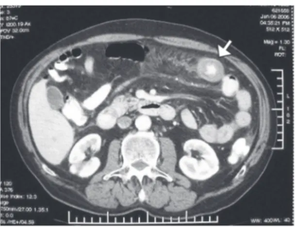
left deviation, activated partial thromboplastin time (APTT) 134 seconds, with relation (R) at 5; and prothrombin time (PT) over 100 seconds - international normalized ratio (INR) greater than 12. Amylase, lipase, arterial gasometry, renal function and electrolytes were normal. Radiographs of the abdomen in the orthostatic, supine and prone posi-tions suggested intestinal obstruction without pneumoperi-toneum (Fig.1).
As the diagnostic hypothesis was small bowel hematoma, clinical treatment consisted of gastric decom-pression, volemic restitution and correction of the coagulopathy with fresh frozen plasma and parenteral vi-tamin K. On June 6th, 2006, the patient was submitted to
On the same day, the patient presented an episode of melena and extensive painful ecchymosis on the upper limbs.
Following initiation of the therapy, the patient showed signs of improvement. On the fourth day, he was given food orally. On June 23rd, 2006, 2.5mg/day sodium warfarin
treatment was reinitiated on alternate days, with adjustment of the dose during hospitalization, in combination with so-dium dalteparin (5.000UI daily). On June 26th, 2006 the
patient was discharged from the hospital.
LITERATURE REVIEW
The MEDLINE, COCHRANE, LILACS and SciELO databases were used in order to review the literature from the previous 25 years.
The inclusion criteria were:
1) Single or multiple spontaneous small bowel hematoma, exclusive or associated with other areas of bleeding; 2) Patients using vitamin K antagonist oral anticoagulants; 3) Presumptive diagnosis by clinical laboratorial findings, with or without documentation for image examination, in those cases that received clinical treatment;
4) Macroscopical diagnosis through inventory of the cav-ity or anatomical-pathological documentation in those submitted to surgical treatment.
Cases were excluded where there was a history of trauma, the use of other forms of anticoagulation (even when used in combination with oral anticoagulation), and where hematoma was present in a condition in which the small bowel was not involved. The evaluated variables were: age and sex, clinical manifestations and length of his-tory, hemoglobin levels and prothrombin time at admission, motive and duration of oral anticoagulation therapy, occur-rence of digestive hemorrhage, diagnostic strategy, presence of small bowel hematoma, the need for surgical interven-tion, associated abdominal findings, and time necessary to resolve the condition.
CRITICAL ANALYSIS
A total of 28 articles were selected, covering the pe-riod from 1991 to 2006. Of these, four were excluded for not having been found full text or abstract in the researched databases. An additional three consisted only of abstracts, which was sufficient cause for their use as case reports, but not in the analysis of variables3,4,5, this encompassed a
to-tal of 57 published cases.
The incidence was higher in males (60%). The aver-age aver-age, described in 40 cases, was 57.6 years (32-78 years). Upon admission to the emergency service, the most frequent clinical condition, also described in 40 cases, was abdominal pain, which was present in 97.5% of the cases,
Figure 3 – Contrast CT in a coronal slice, showing thickening of the jejunum wall, “coiled spring” sign (wide arrow) and contrasted mesenteric vascular branches (narrow arrow).
with nausea associated with half of these cases, and vom-iting in an additional 40%; these latter two items are re-lated to high obstructions involving the duodenum and proximal jejunum. Abdominal pain was the only symptom in 20% of cases, and irradiation in the lumbar region was mentioned in one case, mimicking renal colic.6 The
obstruc-tive condition, characterized by abdominal distension, halted evacuation and elimination of flatulence in 45% of cases, all involving either jejunum or ileum and one includ-ing the ileocecal valve.7 Some cases were related to
extra-intestinal manifestations, among them: epistaxis, hematuria,8 ecchymosis in the upper limbs and face,9
con-junctival hemorrhage10 and mild acute pancreatitis11
(Ta-ble 1).
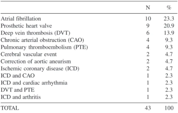
The incidence of digestive hemorrhage was 42.4%, which was exteriorized in the form of melena in 50% of cases, hematemesis associated with melena in 21.4%, only hematemesis in 21.4%, and hematochezia in 7.1% of these cases. The average time from the appearance of symptoms until medical attendance was 2.5 days (3 hours–5 days). The reason for the use of oral anticoagulant was described in 43 cases. Atrial fibrillation was responsible in 23.3% of cases, followed by prosthetic heart valve (20.9%) and deep vein thrombosis (13.9%), as shown in Table 2. The dura-tion of anticoagulant therapy was evaluated in 30 cases, and the average was 25.4 months (18 days - 84 months). There is one case described as “longer than 3 months”6 and
an-other “dose increased ten days ago”.10 In laboratory tests,
the average Hb was 11.5 (5.8-14.3). In one case, it was de-scribed as “normal”,12 and in another, the number of
eryth-rocytes was within the reference value.8
In order to present the results more clearly, we sepa-rated them into two periods, pre- and post-1991.
Until 1991, the results for PT were described in percent-age terms. The observed averpercent-age was 16.8% (5-80%). It was
described as “lower than 15%”(8) in two cases and “lower
than 5%” in an additional three cases(13). After this period,
the values are expressed using the INR. The average obtained was 7.5 (1.1 – 18.4). In two cases, it was “undetectable”,14,15
one result was “greater than 12”, and another two cases were reported PT as 50.2 and 49.7 seconds.10,16
With regard to diagnostic strategies, there are no reports of the use of CT or ultrasound (US) until 1986. Hence, we will present the results of these first five years of research, followed by the last 20 years, when CT and US were avail-able. In the first period, diagnoses were based on clinical and laboratory findings in 33% of cases. Simple radiogra-phy was performed in 6.7% of cases, and this was com-plemented with contrast radiography of the intestinal tran-sit in 20% of cases. Intraoperative diagnosis occurred in 40% of cases, and laparoscopy was used in one case.8
Starting in 1987, there was a reduction in the number of surgically based diagnoses; such operations were per-formed without previous exams in only three (9.7%) out of 31 cases. In fact, it is at this time when CT was adopted as the primary method. Of these three surgeries, section-ing of the intestinal flap was performed in two because of the presence of ischemia and the non-viability of the seg-ment, probably a result of the compressive effect of the hematoma, which impaired local blood irrigation.
The use of US as the only complementary exam oc-curred in 6.45% of cases, and CT in 54.8% of cases. CT alone or in conjunction with other methods, such as US and endoscopy, was responsible for 92% of the diagnoses (Table 3).
Surgery was indicated in distinct situations related to two distinct two periods. From 1981 to 1986, it was used in 40% of cases, always as a diagnostic method. In the sec-ond period (1987-2006), it was performed in 28% of the cases; as a therapeutic measure in 67% of these cases.
Table 1 - Signs and symptoms present upon admission in 40 patients diagnosed with spontaneous small bowel hematoma due to the use of oral anticoagulation.
Signs and symptoms N* %
Abdominal pain 39 97.5
Nausea 20 50
Intestinal obstruction 18 45
Vomiting 16 40
Fever 2 5
Anorexia 2 5
Epistaxis 2 5
Hematuria 2 5
Ecchymosis1 2.5
Conjunctival hemorrhage1 2.5
Source: 21 selected articles and case reports were reviewed
Table 2 - Reason for oral anticoagulation
N %
Atrial fibrillation 10 23.3
Prosthetic heart valve 9 20.9
Deep vein thrombosis (DVT) 6 13.9
Chronic arterial obstruction (CAO) 4 9.3
Pulmonary thromboembolism (PTE) 4 9.3
Cerebral vascular event 2 4.7
Correction of aortic aneurism 2 4.7
Ischemic coronary disease (ICD) 2 4.7
ICD and CAO 1 2.3
ICD and cardiac arrhythmia 1 2.3
DVT and PTE 1 2.3
ICD and arthritis 1 2.3
TOTAL 43 100
Regarding the location of the hematoma in the intesti-nal wall, the jejunum was the most affected (71.6%), fol-lowed by the duodenum (29.8%) and the ileum (15.8%).
The most frequently reported intra-operational findings while inventorying the cavity were: hemoperitoneum (10.3%), necrosis of the intestinal flap (10.3%), hematoma of mesenterium (10.3%), hemoretroperitoneum (5.1%) and cecal hematoma (2.6%).
The resolution time of the condition as shown by in-testinal transit and the acceptance of oral alimentation, de-scribed in 12 cases, was 10.1 days (4-16 days). There is only one report of a late complication, which was described as stenosis and diagnosed after three weeks through barium x-ray.8
DISCUSSION
The first report of intramural intestinal hematoma was given by McLouchlan16 in 1838, following an autopsy on
a man whose cause of death was recorded as “dehydration and bowel obstruction,” attributed to a “false aneurysm.” The first radiological description occurred more than one hundred years later, when Liverud reported a case involv-ing the jejunum.17
This condition rarely occurs spontaneously and is more frequently associated with abdominal trauma. Among non-traumatic causes, the use of oral anticoagulants is the main etiological factor.18 The only reference to the estimated
in-cidence of spontaneous small bowel hematoma related to anticoagulation is that it affects 1:2500 anticoagulation pa-tients/year in research carried out in Switzerland.19 In this
study, both the use of an oral anticoagulant and heparin are named as causes. With regard to the exclusive use of heparin as a cause of the condition, its incidence is even lower and has been the motive for few reports in the lit-erature.19-21
Despite its rarity, the number of reported cases has in-creased, both because of the availability of current image-based diagnostic methods as well as the growing number of patients on anticoagulation therapy. Other known causes
are hemophilia, Von Willebrand disease, idiopathic throm-bocytopenic purpura, lymphoproliferative diseases,22
col-lagenosis, vasculopathies and peptic ulcer. Pancreatic dis-eases, such as pancreatitus, can be either a cause23 or a
con-sequence.16
The small bowel is affected in up to 85% of the occur-rences of spontaneous intramural gastro-intestinal tract hematoma caused by the use of oral anticoagulation, with the jejunum being the most affected region, in contrast with the post-traumatic findings, which affect the duodenum more.18
We also found reports of spontaneous hematoma in the mesenterium, retroperitoneum and abdominal wall.24 More
severe cases may take the form of acute abdomen with necrosis of the flap, requiring emergency surgery.7 In the
Brazilian literature, we found three cases9,16,25 described in
the last 44 years; one occurred in the jejunum (and was diagnosed following necropsy in a patient with a previous history of ischemic stroke), and two occurred in the duo-denum. In one of these, obstruction of the Vater’s papilla resulting from the hematoma led to a mild acute pancre-atic complication.16
Hemorrhaging originates in the submucosa, and, al-though it has not been proven by other studies, it is be-lieved that the progression of the symptoms is due to in-tramural osmotic gradient and the presence of the hematoma, leading to an expansion of the intestinal wall.26
As shown in our results, clinical suspicion is fundamen-tal in making the diagnosis. The clinical condition may vary according to the location of the hematoma, and symptoms of either high or low obstruction may predominate. Ab-dominal pain is present in almost all cases, being either diffuse or predominantly concentrated at the place of the obstruction. Signs of peritoneal irritation appear when there are complications such as necrosis, perforation of the flap or hemoperitoneum. Digestive hemorrhaging occurs in a little over 40% of cases, apparent either through the upper or lower passage, with melena being the most frequent form. Other extra-hemorrhagic manifestations may also be present.
Simple radiography of the abdomen is insufficiently specific and may not show alterations or evidence of in-testinal obstruction, such as gastric dilation, hydro-aero level, dilated and thickened small-bowel flaps and the “pile of coins” sign.
The barium x-ray, widely used until the 1980s8, lost
ground as a diagnostic tool method with the development of CT and US imaging tools. These methods are less inva-sive, more quickly and easily performed and offer a more adequate evaluation regarding the presence or absence of free liquid in the abdominal cavity, as well as the
Table 3 - Diagnostic strategies after 1986
Complementary diagnostic method N %
Computerized tomography 17 54.8
Computerized tomography + US 8 25.8
Intraoperatoive 3 9.7
Ultrasound 2 6.5
Computerized tomography + GE 1 3.2
TOTAL 31 100
US=ultrasound; GE=gastrointestinal endoscopy
retroperitoneum. The combination of these two methods can offer 100% diagnostic accuracy, as demonstrated by Polat and Cols in 2003.27
Where CT is unavailable, US in combination with ab-dominal radiography may be sufficient to diagnose intra-mural hematoma. The indicative findings are tubular or cir-cular images with absence of peristalsis and mucous com-pression enveloped by an anechoic halo corresponding to the thickened intestinal wall. The echogenecity varies ac-cording to stage of maturation of the hematoma.28
Despite the diagnostic viability of US, CT is the diag-nostic exam of choice and has been proven to be extremely sensitive, showing alterations suggestive of the presence of hematoma in almost 100% of cases.10,12,14,15,18,27
Initially, CT should be performed without contrast, as this may mask the presence of intramural hemorrhage. The findings consist of a thickening of the wall greater than 1cm, with partial reduction to total obstruction of the pas-sage, the “pseudo-kidney” and “coiled spring” signs, gen-erally, in short segments (average 23cm) with profuse di-lation of the flaps.18 In the first ten days, hyperdensity is
seen, varying from 50 to 80 Hounsfield units depending on the time elapsed between the onset of the event and the exam;22,28 it then evolves towards hypodensity, with
re-ab-sorption and resolution of the radiological condition in a few weeks(6). Particular attention should be given to the
in-terpretation of the thickened intestinal wall, which occurs in other afflictions, such as inflammatory, infectious and neoplasic diseases as well as intestinal ischemia.22
Cur-rently, in the absence of anatomopathological confirmation, the diagnosis is considered confirmed with the presence of clinical and radiological findings combined with resolution of the condition following control CT.(18)
Due to the rarity of this entity, there are no studies that contain sufficient evidence to standardize treatment. There are only case reports of this condition in the literature, the largest of which includes eight patients using oral antico-agulation.29
The appearance as acute obstructive abdomen may lead
to operative treatment, although the use of US and CT in diagnosis has contributed to changes in conduct. Surgical exploration, which was previously used as a diagnostic method, has now become primarily a treatment method re-served for cases with complications that include intra-ab-dominal hemorrhage, suspected ischemia with or with-out perforation and peritonitis due to diagnosis doubt or to late complications. Clinical treatment should be insti-tuted in the absolute majority of cases.
The first measure is the immediate suspension of oral anticoagulants, and when possible, of medications used to enhance anticoagulation. Clinical treatment consists of gas-tric decompression in the presence of vomiting, the cor-rection of electrolyte disturbances, vitamin K administra-tion and the transfusion of fresh frozen plasma and blood when indicated. Total parenteral nutrition may be neces-sary in cases of prolonged fasting. In this review, we found one case treated with prothrombin, proconvertin, factor X and globulin B anti-hemophiliacs and heparin.30
In cases where there are no complications and surgery is unnecessary, resolution tends to occur within a few days. The recurrence of bleeding, as well as late sequelae such as stenosis, is unusual. Death during the presence of an in-tramural hematoma has occurred as a result of secondary complications such as digestive hemorrhage due to ulcer.28
CONCLUSION
Given the rarity of cases and the high incidence of pain-ful and obstructive abdominal syndromes caused by highly prevalent affections often seen in emergency services, the diagnostic possibility of spontaneous intestinal intramural hematoma should be remembered when there are indica-tions suggesting coagulation or other hemorrhagic mani-festations, mainly in patients who use oral anticoagulants. Non-invasive investigative methods offer high accuracy, permitting the adoption of clinical procedures that avoid unnecessary surgery, with complete resolution of the con-dition in a short period in nearly 100% of cases.
REFERENCES
1. Brewer RJ, Golden GT, Hitch DC. Abdominal pain: An analysis of 1000 consecutive cases in a hospital emergency room. Am J Surg. 1976;131:219-223.
2. Lohn, JWG, Austin, RCT, Winslet, MC. Unusual causes of small-bowel obstruction. J R Soc Méd. 2000;93:365-368.
3. Konan AV, Rajhi H, Chatti K, Mnif N, Salem A, Hamza R. Intramural small-bowel hematoma due to anticoagulant therapy: a radiologic study; with regard to two cases. Tunis Med. 2005;83:233-6.
4. Cappelli J, Lenaerts A, Lamy V, Ramdani B, Moisse R. Clinical case of the month. Intramural hematoma of the small intestine associated with anticoagulants, potentiated by interaction with cimetidine. Rev Med Liege. 1997;52:753-5.
6. Avent ML, Canaday BR, Sawyer WT. Warfarin-induced intramural hematoma of the small intestine. Clin Pharm. 1992;11:632-5. 7. Hsiao CW, Chao PC. Warfarin-induced intramural haematoma of the
ileocecal valve with obstruction. ANZ J Surg. 2004;74(9):810-1. 8. Vinard JL, Bouchet C, Aubert H, Meullenet J, Ohanessian JH, Aubert
M et al. Intramural hematomas of the small bowel (duodenum excluded) during long-term anticoagulant treatment. Report on 6 cases of which 2 required operation (author’s transl). J Chir (Paris). 1981;118:307-14. 9. Faria J, Pessoa R, Hudson M, Vitoi S, Villela O, Torres J et al. Hematoma intramural duodenal como complicação de terapia anticoagulante com Warfarin: relato de caso e revisão da literatura. Radiol Bras. 2004;37:461-463.
10. Shah P, Kraklow W, Lamb G. Unusual complication of coumadin toxicity. Wis Med J. 1994;93:212-4.
11. Meier R, Wyss-Meyer H, Jubin E, Gyr K. Intramural hematoma of the duodenum as complication in anticoagulant therapy. Diagnosis and therapy. Schweiz Rundsch Med Prax. 1990;79:517-20.
12. Secil M, Ucar G. Spontaneous duodenal hematoma. J Emerg Med. 2004;27:291-3.
13. Giesbers AA, Voets AJ, de Smet HL, van Wilderen LJ. Intramural hematomas of the small intestine during use of oral anticoagulants. Ned Tijdschr Geneeskd. 1986;130:113-7.
14. Acea Nebril B, Taboada Filgueira L, Sanchez Gonzalez F, Freire Rodriguez D, Fraguela Marina J, Aguirrezabalaga Gonzalez J et al. Acute abdomen in anticoagulated patients. Its assessment and the surgical indications. Rev Clin Esp. 1995;195:463-7.
15. Rios R, Garaulet P, Rodriguez M, Leon C, Limones M. Spontaneous intramural hematoma of the small intestine. Cir Esp. 2005;78:275. 16. Fahrhoud S, Stephani SM, Bromberg SH. Pancreatite aguda devida a
hematoma intramural do duodeno por uso de anticoagulante. Arq. Gastroenterol. 2001;38:53-56.
17. Liverud, K. Hematoma of the jejunum with subileus. Arch Radiol 1948;30:163; in aput Jones WR, Hardin WJ, Davis JT, Hardy JD. Intramural hematoma of the duodenum: a review of the literature and case report. Ann Surg. 1971;173:534–544.
18. Abbas MA, Collins JM, Olden KW. Spontaneous intramural small-bowel hematoma: imaging findings and outcome. AJR Am J Roentgenol. 2002;179(6):1389-94.
19. Bettler S, Montani S, Bachmann F. Incidence of intramural digestive system hematoma in anticoagulation. Epidemiologic study and clinical aspects of 59 cases observed in Switzerland (1970-1975). Schweiz Med Wochenschr. 1983;113(17):630-6.
20. Shaw PH, Ranganathan S, Gaines B. A spontaneous intramural hematoma of the bowel presenting as obstruction in a child receiving low-molecular-weight heparin. J Pediatr Hematol Oncol. 2005;27:558-60.
21. Hill H, Deppe H, Huchzermeyer H, Dormann AJ. Duodenal ileus due to an intramural duodenal haematoma. Conservative therapy using a multiple lumen intestinal probe. Dtsch Med Wochenschr. 2005;130:92-4.
22. Lane MJ, Katz DS, Mindelzun RE, Jeffrey RB Jr. Spontaneous intramural small bowel haemorrhage: importance of non-contrast CT. Clin Radiol. 1997;52:378-80.
23. Dubois J, Guy F, Porcheron J. A pancreatic-induced intramural duodenal hematoma: a case report and literature review. Hepatogastroenterology. 2003;50:1689-92.
24. Dineen RA, Lewis NR, Altaf N. Small bowel infarction complicating rectus sheath haematoma in an anticoagulated patient. Med Sci Monit. 2005;11:CS57-9.
25. de Mello G, Choma L, Santos HR, de Paola. Intraparietal hematoma of the small intestine as a complication of anticoagulant therapy. Presentation of 1 case with anatomo-pathological confirmation. Hospital (Rio J). 1963;63:121-7.
26. Judd DR, Taybi H, King H. Intramural hematoma of the small bowel; a report of two cases and a review of the literature. Arch Surg. 1964;89:527-35.
27. Polat C, Dervisoglu A, Guven H, Kaya E, Malazgirt Z, Danaci M et al. Anticoagulant-induced intramural intestinal hematoma. Am J Emerg Med. 2003;21:208-11.
28. Lorente-Ramos RM, Santiago-Hernando A, Del Valle-Sanz Y, Arjonilla-Lopez A. Sonographic diagnosis of intramural duodenal hematomas. J Clin Ultrasound. 1999;27:213-6.
29. Abbas MA, Collins JM, Olden KW, Kelly KA. Spontaneous intramural small-bowel hematoma: clinical presentation and long-term outcome. Arch Surg. 2002;137:306-10.