Original Research Article doi:10.12980/JCLM.3.2015JCLM-2015-0013 ©2015 by the Journal of Coastal Life Medicine. All rights reserved.
Antimicrobial and antioxidant activities of Cystoseira crinita Duby and Ulva intestinalis Linnaeus from the
coastal region of Sinop, Turkey
İsmet Berber, Cumhur Avşar*
, Hilal Koyuncu
Department of Biology, Faculty of Science and Arts, Sinop University, Sinop 57000, Turkey
Journal of Coastal Life Medicine
*Corresponding author: Cumhur Avşar, Department of Biology, Faculty of Science and Arts, Sinop University, Sinop 57000, Turkey.
Tel: +90 5384533639 Fax: +90 3682715524
E-mail: cumhur.avsar@gmail.com
1. Introduction
The marine world, due to its phenomenal biodiversity, is an important natural resource of many bioactive substances such as polyunsaturated fatty acids, sterols, proteins, polysaccharides, antioxidants and pigments[1]. Seaweeds known as macroalgae produce many biologically active phytochemicals such as carotenoids, terpenoids, xanthophylls, chlorophylls, phycobilins, polyunsaturated fatty acids, polysaccharides, vitamins, sterols, tocopherols and phycocyanins[2]. Nowadays seaweeds are used as dietary supplements in daily life and metabolize human health[3]. Marine macroalgae are also important ecologically and commercially to many regions of the world, especially in Asian countries such as China, Japan and Korea. The Japanese and
Chinese use brown algae in the treatment of hyperthyroidism and other glandular disorders.
Lipids, proteins and nucleic acids involve in reactive oxygen species that cause oxidative damage. It may trigger various chronic diseases such as coronary heart disease, atherosclerosis, cancer, and aging[4]. Epidemiological studies have indicated that intake of certain vitamins, minerals and other nutrients help to protect the body against heart disease, cancer and aging process. Antioxidants may have a protective effect in preventing or reducing the severity of these diseases[5]. The synthetic antioxidants, such as butylated hydroxyanisol and butylated hydroxytoluene have been suspected to use theirs effects of carcinogenesis and liver damage[6]. Concerns about the reliability of synthetic antioxidants have increased the interest in plants and algae commonly present in natural antioxidants[7,8].
Cystoseira crinita (C. crinita) and Ulva intestinalis (U. intestinalis) are plentiful and extensive seaweed species along the northern of Black Sea seacoast of Turkey[9]. Two species of marine algae are highly valuable marine resources in Asian A RT I C L E I N F O A B S T R AC T
Objective: To evaluate in vitro antimicrobial, antioxidant activities and total phenol contents of
Cystoseira crinita and Ulva intestinalis species collected from the coastal region of Sinop.
Methods: The antimicrobial activity of each methanolic algae sample was screened by using
disc diffusion method against to 15 bacteria and 3 yeasts. The antioxidant potential of the extracts on the stable radical 1,1-diphenyl-2-picrylhydrazylwas determined. The total phenolic content of the 3 methanolic extracts of the seaweed samples were determined by the Folin-Ciocalteu method.
Results: The results of antimicrobial assay showed that extracts were more effective against Gram-positive bacteria rather than Gram-negative. In addition, while the algal extracts had antifungal efficacy against Candida krusei, the other yeast strains were not affected at all. According to the findings of antioxidant activity, all methanolic extracts displayed good free radical scavenging activity ranging from IC50 = (32.19 ± 0.08) mg/mL to the IC50 = (37.57 ±
0.11) mg/mL. The total phenols content of the macroalgal extracts were found as between (5.10 ± 0.16) mg gallic acid equivalent/g and (87.70 ± 1.03) mg gallic acid equivalent/g. In this sense, our findings confirmed that there was a positive linear correlations (r = 0.86) between total phenol contents and the IC50 values.
Conclusions: The data gathered from this study suggested that the seaweeds can be used as a potential natural seafood sources owing to the antimicrobial efficiency and good antioxidant activity.
Article history: Received 4 Mar 2015
Received in revised form 26 Apr 2015
Accepted 30 Apr 2015 Available online 5 May 2015
Keywords: Antimicrobial Antioxidant activity Cystoseira crinita Ulva intestinalis
country and Europe due to using human-edible, food additives, fertilizers and animal feed[10]. They are also too sensitive against anthropogenic effect and pollution[11,12]. In recent years, many microorganisms are developing resistance against to synthetic and semi-synthetic drugs used for microbial infections treatment. However, the synthetic antimicrobial agents have the side effects. Thus, numerous scientists supposed that there is an urgent need to develop or discover new antimicrobial substances[13,14]. The results of numerous studies indicated that seaweed extracts had selective and effective antimicrobial activities against bacteria, fungi and virus in the parts of the world[15]. However, our knowledge obtained from literature indicated that data related to antioxidant and antimicrobial activity of seaweeds throughout Black Sea costal line of Turkey are very limited. Hence, the aim of the present study was to investigate the activity of the antioxidant and antimicrobial of two common species of seaweeds in Sinop Peninsula coast of the Black Sea, Turkey.
2. Materials and methods
2.1. Samples collection
The samples of C. crinita and U. intestinalis were collected from Sinop Peninsula coast of the Black Sea, Turkey, in December 2013. After collection, the seaweeds were washed with fresh water to remove associated debris and epiphytes. The cleaned material was then air dried to dryness in the shade at 30°°C. The dried samples
were finely powdered and stored at -20°°C until the next analysis
were carried out.
2.2. Preparation of algae extracts
About 10 g of dried samples were extracted by 100 mL of methanol for 24 h in a shaking water bath. The combined extracts were filtered and concentrated under vacuum to obtain a crude extract.
2.3. Microorganisms
The test microorganisms, 15 Gram-positive bacteria [Bacillus subtilis, Bacillus megaterium ATCC 1842 (B. megaterium), Bacillus thuringiensis var. israelensis, Bacillus cereusATCC 7064, Bacillus sphaericus (B. sphaericus), B. sphaericusATCC 2362, B. megateriumMRS 400, B. megateriumDSM 32, Bacillus licheniformis, Micrococcus luteusATCC 9345, Staphylococcus aureus (S. aureus) (clinical sp.), S. aureus ATCC 25923, Staphylococcus epidermidis (S. epidermidis), vancomycin resistant Enterococcus, Enterococcus faecalisATCC 51299 (E. faecalis)], 3 Gram-negative bacteria [Escherichia coliATCC 11293 (E. coli), E. coli extended spectrum beta-lactamase-positive (ESBL+), Klebsiella pneumonia (K. pneumonia)] and 3 yeast species [Candida albicans ATCC 14053 (C. albicans), Candida kruseiATCC 6258 (C. krusei) and Candida parapsilosisATCC 22019 (C. parapsilosis)] supplied
from the Molecular Biology and Microbiology Laboratory, Department of Biology, Faculty of Arts and Science, Sinop University, Turkey.
2.4. Antimicrobial assay
The antimicrobial activity of each methanolic algae sample was screened by using disc diffusion method[16]. All the microorganisms were maintained at -20 °C in nutrient agar for Bacillus spp.,
Mueller-Hinton agar for Staphylococcus spp., Luria agar for Enterococcus spp. and E. coli and Sabouraud dextrose agar for yeast (Difco) containing 17% (v/v) glycerol. Before testing, the microorganisms were transferred in nutrient broth for Bacillus spp., Mueller-Hinton broth for Staphylococcus spp., Luria broth for Enterococcus spp. and E. coli and Sabouraud dextrose broth for fungus (Difco) and cultured overnight at 37 °C. Then, the turbidity was adjusted equivalent to
0.5 McFarland standards and 100 μL of microorganisms was spread over the surface of an agar plate. The filter paper discs (6 mm) were loaded with methanolic algae extracts (2 mg/disc) and was allowed to dry completely. Afterwards, it was placed on the surface of the freshly inoculated medium. The plates were incubated for 24 h at 37 °C. Penicillin G (10 mg/mL), chloramphenicol (10 mg/mL),
erythromycin (10 mg/mL) and cycloheximide (10 mg/mL) were used as positive control and negative control was 12.5% dimethyl sulfoxide. The antimicrobial activity was evaluated by measuring the diameter of inhibition zone.
2.5. Determination of free radical scavenging activity by
1,1-diphenyl-2-picrylhydrazyl (DPPH) method
The antioxidant potential (free radical scavenging activity) of the extracts on the stable radical DPPH was determined by Blois[17]
and Kumar et al.[18] methods. The seaweed samples were dissolved as 1 000 µ g/mL in ethanol. Then, the solutions at different concentrations such as 1 000, 500, 250, 125 and 62.5 µg/mL were prepared with serial dilution technique. About 1 mL of the ethanol solution of each concentration containing the seaweed extract was mixed with 4 mL of a DPPH-ethanol solution (0.1 mmol/L). The samples were shaken well and kept in dark for 30 min at room temperature. The absorbance was measured at 517 nm. The free radical scavenging activity was calculated by using the following equation:
%Inhibition = AB - AS AB
× 100
Where AB is the absorbance of the control reaction and AS is
the absorbance of the test compound in this equation. Ascorbic acid was used as a standard or positive control. Not contained compound/standard was used as the negative control.
2.6. Determination of total phenolic contents (TPCs)
About 100 μL of diluted sample was added to 2 mL of 2% Na2CO3
reagent. After 2 min room temperature incubation, 100 μL of 50% Folin-Ciocalteu reagent was added. After 30 min of incubation at room temperature in the dark, the absorbance was measured at 720 nm by spectrophotometer (Thermo Scientific, Helios Alpha). Gallic acid (0.05 to 1 mg/mL) was used for the standard calibration curve. The results were expressed as gallic acid equivalent (GAE)/g dry weight of extracts.
2.7. Statistical analysis
All the experiments were carried out in triplicates and values were expressed as mean ± SD. Graphics were made using Microsoft Office Excel 2007. In addition, extract IC50 was
calculated from the graph plotting inhibition percentage against extract concentration in Excel 2007. Pearson’s correlation coefficient was calculated using Excel 2007.
3. Results
3.1. Antioxidant activity
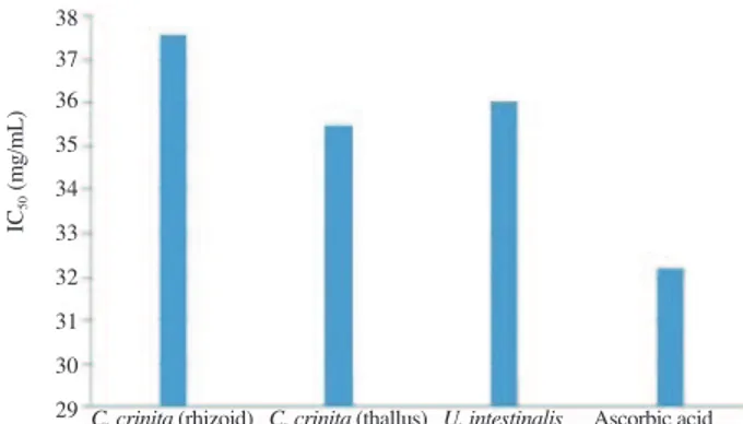
The percentages of free radical scavenging activity of methanolic algae extracts are given in Figure 1. The IC50 values
of the extracts are shown in Figure 2. The IC50 values of seaweed
extracts were found ranging from (32.19 ± 0.08) mg/mL to (37.57 ± 0.11) mg/mL and this value for ascorbic acid was determined as (32.19 ± 0.08) mg/mL.
According to determined IC50 values, the extract of C. crinita
(thallus) exhibited strong antioxidant activity with value IC50
(35.46 ± 0.09) mg/mL, compared to control and other seaweed extracts (Figure 2).
C. crinita (rhizoid) C. crinita (thallus) U. intestinalis Ascorbic acid 1 000 500 250 125 62.5
Concentrations (μg/mL) 100
80
60
40
20
0
Inhibition ef
fects (%)
Figure 1. Free radical scavenging activity of methanolic seaweed extracts
and ascorbic acid.
Figure 2. IC50 values of methanolic extracts of seaweed and ascorbic acid.
38 37 36 35
34 33 32 31
30 29
IC50
(m
g/
m
L
)
C. crinita (rhizoid) C. crinita (thallus) U. intestinalis Ascorbic acid
3.2. Antimicrobial activity
The antimicrobial activities of the seaweed extracts against 18
Table 1
Antimicrobial activity of the seaweed extracts against tested microorganisms and some standard drugs.
Microorganisms Inhibition zone (mm)
C. crinita
(thallus)
C. crinita
(rhizoid)
U. intestinalisPenicillin G Chloramphenicol Erythromycin Cycloheximide Dimethyl sulfoxide (12.5%)
Bacillussubtilis 9 10 10 10 15 18 *
-B. megaterium ATCC 1842 - 10 10 16 20 24 *
-Bacillus thuringiensis var. israelensis - 13 - 12 22 30 *
-Bacillus cereus ATCC 7064 12 12 11 6 25 31 *
-B. sphaericus 10 11 10 - - 35 *
-B. sphaericus ATCC 2362 - 10 11 18 24 36 *
-B. megaterium MRS 400 - 12 - 20 28 35 *
-B. megaterium DSM 32 - 11 11 16 25 28 *
-Bacillus licheniformis - 13 - 16 - - *
-K. pneumonia ESBL+ - 10 10 11 26 22 *
-E. faecalis ATCC 51299 - 9 - 10 12 9 *
-Micrococcus luteus ATCC 9345 - 15 - 12 23 21 *
-S. aureus ATCC 25923 - 17 - 12 28 40 *
-S. aureus (clinical sp.) - 20 11 11 12 37 *
-S. epidermidis 20 17 15 11 11 24 *
-Vancomycin resistant Enterococcus 11 12 13 - 22 - *
-E. coli ESBL+ - - - 11 24 11 *
-E. coli ATCC 11293 - - - 11 14 18 *
-C. krusei ATCC 6258 12 11 12 * * * 40
-C. albicans ATCC 14053 - - - * * * 41
-C. parapsilosis ATCC 22019 - - - * * * 42
bacterial strains and 3 Candida species are shown in Table 1. In the disc diffusion assays, the rhizoid extract of C. crinita displayed the antimicrobial activity at the different levels towards all tested microorganisms. The efficacy of C. crinita (rhizoid) was also found high against staphylococcal strains compared to other strains. On the other hands, there was no activity against to E. coliESBL+ and E. coliATCC 11293. All marine algal extracts were found more effective against Gram-positive bacteria than Gram-negative. In addition, it was determined that the tested extracts had weakly inhibitor effect on the growth of C. krusei, although there was no activity against C. albicans and C. parapsilosis. The antibacterial activity of extracts was found more effective than some of commercial antibiotics (Table 1).
3.3. TPCs
TPCs of the macroalgae extracts were calculated from the regression equation of calibration curve (y = 0.001 8x + 0.015 9; R2
= 0.999) and expressed as mg GAE/g in dried weight. TPC of the methanol extracts are given in Table 2. The TPCs of algal methanolic extracts were found ranging from (5.10 ± 0.16) mg GAE/g to (87.70 ± 1.03) mg GAE/g. The highest amounts of the TPC were determined in the rhizoid extract of C. crinita with value (87.70 ± 1.03) mg
GAE/g in dried sample.
Table 2
TPCs of 2 macro algae.
Algae species TPCs (mg GAE/g)
C. crinita (rhizoid) 87.70 ± 1.03
C. crinita (thallus) 27.70 ± 0.83
U. intestinalis 5.10 ± 0.16
Our findings validated that there was a positive linear correlation (r = 0.86) between the TPCs and IC50 values.
4. Discussion
Many marine seaweed species include many phytochemicals such as flavonoids, terpenes and polyphenolic compounds[20]. Ethnopharmacologic studies proposed that such substances produced by plants have been used to treat adverse effects of several bacterial, fungal and viral infections due to high levels of antimicrobial activity against microorganisms[21]. Multi-drug resistant microorganisms pose a serious challenge to the medical community in many countries over the world and there is an urgent need to develop new agents[22].
Naturally growing seaweeds contain novel possible antioxidant compounds which scavenge side effects of the free radical generated by metabolic reactions. Several valuable studies suggested that brown algae have significantly high phenolic content[23]. Nakamura et al.reported that the members of Phaeophyta include a kind of polyphenol called phlorotannins[24]. This polyphenol had been reported as potential antioxidant, anticancer, antibacterial and antifungal compounds. A few researches were investigated
antioxidant properties of three brown algae Sargassum wightii, C. crinita, Cystoseira myrica and a green algae Ulva lactuca[25-27]. The results of the above mentioned studies offered that Sargassum wightii, C. crinita and Cystoseira myrica had higher antioxidant activity than Ulva lactuca. According to the results of our study, the thallus extract of C. crinita showed good antioxidant activity with IC50 value (35.46 ± 0.09) mg/mL compared to U. intestinalis extract
and the rhizoid extract of C. crinita.
Some studies suggested that brown algae species had good antioxidant efficiency due to high phenolic compound contents[28]. The data gathered from this research demonstrated that the TPCs of algal extracts were ranging from (5.10 ± 0.16) mg GAE/g to (87.70 ± 1.03) mg GAE/g. The rhizoid extract of C. crinita had the highest phenolic content with value (87.70 ± 1.03) mg GAE/g, compared to other algal methanolic extracts. In this case, our results were coherent with the previous studies. Our findings also showed that there was a positive linear correlations (r = 0.86) between TPCs and the IC50 values.
A vast number of studies have been conducted about the antimicrobial effects of marine organisms, such as sponge, see grass and seaweeds in recent years[14,26,28]. In a study done by Mhadhebi et al.,C. crinita extracts showed low activity against tested Gram-positive and Gram-negative bacteria but was no activity against yeast[26]. According to Tüney et al., the methanol extracts of Ulva rigida and Ulva linza were no activity against E. faecalis and E. coli[29]. On the other hand, Taskin et al.determined that a pathogenic bacterium E. coli O157:H7 was sensitive against Coronosphaera mediterranea[30]. In the present study, it was determined that the rhizoid extract of a brown algae C. crinita showed high antimicrobial activity compared to other extracts and some standard antibiotics. Thallus extract of C. crinita was no activity against Gram-negative bacteria such as E. coliESBL and K. pneumonia whereas showed high antibacterial efficiency against Gram-positive bacteria especially S. epidermidis and S. aureus. In this case, it can be said that the rhizoid extract of C. crinita had good antimicrobial efficacy rather than its thallus extract.
activity.
Our findings supposed that two marine algae called C. crinita and U. intestinalis species harvested from Sinop coastal line can be used as a potential natural seafood source owing to the antimicrobial efficiency and good free radical scavenging activity. We also put forward to an idea about the determination of these two seaweed species including important bioactive substance in terms of medicine and pharmaceutics.
Conflict of interest statement
We declare that we have no conflict of interest.
References
[1] Lordan S, Ross RP, Stanton C. Marine bioactives as functional food ingredients: potential to reduce the incidence of chronic diseases. Mar Drugs 2011; 9: 1056-100.
[2] de Almeida CL, Falcão H de S, Lima GR, Montenegro C de A, Lira NS, de Athayde-Filho PF, et al. Bioactivities from marine algae of the genus
Gracilaria. Int J Mol Sci 2011; 12: 4550-73.
[3] Ganesan P, Kumar CS, Bhaskar N. Antioxidant properties of methanol extract and its solvent fractions obtained from selected Indian red seaweeds. Bioresour Tech 2008; 99: 2717-23.
[4] Finkel T, Holbrook NJ. Oxidants, oxidative stress and the biology of ageing. Nature 2000; 408: 239-47.
[5] Li HB, Cheng KW, Wong CC, Fan KW, Chen F, Jiang Y. Evaluation of antioxidant capacity and total phenolic content of different fractions of selected microalgae. Food Chem 2007; 102: 771-6.
[6] Foon TS, Ai LA, Kuppusamy P, Yusoff MM, Govindan N. Studies on in-vitro antioxidant activity of marine edible seaweeds from the east coastal region of Peninsular Malaysia using different extraction methods. J Coast Life Med 2013; 1: 193-8.
[7] Duan XJ, Zhang WW, Li XM, Wang BG. Evaluation of antioxidant property of extract and fractions obtained from a red alga, Polysiphonia urceolata. Food Chem 2006; 95: 37-43.
[8] Kelman D, Posner EK, McDermid KJ, Tabandera NK, Wright PR, Wright AD. Antioxidant activity of Hawaiian marine algae. Mar Drugs
2012; 10: 403-16.
[9] Karaçuha A, Karaçuha ME. Changes of macroalgae biomass in Sinop peninsula coast of the Black Sea, Turkey. Turk J Fish Aquat Sci 2013;
13: 725-36.
[10] Devi KP, Suganthy N, Kesika P, Pandian SK. Bioprotective properties of seaweeds: in vitro evaluation of antioxidant activity and antimicrobial activity against food borne bacteria in relation to polyphenolic content.
BMC Complement Altern Med 2008; 8: 38.
[11] Mhadhebi L, Laroche A, Robert J, Bouraoui A. Anti-inflammatory, antiproliferative and antioxidant activities of organic extracts from the Mediterranean seaweed, Cystoseira crinita. Afr J Biotech 2011; 10: 16682-90.
[12] Sales M, Ballesteros E. Seasonal dynamics and annual production of
Cystoseira crinita (Fucales: Ochrophyta)-dominated assemblages from the Northwestern Mediterranean. Sci Mar 2012; 76: 391-401.
[13] Parekh J, Chanda SV. In vitro antimicrobial activity and phytochemical analysis of some Indian medicinal plants. Turk J Biol 2007; 31: 53-8. [14] Nwinyi OC, Chinedu NS, Ajani OO, Ikpo CO, Ogunniran KO.
Antibacterial effects of extracts of Ocimum gratissimum and Piper guineense on Escherichia coli and Staphylococcus aureus. Afr J Food Sci 2009; 3: 77-81.
[15] Indira K, Balakrishnan S, Srinivasan M, Subramanian BS, Thamgavel B. Evaluation of in vitro antimicrobial property of seaweed (Halimeda tuna) from Tuticorin coast, Tamil Nadu, southeast coast of India. Afr J Biotechnol 2013; 12: 284-9.
[16] Bauer AW, Kirby WM, Sherris JC, Turck M. Antibiotic susceptibility testing by a standardized single disk method. Am J Clin Pathol 1966; 45: 493-6.
[17] Blois MS. Antioxidant determinations by the use of a stable free radical.
Nature 1958; 181: 1199-200.
[18] Kumar LS, Prasad KS, Revanasiddappa HD. Synthesis, characterization, antioxidant, antimicrobial, DNA binding and cleavage studies of mononuclear Cu(II) and Co(II) complexes of 3-hydroxy-N’-(2-hydroxybenzylidene)-2-naphthohydrazide. Eur J Chem 2011; 2: 394-403.
[19] Taga MS, Miller EE, Pratt DE. Chia seeds as a source of natural lipid antioxidants. J Am Oil Chem Soc 1984; 61: 928-31.
[20] Heo SJ, Cha SH, Lee KW, Cho SK, Jeon YJ. Antioxidant activities of chlorophyta and phaeophyta from Jeju Island. Algae 2005; 20: 251-60. [21] Berber İ, Avşar C, Çine N, Bozkurt N, Elmas E. [Determination of
antibacterial and antifungal activities of methanolic extracts of some plants growing in Sinop]. Karaelmas Sci Eng J 2013; 3: 10-6. Turkish. [22] Berber İ, Özgökçe F, Şeker A. [Determination of antimicrobial activity
of some plants growing in Van Region]. Y.Y.Ü Fen Bil Derg 2009; 14: 117-21. Turkish.
[23] Jiménez-Escrig A, Jiménez-Jiménez I, Pulido R, Saura-Calixto F. Antioxidant activity of fresh and processed edible seaweeds. J Sci Food Agr 2001; 81: 530-4.
[24] Nakamura T, Nagayama K, Uchida K, Tanaka R. Antioxidant activity of phlorotannins isolated from the brown alga Eisenia bycyclis. Fish Sci
1996; 62: 923-6.
[25] Meenakshi S, Umayaparvathi S, Arumugam M, Balasubramanian T. In vitro antioxidant properties and FTIR analysis of two seaweeds of Gulf of Mannar. Asian Pac J Trop Biomed 2011; 1: S66-70.
[26] Mhadhebi L, Chaeib K, Bouraoui A. Evaluation of antimicrobial activity of organic fractions of six marine algae from Tunisian Mediterranean coasts. Int J Pharm Pharm Sci 2012; 4: 534-7.
[27] Kokabi M, Yousefzadi M, Ahmadi AA, Feghhi MA, Keshavarz M. Antioxidant activity of extracts of selected algae from the Persian Gulf, Iran. J Persian Gulf (Mar Sci)2013; 4: 45-50.
[28] Nagai T, Yukimoto T. Preparation and functional properties of beverages made from sea algae. Food Chem 2003; 81: 327-32.
[29] Tüney İ, Çadirci BH, Ünal D, Sukatar A. Antimicrobial activities of the extracts of marine algae from the coast of Urla (Ä°zmir, Turkey). Turk J Biol 2006; 30: 171-5.