BMP-2 and titanium particles
synergistically activate
osteoclast formation
S.X. Sun
1, H.H. Guo
2, J. Zhang
3, B. Yu
2, K.N. Sun
1and Q.H. Jin
11Department of Orthopedics, Affiliated Hospital of Ningxia Medical University, Yinchuan, Ningxia Hui Autonomous Region, China 2Ningxia Medical University, Yinchuan, Ningxia Hui Autonomous Region, China 3Institute of Pathology, Xi’an Jiaotong University, Xi’an Shaanxi, China
Abstract
A previous study showed that BMP-2 (bone morphogenetic protein-2) and wear debris can separately support osteoclast formation induced by the receptor activator of NF-kB ligand (RANKL). However, the effect of BMP-2 on wear debris-induced osteoclast formation is unclear. In this study, we show that neither titanium particles nor BMP-2 can induce osteoclast formation in RAW 264.7 mouse leukemic monocyte macrophage cells but that BMP-2 synergizes with titanium particles to enhance osteoclast formation in the presence of RANKL, and that at a low concentration, BMP-2 has an optimal effect to stimulate the size and number of multinuclear osteoclasts, expression of osteoclast genes, and resorption area. Our data also clarify that the effects caused by the increase in BMP-2 on phosphorylated SMAD levels such as c-Fos expression increased throughout the early stages of osteoclastogenesis. BMP-2 and titanium particles stimulate the expression of p-JNK, p-P38, p-IkB, and P50 compared with the titanium group. These data suggested that BMP-2 may be a crucial factor in titanium particle-mediated osteoclast formation.
Key words: Bone morphogenetic protein; Titanium; Osteoclasts; SMAD; RANKL
Introduction
The biological response to wear particles at the bone-implant interface is considered the main cause of aseptic loosening and osteolysis (1,2) by increasing osteoclasto-genesis and bone resorption (3,4). Macrophages phago-cytize particles and secrete proinflammatory factors such as tumor necrosis factor-a(TNF-a), which are thought to mediate the differentiation of osteoclast precursor cells and play a fundamental role in this pathologic response (5,6). A recent study showed that bone morphogenetic protein-2 (BMP-2), a member of the superfamily of transforming growth factor beta (TGF-b) ligands, could also affect osteoclastogenesis (7,8). It seems to have a double effect on osteoclast formation. In the presence of osteoblasts, BMPs can indirectly suppress osteoclasto-genesis by decreasing the receptor activator of NF-kB ligand (RANKL) released by osteoblasts (9,10). In the absence of osteoblasts, BMP-2 appears to directly increase osteoclast formation and survival (11-13).
However, neither wear particles nor BMP-2 alone can induce osteoclast recruitment without RANKL, which is
essential for osteoclastogenesis (14,15). RANKL is a member of the TNF superfamily and is expressed on the surface of osteoblast-lineage cells. It functions by inter-acting with its receptor, RANK, on osteoclast precursors through the RANK/RANKL/osteoprotegerin axis, which regulates osteoclast maturation and activation (16,17).
Mitogen-activated protein kinase (MAPK) family mem-bers (JNK, p38, and ERK) and AP-1 (c-Fos and c-Jun) are well known to be essential to osteoclast formation, and inhibitors of these pathways have been shown to reduce osteoclast formation (18,19). At the molecular level, MAPK and c-Fos have been shown to become activated following wear debris treatment of macrophages (20,21). NF-kB is also important in osteoclastogenesis, and wear debris can activate NF-kB in cultured macrophages (22). A deficiency of NF-kB1 in mice protects against titanium-induced calvarial osteolysis (23).
Although these pathways have been well established to exert multiple effects to promote osteoclast formation, the significance of BMP signaling in wear debris-induced
Correspondence: Q.H. Jin, Department of Orthopedics, Affiliated Hospital of Ningxia Medical University, Yinchuan 750004, Ningxia Hui Autonomous Region, China. Fax: ++86-095-1408-2981. E-mail: [email protected]
osteoclast formation is not clear. Some reports have shown that the activity of TGF-b is not exclusive to the SMAD signaling pathway and that it can regulate other signaling pathways, such as p38, JNK, AP-1, and NFkB (24-27). Similar to TNF, TGF-b may synergistically potentiate the action of RANKL by such pathways. Interestingly, blocking the action of TGF-b also blocks RANKL-dependent osteoclastogenesis (28). This obser-vation implies that TGF-bmay be an essential factor for osteoclast formation.
Although the application of BMPs during orthopedic procedures has been found to promote a transient increase in osteoclast numbers and osteoclastic bone resorption (13), the effect of BMP-2 on wear debris-induced osteo-clast formation is unclear. The aims of this study were to determine the effect of BMP-2 on wear debris-induced osteoclast formation, and to determine whether the activity of BMP-2 involves the SMAD pathway and/or SMAD-independent pathway. Because osteoclasts play a key physiological and pathological role in osteoporosis, we used RAW 264.7 macrophage-like cells, which are osteoclast precursors, to perform this study (29).
Material and Methods
Cultured cells and reagents
Mouse monocyte/macrophage RAW 264.7 cells (American Type Culture Collection, Cell Bank of Chinese Academy of Sciences, China) were cultured in Dulbecco’s modified Eagle’s medium with glutaMAX (Gibco Invitrogen, USA) containing 10% fetal bovine serum (Hyclone, USA) and 1% antibiotic-antimycotic (Gibco Invitrogen) in a humidified atmosphere of 5% CO2at 376C. RAW 264.7 cells were grown to 60-70%
confluency, counted and cultured on 6-well tissue culture plates (46105cells/well). The cells were pretreated with
30 ng/mL RANKL (R&D Systems, USA) for 1 h and stimulated with titanium particles alone, 50 ng/mL BMP-2 alone (R&D Systems), or 50 ng/mL BMP-2 and 150mg/
mL titanium particles for the indicated times. The super-natants or cells were collected for further analysis.
Titanium particles
The titanium particles, average diameter 5mm, were kindly provided by the Zimmer Company (USA). The particles were rinsed in ethanol three times and then in PBS three times. Next, the particles were mixed with the culture medium at concentrations of 10, 50, 100, 150, and 200mg/mL. The titanium suspensions were sonicated for
30 min before using.
Cell viability assay
The cells were plated on 96-well plates (Corning Incorporated, USA) at a density of 104 cells/well. After treatment with 30 ng/mL RANKL, the cells were incubated with 150mg/mL titanium particles alone or 50 ng/mL BMP-2
and 150mg/mL titanium particles for 3 days. Osteoclast
viability was assessed using an MTT assay (Sigma, USA) according to the manufacturer’s instructions.
Tartrate-resistant acid phosphatase-positive (TRAP) staining assay
For differentiation of RAW 264.7 cells into osteoclasts, the cells were seeded and cultured on 96-well plates (104 cells/well). Multinucleated osteoclasts were observed at day 8. The TRAP-staining kit (Sigma) was used to evaluate TRAP expression. The TRAP(++) multinucleated cells that contained three or more nuclei were counted as osteoclasts using optical microscopy, and ImagePro Plus was used to quantify the data.
Pit resorption assay
The effects of osteoclastic modulators were tested with the 24-well Corning Osteo Assay (Corning Incorporated). The RAW 264.7 cells (approximately 5000 cells per well) were transferred to each well of a 24-well Corning Osteo Assay plate. The plates were incubated at 376C in a humidified atmosphere with 5% CO2 for 8 days with a
change of medium on day 3. After 8 days, the plates were washed with 6% sodium hypochlorite to remove the cells. Individual pits or multiple pit clusters were observed using a microscope at 106magnification.
Enzyme-linked immunosorbent assay (ELISA)
To examine TNF-a secretion, the RAW 264.7 cells were plated on 96-well plates (104cells/well) with 30 ng/ mL RANKL for 1 h and then incubated for 1 day with 50 ng/mL BMP-2 alone or 50 ng/mL BMP-2 and 150mg/
mL titanium particles in the presence of 30 ng/mL RANKL for 1 day. The levels of TNF-a secreted into the cell culture supernatant were measured using ELISA kits (Shanghai Lianshuo Biological Technology, China) according to the manufacturer’s instructions.
Quantitative real-time PCR
Total RNA was isolated from cells using the TRIzol reagent (Gibco Invitrogen) and quantified by NanoDrop 2000 (Thermo Scientific, USA). The RNA was reverse transcribed to cDNA with the RevertAid First-Strand cDNA synthesis kit (Fermentas, China). Quantitative real-time PCR was then performed in triplicate using the Bio-Rad iQ5 gradient real-time PCR system. All reactions were performed in a 25-mL reaction volume containing 12.5mL SYBR Premix Ex TaqTM II (TaKaRa, Japan)
CAAGG-39 and (reverse) 59-ACTCCACGACATACTCA GC-39.
Western blot analysis
Whole cells were extracted in radioimmunoprecipitation assay buffer containing protease and phosphatase inhibi-tors (Sigma-Aldrich, USA). The protein concentration was measured with a bicinchoninic acid protein assay kit following the manufacturer’s instructions. The proteins were resolved by SDS-PAGE, transferred to a polyvinylidene fluoride membrane and incubated overnight at 46C with primary antibodies against phospho (P)-SMAD 1/5/8 (Cell Signaling, USA), SMAD 1/5/8 (Abcam, USA), NFKB1 (Cell Signaling), c-Fos (Cell Signaling), p-P38 (Cell Signaling), p-JNK and p-IkB (Anbobio, China). The same membranes were probed with anti-b-actin antibodies (Sigma) to normal-ize protein loading in each lane. Subsequently, the blots were washed in Tris-buffered saline with Tween (10 mM Tris-HCl, 50 mM NaCl, and 0.25% Tween-20) and incu-bated with the secondary antibody. Immunoreactive bands were visualized using an ECL Plus substrate (GE Healthcare Life Sciences, China).
Statistical analysis
All experiments were performed in triplicate. Significant differences were determined using one-way ANOVA with a least-significant difference test. Differences of P,0.05 were considered to be significant.
Results
Effect of titanium particles on RAW 264.7 cell differentiation
To evaluate the effects of the titanium particles on the
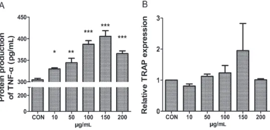
RANKL-mediated osteoclast differentiation of RAW 264.7 cells, we cultured the cells in the presence of suboptimal levels of 30 ng/mL RANKL and titanium particles. After 1 day, we measured TNF-aprotein expression, which plays a major role in activating osteoclast recruitment (5,6), and after 5 days, we measured TRAP gene expression, a hallmark of osteoclasts (30). The titanium particles increased TNF-aexpression in a dose-dependent manner and showed an optimum effect at a concentration of 150mg/mL (Figure 1A). There were no significant
differences in TRAP expression between the different concentrations of titanium particles and the control (Figure 1B). The TRAP stain also failed to produce any differentiated osteoclasts in each group (data not shown). This result indicated that the titanium particles failed to induce the differentiation of osteoclast precursors in the presence of RANKL.
Effect of BMP-2 and titanium particles on the differentiation of osteoclast precursors
BMP-2 is another cytokine that was reported recently to stimulate osteoclast differentiation (8). Therefore, we hypothesized that BMP-2 could affect the titanium particle-induced osteoclast formation of RAW 264.7 cells in the presence of RANKL. We used real-time PCR to examine the expression of the osteoclast marker genes
NFATc1,TRAP, andCathepsin K(31-33). The cells were pretreated with RANKL (30 ng/mL) for 1 h and subse-quently treated or not with 50 ng/mL BMP-2 alone, 150mg/mL titanium particles alone, or BMP-2 and
titanium for 5 days. The PCR analysis showed that neither BMP-2 nor the titanium particles can significantly enhance the expression of all these osteoclastogenesis-related genes in the presence of RANKL (Figure 2).
Surprisingly, however, we found that adding BMP-2 and titanium particles together greatly enhanced the ex-pression of theTRAP, Cathepsin K and NFATc1genes (Figure 2). TRAP staining confirmed these results. Many large, multinucleated TRAP-positive cells were detected in the cultures treated with both BMP-2 and titanium particles. Only small, mononucleated cells were observed in the titanium particles and RANKL treatment (Figure 3).
To further confirm this synergy between BMP-2 and the titanium particles, we prolonged the culture time from 5 to 7 days. We treated the RAW 264.7 cells with RANKL
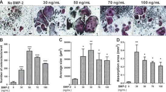
or not and then added the titanium particles and different concentrations of BMP-2. After 7 days of culture, we performed real-time PCR to measure TRAP gene expression. We found that BMP-2 greatly enhanced osteoclast formation, with a peak at 50 ng/mL and to a lesser extent at higher concentrations. There was no expression of theTRAPgene in the absence of RANKL (Figure 4). TRAP staining showed that abundant TRAP-positive multinucleated cells appeared when the cells were treated with BMP-2 from low to high doses in the presence of RANKL (Figure 5A). No TRAP-positive
Figure 2.BMP-2 and titanium (Ti) particles synergistically activated osteoclast formation. Gene relative expression was determined by real-time PCR; RAW 264.7 cells were cultured with suboptimal (30 ng/mL) RANKL, BMP-2 (50 ng/mL) or Ti particles (150mg/mL) as indicated for 5 days.A,TRAPgene expression.B,NFATc1gene expression.C,Cathepsin Kgene expression. Data are reported as means±SD. *P,0.01; **P,0.0001 (one-way ANOVA).
multinucleated cells were observed in the absence of RANKL (data not shown). These results are quantified in Figure 5B and C, and show that BMP-2 greatly increased the number and average size of the osteoclasts in the RAW 264.7 cells to levels several-fold greater than by the stimulation with titanium particles and RANKL. In addition, the pit formation assay revealed that the bone resorptive activity of mature osteoclasts was significantly acceler-ated when treacceler-ated with BMP-2 (Figure 5D). These data
indicated that BMP-2, even at a low concentration, synergized with titanium particles and RANKL to enhance the differentiation of osteoclast precursors. BMP-2 may be a crucial factor in the titanium particle-mediated osteoclast formation.
Effect of BMP-2 and titanium particles on cell viability
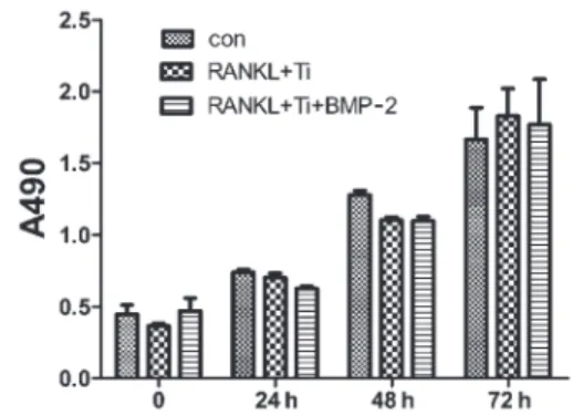
The MTT assay was used to determine whether BMP-2 and titanium particles induced the enhancement of proliferation and viability of RAW 264.7 cells. The cells treated with 50 ng/mL BMP-2 and titanium particles showed a slight, nonsignificant decrease of cell viability. However, neither BMP-2 nor the titanium particles showed any cytotoxic effects on the RAW 264.7 cells compared with the control (Figure 6). Based on these findings, we concluded that neither BMP-2 nor the titanium particles increased RAW 264.7 macrophage-like cell proliferation.
Expression of BMP-2 signaling during osteoclast differentiation
To confirm that BMP-2 acted directly upon the osteoclast precursors, we performed Western blotting against c-Fos and p-SMAD 1/5/8. c-Fos is well known to be essential for RANKL-mediated osteoclast differentia-tion and to be expressed in the early stages of osteoclast formation (34). Therefore, we observed it from days 2 to 5. As shown in Figure 7, the expression of c-Fos was strongly detected from days 2 to 5 when treated with BMP-2. In contrast, in the absence of BMP-2, the expression of c-Fos decreased gradually over time.
Figure 4. Effects of BMP-2 on titanium particles-mediated osteoclast formation. Real-time PCR showed the expression of TRAP. The RAW 264.7 cells were pretreated with 150mg/mL titanium particles and different concentrations of BMP-2 for 1 h, then treated or not with 30 ng/mL RANKL for 7 days. CON: control. Data are reported as means±SD. ***P,0.001 (one-way ANOVA).
During the expression of c-Fos, p-SMAD 1/5/8 was constantly expressed at high levels throughout osteoclast differentiation when treated with BMP-2, and p-SMAD 1/5/ 8 was only weakly detected when BMP-2 was absent (Figure 7). These results strongly indicated that BMP-2 signaling was involved in osteoclast differentiation and that BMP signal transduction may play a crucial role in the differentiation of osteoclasts.
Effect of titanium particles on BMP-induced SMAD phosphorylation during osteoclastogenesis
Previous reports have shown that the BMP-2/SMAD and TNF-a/NF-kB systems appear to exert antagonistic effects (35). Thus, we hypothesized that the titanium particles could inhibit BMP-induced SMAD phosphoryla-tion. As shown in Figure 8A, the RAW 264.7 cells were pretreated with BMP-2 for 1 h and then treated with or without titanium particles. BMP-2 increased p-SMAD 1/5/ 8 protein expression, which peaked at 12 h in both the titanium particle and nonparticle groups; there was no significant difference between the two groups. This result showed that the secretion of TNF-a induced by the titanium particles did not affect BMP-induced SMAD phosphorylation in RAW 264.7 cells (Figure 8A).
Secretion of TNF-afrom RAW 264.7 cells treated with BMP-2 and titanium particles
Hong et al. (36) reported that BMP-6 induced the expression of TNF-a of RAW 264.7 cells. To determine whether BMP-2 had the same effect, we performed ELISA to observe the expression of TNF-a. As shown in Figure 8B, although TNF-awas greatly increased in the titanium particle and titanium particle-BMP-2 groups, there was no significant difference between these two groups, demon-strating that BMP-2 did not increase TNF-aexpression.
BMP-2 regulates the expression of SMAD-independent pathways in titanium particle-meditated osteoclast differentiation
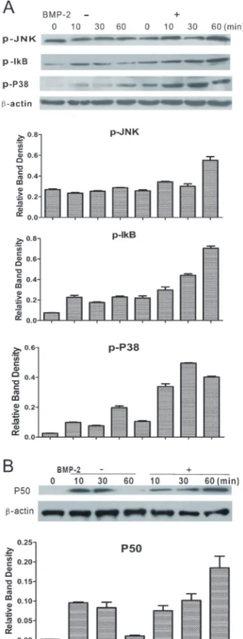
We further investigated signaling pathways other than the SMAD pathways that could be activated by BMP-2. We detected the expression of p-P38, p-IkB, and p-JNK by Western blotting, and found that the RAW 264.7 cells expressed elevated levels of p-P38 and p-IkB in a time-dependent manner in the presence of BMP-2 and that the expression of these proteins was lowered without BMP-2 treatment (Figure 9A). There was no significant difference in the expression of p-JNK between the two groups. To further confirm the effect of BMP-2 on the activation of p-IkB, we evaluated the expression of P50, an NF-kB subunit. The result showed that BMP-2 increased nuclear translocation of P50 compared with the group without BMP-2 treatment (Figure 9B). These results demonstrate that BMP-2 synergized with the titanium particles in osteoclastogenesis by directly stimulating the expression of p-JNK, p-P38, p-IkB, and P50.
Discussion
Not all RAW 264.7 cell lines or passages will form osteoclasts (30). In this study, we confirmed that titanium particles at different concentrations could not induce osteoclast differentiation in the presence of RANKL.
Although the role of BMP-2 in promoting osteoblast differentiation has been extensively investigated, and it has been approved by the FDA as an alternative to bone grafts in long bone fractures and healing in a number of orthopedic and maxillofacial applications (37), its function in particle-induced aseptic loosening and osteolysis has
Figure 6. Neither the BMP-2 nor titanium (Ti) particles affect RAW 264.7 cell proliferation. RAW 264.7 cells (control, con) were pretreated with 30 ng/mL RANKL and 150mg/mL Ti particles for 1 h, then treated or not with 50 ng/mL BMP-2 for 3 days. Data are reported as means±SD cell proliferation as the absorbance ratio at 490 nm.
not yet been elucidated. Jager et al. (38) showed that a local BMP-2/mesenchymal stem cell application can promote bone formation within a wear debris-induced periprosthetic osteolytic area, but there was no evidence that the BMP-2 application was solely responsible for healing the severe osteolysis. Quinn et al. (39) reported that TGF-bgreatly increased osteoclast formation in RAW 264.7 macrophage-like cells to levels that were several-fold greater than those attainable with maximal stimulation by RANKL or TNF-a. In this study, we demonstrated that BMP-2 was not sufficient to increase osteoclast formation. However, BMP-2 greatly enhanced titanium particle-meditated osteoclast formation in the presence of RANKL. This treatment increased the size and number of multinuclear osteoclasts, the expression of osteoclast genes (TRAPandNFATc1), and the resorption area. At a low concentration (50 ng/mL), BMP-2 appears to have an optimal effect on stimulation of osteoclast formation. However, no multinucleated cells were observed in the absence of RANKL.
Although RANKL and titanium particles could induce c-Fos expression, more c-Fos was detected in the group treated with BMP-2 and titanium particles from days 2 to 5
in the presence of RANKL. Our data also showed that BMP-2 treatment increased SMAD phosphorylation fol-lowing the high expression of c-Fos throughout the early stages of osteoclastogenesis. Moreover, the phosphory-lated SMAD levels were negligible in the titanium particle groups. All these findings imply that BMP-2 activated osteoclast formation by increasing c-Fos expression and
Figure 8.Titanium (Ti) particles do not affect phospho-SMAD (p-SMAD) 1/5/8 expression. BMP-2 does not affect expression of TNF-a.A, RAW 264.7 cells were treated with 30 ng/mL RANKL and 50 ng/mL BMP-2 for 1 h, then treated with or without 150 mg/mL Ti particles at indicated times. B, ELISA showed the expression of TNF-a. RAW 264.7 cells (control) were treated with 30 ng/mL RANKL and 150mg/mL Ti particles for 1 h, then treated or not with 50 ng/mL BMP-2 for 24 h. Data are reported as means±SD. *P,0.05 (one-way ANOVA).
that the BMP-dependent pathway was at least implicated in titanium particle-induced osteoclastogenesis.
Macrophages are well known to respond to the titanium particle challenge in two distinct ways. First, the wear debris activates proinflammatory signaling. Second, it activates MAPK cascades as well as NF-kB and other transcription factors. Both actions contribute to an increase of osteoclast recruitment. In our study, we first demon-strated that BMP-2 did not increase the expression of TNF-a, the major proinflammatory factor in inflammatory osteolysis. Then, our data showed that BMP-2 stimulated the expression of p-JNK, p-P38, and p-IkB, and the increase in p-IkB explains the progressive increase in P50. In addition to the canonical SMAD-dependent path-way, BMP has been reported to activate MAPK pathways in some systems (40). Nakashima et al. (22) reported that titanium particles can induce the rapid activation of MAPK
family members. Our data showed that titanium particles did not affect the expression of BMP-induced SMAD phosphorylation. Although the titanium particles slightly enhanced the levels of these signaling pathways, they were insufficient to induce osteoclast formation unless BMP-2 was also added. This result suggests that BMP-2 did not increase the titanium particle-induced osteoclasto-genesis by activating the expression of proinflammatory cytokines but instead acted upon downstream signaling pathways, such as p-JNK, p-P38, and p-IkB. Thus, the effect of BMP-2 in the wear debris-induced osteoclasto-genesis has been underestimated.
In summary, this study suggests that synergy of BMP-2 and titanium particles enhanced osteoclast formation in the presence of RANKL by increasing the expression of p-JNK, p-P38, and p-IkB. BMP-2 may be a crucial factor in titanium particle-mediated osteoclast formation.
References
1. Jacobs JJ, Roebuck KA, Archibeck M, Hallab NJ, Glant TT. Osteolysis: basic science.Clin Orthop Relat Res2001; 393: 71-77, doi: 10.1097/00003086-200112000-00008. 2. Ingham E, Fisher J. The role of macrophages in osteolysis
of total joint replacement. Biomaterials 2005; 26: 1271-1286, doi: 10.1016/j.biomaterials.2004.04.035.
3. Haynes DR, Crotti TN, Potter AE, Loric M, Atkins GJ, Howie DW, et al. The osteoclastogenic molecules RANKL and RANK are associated with periprosthetic osteolysis.J Bone Joint Surg Br2001; 83: 902-911, doi: 10.1302/0301-620X. 83B6.10905.
4. Kobayashi A, Freeman MA, Bonfield W, Kadoya Y, Yamac T, Al-Saffar N, et al. Number of polyethylene particles and osteolysis in total joint replacements. A quantitative study using a tissue-digestion method.J Bone Joint Surg Br1997; 79: 844-848, doi: 10.1302/0301-620X.79B5.7602.
5. Merkel KD, Erdmann JM, McHugh KP, Abu-Amer Y, Ross FP, Teitelbaum SL. Tumor necrosis factor-alpha mediates orthopedic implant osteolysis.Am J Pathol1999; 154: 203-210, doi: 10.1016/S0002-9440(10)65266-2.
6. Maloney WJ, James RE, Smith RL. Human macrophage response to retrieved titanium alloy particlesin vitro. Clin Orthop Relat Res1996; 322: 268-278.
7. Liu F, Ventura F, Doody J, Massague J. Human type II receptor for bone morphogenic proteins (BMPs): extension of the two-kinase receptor model to the BMPs.Mol Cell Biol 1995; 15: 3479-3486.
8. Jensen ED, Pham L, Billington CJ Jr, Espe K, Carlson AE, Westendorf JJ, et al. Bone morphogenic protein 2 directly enhances differentiation of murine osteoclast precursors.J Cell Biochem2010; 109: 672-682.
9. Lacey DL, Timms E, Tan HL, Kelley MJ, Dunstan CR, Burgess T, et al. Osteoprotegerin ligand is a cytokine that regulates osteoclast differentiation and activation. Cell 1998; 93: 165-176, doi: 10.1016/S0092-8674(00)81569-X. 10. Matsuo K. Cross-talk among bone cells.Curr Opin Nephrol
Hypertens2009; 18: 292-297, doi: 10.1097/MNH.0b013e3 2832b75f1.
11. Itoh K, Udagawa N, Katagiri T, Iemura S, Ueno N, Yasuda
H, et al. Bone morphogenetic protein 2 stimulates osteoclast differentiation and survival supported by receptor activator of nuclear factor-kappaB ligand.Endocrinology2001; 142: 3656-3662.
12. Falany ML, Thames AM III, McDonald JM, Blair HC, McKenna MA, Moore RE, et al. Osteoclasts secrete the chemotactic cytokine mim-1. Biochem Biophys Res Commun2001; 281: 180-185, doi: 10.1006/bbrc.2001.4307. 13. Toth JM, Boden SD, Burkus JK, Badura JM, Peckham SM, McKay WF. Short-term osteoclastic activity induced by locally high concentrations of recombinant human bone morphogenetic protein-2 in a cancellous bone environment. Spine 2009; 34: 539-550, doi: 10.1097/BRS.0b013e318 1952695.
14. Troen BR. Molecular mechanisms underlying osteoclast formation and activation.Exp Gerontol2003; 38: 605-614, doi: 10.1016/S0531-5565(03)00069-X.
15. Roodman GD. Regulation of osteoclast differentiation.Ann N Y Acad Sci 2006; 1068: 100-109, doi: 10.1196/annals. 1346.013.
16. Boyce BF, Yamashita T, Yao Z, Zhang Q, Li F, Xing L. Roles for NF-kappaB and c-Fos in osteoclasts. J Bone Miner Metab 2005; 23 (Suppl): 11-15, doi: 10.1007/ BF03026317.
17. Fuller K, Wong B, Fox S, Choi Y, Chambers TJ. TRANCE is necessary and sufficient for osteoblast-mediated activation of bone resorption in osteoclasts. J Exp Med1998; 188: 997-1001, doi: 10.1084/jem.188.5.997.
18. Abbas S, Clohisy JC, Abu-Amer Y. Mitogen-activated protein (MAP) kinases mediate PMMA-induction of osteo-clasts.J Orthop Res2003; 21: 1041-1048, doi: 10.1016/ S0736-0266(03)00081-0.
19. Grigoriadis AE, Wang ZQ, Cecchini MG, Hofstetter W, Felix R, Fleisch HA, et al. c-Fos: a key regulator of osteoclast-macrophage lineage determination and bone remodeling. Science 1994; 266: 443-448, doi: 10.1126/science.7939 685.
vascular endothelial growth factor from human monocyte/ macrophages by orthopaedic biomaterial particles.J Bone Miner Res 2003; 18: 1573-1583, doi: 10.1359/jbmr.2003. 18.9.1573.
21. Boyle WJ, Simonet WS, Lacey DL. Osteoclast differentia-tion and activadifferentia-tion. Nature 2003; 423: 337-342, doi: 10.1038/nature01658.
22. Nakashima Y, Sun DH, Trindade MC, Maloney WJ, Goodman SB, Schurman DJ, et al. Signaling pathways for tumor necrosis factor-alpha and interleukin-6 expression in human macrophages exposed to titanium-alloy particulate debrisin vitro.J Bone Joint Surg Am1999; 81: 603-615, doi: 10.1302/0301-620X.81B1.8884.
23. Schwarz EM, Lu AP, Goater JJ, Benz EB, Kollias G, Rosier RN, et al. Tumor necrosis factor-alpha/nuclear transcription factor-kappaB signaling in periprosthetic osteolysis.J Orthop Res2000; 18: 472-480, doi: 10.1002/jor.1100180321. 24. Liberati NT, Datto MB, Frederick JP, Shen X, Wong C,
Rougier-Chapman EM, et al. Smads bind directly to the Jun family of AP-1 transcription factors.Proc Natl Acad Sci U S A1999; 96: 4844-4849, doi: 10.1073/pnas.96.9.4844. 25. Boyce BF, Xing L, Franzoso G, Siebenlist U. Required and
nonessential functions of nuclear factor-kappa B in bone cells. Bone 1999; 25: 137-139, doi: 10.1016/S8756-3282 (99)00105-2.
26. Derynck R, Zhang YE. dependent and Smad-independent pathways in TGF-beta family signalling. Nature2003; 425: 577-584, doi: 10.1038/nature02006. 27. Moustakas A, Heldin CH. Non-Smad TGF-beta signals.J
Cell Sci2005; 118: 3573-3584, doi: 10.1242/jcs.02554. 28. Fuller K, Lean JM, Bayley KE, Wani MR, Chambers TJ. A
role for TGFbeta(1) in osteoclast differentiation and survival. J Cell Sci2000; 113 (Part 13): 2445-2453.
29. Valles G, Gonzalez-Melendi P, Gonzalez-Carrasco JL, Saldana L, Sanchez-Sabate E, Munuera L, et al. Differential inflammatory macrophage response to rutile and titanium particles.Biomaterials 2006; 27: 5199-5211, doi: 10.1016/j.biomaterials.2006.05.045.
30. Halleen JM. Tartrate-resistant acid phosphatase 5B is a specific and sensitive marker of bone resorption.Anticancer Res2003; 23: 1027-1029.
31. Takayanagi H, Kim S, Koga T, Nishina H, Isshiki M, Yoshida
H, et al. Induction and activation of the transcription factor NFATc1 (NFAT2) integrate RANKL signaling in terminal differentiation of osteoclasts. Dev Cell 2002; 3: 889-901, doi: 10.1016/S1534-5807(02)00369-6.
32. Novack DV, Teitelbaum SL. The osteoclast: friend or foe? Annu Rev Pathol2008; 3: 457-484, doi: 10.1146/annurev. pathmechdis.3.121806.151431.
33. Hsu H, Lacey DL, Dunstan CR, Solovyev I, Colombero A, Timms E, et al. Tumor necrosis factor receptor family member RANK mediates osteoclast differentiation and activation induced by osteoprotegerin ligand. Proc Natl Acad Sci U S A1999; 96: 3540-3545, doi: 10.1073/pnas. 96.7.3540.
34. Arai A, Mizoguchi T, Harada S, Kobayashi Y, Nakamichi Y, Yasuda H, et al. Fos plays an essential role in the upregulation of RANK expression in osteoclast precursors within the bone microenvironment. J Cell Sci2012; 125: 2910-2917, doi: 10.1242/jcs.099986.
35. Yamazaki M, Fukushima H, Shin M, Katagiri T, Doi T, Takahashi T, et al. Tumor necrosis factor alpha represses bone morphogenetic protein (BMP) signaling by interfering with the DNA binding of Smads through the activation of NF-kappaB. J Biol Chem 2009; 284: 35987-35995, doi: 10.1074/jbc.M109.070540.
36. Hong JH, Lee GT, Lee JH, Kwon SJ, Park SH, Kim SJ, et al. Effect of bone morphogenetic protein-6 on macrophages. Immunology 2009; 128: e442-e450, doi: 10.1111/j.1365-2567.2008.02998.x.
37. Gautschi OP, Frey SP, Zellweger R. Bone morphogenetic proteins in clinical applications.ANZ J Surg2007; 77: 626-631, doi: 10.1111/j.1445-2197.2007.04175.x.
38. Jager M, Emami R, Thorey F, Krauspe R. Saving implants BMP-2 application in revision total hip surgery.Int J Biomed Sci2006; 2: 187-195.
39. Quinn JM, Itoh K, Udagawa N, Hausler K, Yasuda H, Shima N, et al. Transforming growth factor beta affects osteoclast differentiation via direct and indirect actions.J Bone Miner Res 2001; 16: 1787-1794, doi: 10.1359/jbmr.2001.16. 10.1787.