SHORT COMMUNICATION / COMUNICAÇÃO
Colonization dynamics of
Acidovorax
citrulli
in melon
Aldenir de Oliveira Alves1, André da S. Xavier1, Ivanise O. Viana1, Rosa de L. R. Mariano1 & Elineide
Barbosa da Silveira2
1Departamento de Agronomia/Fitossanidade; 2Departamento de Biologia/Microbiologia, Universidade Federal Rural de Pernambuco, 52171-900, Recife, PE, Brazil
Author for correspondence: Elineide Barbosa da Silveira, e-mail: elineidebs@yahoo.com.br
Part of the Master Thesis of the first author. Universidade Federal Rural de Pernambuco. Recife PE. 2010.
ABSTRACT
The aim of this work was to investigate the ability of Acidovoraxcitrulli,the causal agent of bacterial fruit blotch (BFB) of cucurbits, to colonize melon tissues. Under greenhouse conditions leaves and seeds were inoculated with a spontaneous mutant of A.
citrulli (group I) resistant to rifampicin (Ac1Rif), and samples of hypocotyls, cotyledonary leaves, roots, leaves and stems were processed
at three-day intervals. When the 1st pair of melon leaves had been inoculated, A.citrulli was only detected until the 10th pair of leaves (1.3
x 103 CFU g-1 of leaf) and the consecutive stem (3.3 x 10³ CFU g-1 of stem) on the 30th day. However, after seed inoculation A. citrulli
colonized the hypocotyl, roots, cotyledonary leaves, leaves and stems and was only detected until the 4th pair of leaves (1.62 x 10² CFU g-1
of leaf) and the consecutive stem (9.1 x 101 CFU g-1 of stem) on the 24th day post-inoculation. In conclusion, A. citrulli colonized different
parts of the melon plant over time, depending on its initial location, leaves or seed. This confirms what has been observed in the field, that expanded leaves and stems are the main inoculum sources for melon blossoms and fruit, therefore providing scientific bases for developing more effective strategies for BFB management.
Key words: Cucumis melo, ecology, plant pathogenic bacteria, bacterial fruit blotch. RESUMO
Dinâmica de colonização de Acidovorax citrulli em meloeiro
Este trabalho investigou a habilidade de Acidovoraxcitrulli, agente causal da mancha-aquosa em cucurbitáceas, colonizar plantas de meloeiro. Em condições de casa de vegetação, folhas e sementes foram inoculadas com um mutante espontâneo de A. citrulli (grupo I) resistente a rifampicina (Ac1Rif) e a intervalos de três dias, amostras de hipocótilo, folhas cotiledonares, raízes, folhas ou ramos foram
processadas. Após a inoculação no 1º par de folhas, A. citrulli foi detectada até o 10º par de folhas (1,3 x 10³ UFC g-1 de folha) e no ramo
consecutivo (3,3 x 10³ UFC g-1 de ramo) aos 30 dias. A partir da inoculação das sementes A. citrulli colonizou efetivamente o hipocótilo,
raízes, folhas cotiledonares, folhas e ramos, sendo a bactéria detectada até o 4º par de folhas (1,62 x 10² UFC g-1 de folha) e no ramo
consecutivo (9,1 x 101 UFC g-1 e ramo) aos 24 dias após a inoculação. Pode-se concluir que A. citrulli colonizou diferentes partes do
meloeiro ao longo do tempo, dependendo da localização do inóculo inicial, folhas ou sementes. Isto confirma o que tem sido observado nas condições de campo, ou seja, que folhas expandidas e ramos são fontes de inóculo para flores e frutos de melão, dando, portanto, suporte científico para o desenvolvimento de estratégias mais efetivas de manejo da mancha-aquosa do meloeiro.
Palavras-chave: Cucumis melo, ecologia, fitobactéria, mancha-aquosa.
Bacterial fruit blotch (BFB) caused by Acidovorax
citrulli (Schaad et al.) Schaad et al. is one of the main
problems facing melon crops in northeastern Brazil, especially in the rainy season. The main symptoms are found in the fruit as small, oily blotches, with or without halo, which rapidly progress and coalesce, becoming aqueous, light or dark brown blotches. Internally, the bacterium causes dry rot in the fruit pulp (Medeiros et al., 2009).
In the BFB cycle, contaminated seeds produce diseased plantlets. The bacterium spreads among the plantlets and infects a significant proportion of the seedlings. As the plants grow in the field, the pathogen spreads to new
leaves and neighboring plants. Lesions on the leaves are the main source of inoculum for immature fruit. Ripe fruits exhibit the typical disease symptoms and seeds become infected (Mariano et al., 2001). Molecular, biochemical and host-range analysis of A. citrulli isolates showed the existence of two distinct groups: group I includes strains isolated mainly from non-watermelon plants, while group II includes strains isolated mostly from watermelon (Walcott et al., 2000, 2004; Bahar et al., 2010).
I cells are preferentially located in protected sites of the phylloplane, such as depressions between epidermal cells, bases of the trichomes and around and within the stomata. Moreover, epiphytic and endophytic colonization was observed in melon seeds, in external and internal teguments, embryo and endosperm (Silva Neto et al., 2006). As far as we know, the other authors studying melon colonization by A. citrulli are Bahar and co-authors in Israel. According to them, A. citrulli group I has the ability to colonize and move through the xylem vessels of melon seedlings (Bahar et al., 2009).
Investigations into the ability of A. citrulli to colonize seeds, leaves and stems of melon plants and serve as inoculum source for fruit infection in the field are of great importance to a better understanding of the bacterial colonization dynamic, leading to more effective strategies for BFB management. Since we were not able to find any published studies on the quantification of A. citrulli colonization in melon plants, this was the aim of the present work.
Melon plants of a hybrid Yellow AF-646 (Sakata®) were grown in pots containing 3.5 kg of sterilized soil + substrate Basaplant (Base – Soluções em Substratos, Holambra. São Paulo, Brazil) (2:1) and kept in a greenhouse. Plants were conducted as single vines, tutored and the pots were placed in saucers which were maintained with water (subirrigation). All colonization studies were performed with an A. citrulli (group I) spontaneous mutant resistant to 100 ppm of rifampicin, named Ac1Rif, which showed growth in liquid medium and pathogenicity similar to the wild strain. Leaf inoculations were conducted between 07:00 and 10:00 h.
To study colonization of A. citrulli in leaves and stems the pathogen was cultivated on NYDA medium (Pusey & Wilson, 1984) for 36-48 h. The suspension was prepared in distilled water amended with Tween 20 (0.05%), and adjusted in a spectrophotometer (Analyser, model 500M, São Paulo, Brazil) to A570nm= 0.36, which corresponds to 3.4 x 10⁷ CFU mL-1. The 1st pair of leaves of the 20 day-old melon plants was sprayed with the suspension and the plants were incubated in a moist chamber for 24 h prior to and post-inoculation (Silveira et al., 2003). Sampling of leaves and stems toward the plant apex was carried out at three-day intervals until the bacterium could not be detected in two successive samples. The 1st pair of leaves was analyzed on Day Zero (2 h following inoculation) and on Day 3. The experimental design was completely randomized, with four replications. The experimental unit was either the pair of leaves or its consecutive stem, to be analyzed at each sampling time. The experiment was carried out twice. At each evaluation, 0.5 g of fragmented samples were added to 4.5 mL of sterilized distilled water (SDW) in tubes, sonicated (Thorton T-7, Thorton Inpec Electronic LTDa, Vinhedo, São Paulo, Brazil) for five minutes at power 10, diluted until 10 -3, and 10 µL of the suspensions plated on medium NYDA + Rif (NYDARif) with three replications. Incubation was
carried out for 36 hours at 30ºC in B.O.D. (Biochemical Oxygen Demand) (Tecnal, TE-391, Campinas, São Paulo, Brazil), and the bacterial population of each sample was determined and expressed in CFU g-1.
To analyze the colonization of A. citrulli in the hypocotyls, roots, cotyledonary leaves, leaves and stems Yellow AF-646 (Sakata®) melon seeds were rinsed in running water for 10 minutes, dried at room temperature (25 ± 2ºC), immersed in the bacterial suspension (3.4 x 10⁷ CFU mL-1) and submitted twice to vacuum infiltration (450 mg of Hg) for two minutes. Seeds were dried at room temperature for 16 h, planted as already described, and kept in greenhouse. Seeds were assessed on Day Zero (two hours after inoculation); hypocotyls were assessed on Day 6; roots were assessed on Day 9; cotyledonary leaves were assessed on Day 12; and leaves and consecutive stems were assessed from Day 15 until the bacterium could not be detected in two successive samples. At each evaluation, samples were processed as described above. The experimental design was completely randomized, with four replications. The experimental unit was the melon plant organ to be analyzed at each sampling time. The populations were transformed into log10 CFU g-1 of sample and analyzed using linear regression by the SAEG® 9.0 (Statistical and Genetic Analysis System, Universidade Federal de Viçosa, Minas Gerais, Brazil, 2005). Standard deviations of the means were also calculated.
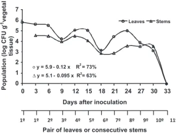
In the 1st pair of leaves inoculated with the Ac1Rif suspension at 3.4 x 10⁷ CFU mL-1, an initial population of 6.3 x 10⁵ CFU g-1 of leaf was detected two hours after inoculation (Day Zero) (Figure 1). Three days after inoculation the size of the population on the 1st pair of leaves remained stable (3.98 x 10⁵ CFU g-1 of leaf) and there was colonization of the consecutive stem, with 3.46 x 10⁴ CFU g-1 of stem (Figure 1).
FIGURE 1 - Colonization of Acidovorax citrulli resistant to
rifampicin (Ac1Rif) in leaves and consecutive stems following
inoculation in the 1st pair of leaves, performed 20 days after
sowing. The 1st pair of leaves was analyzed on Day Zero (2 h after
inoculation) and on Day 3.
0 1 2 3 4 5 6 7
0 3 6 9 12 15 18 21 24 27 30 33
Days after inoculation
Leaves Stems
1º 1º 2º 3º 4º 5º 6º 7º 8º 9º 10º 11º
Pair of leaves or consecutive stems
Population
(log
CFU
g
vegetal
tissue)
Throughout the evaluations, a general decrease in population size was observed in the leaves, with a drastic reduction in the 3rd and 6th pairs of leaves and consecutive stems at the 9th and 18th days after inoculation (Figure 1). Ac1Rif colonization was detected within 30 days after inoculation in the 10th pair of leaves, with a population of 1.3 x 10³ CFU g-1 of leaf. In this same time period, colonization in the consecutive segment of stem was 3.3 x 10³ CFU g-1 of stem. Bacterial populations were not detected in the 11th and 12th pair of leaves after 33 days.
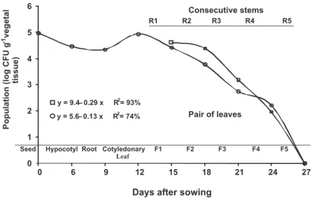
Seeds inoculated with Ac1Rif through vacuum infiltration (3.4 x 10⁷ CFU mL-1) showed an initial population of 1 x 10⁵ CFU g-1 of seed two hours after inoculation (Day Zero) (Figure 2). The bacterium colonized hypocotyl and roots, with populations of 2.81 x 10⁴ CFU g-1 and 2.18 x 10⁴ CFU g-1 of tissue, respectively. In cotyledonary leaves
A. citrulli population reached 8.7 x 10⁴ CFU g-1, from
which point there was colonization of leaves and stems until reaching undetectable levels at the 5th pair of leaves and consecutive stem 27 days after inoculation. The last populations detected (24 days after inoculation) were 1.62 x 10² CFU g-1 of leaf in the 4th pair of leaves and 9.1 x 101 CFU g-1 of stem in the consecutive stem (Figure 2).
The colonization dynamics of Ac1Rif in leaves and stems of the melon plants shows that inoculation in the 1st pair of leaves maintained higher A. citrulli populations throughout the evaluations (Figures 1 and 2) than when the bacterium was seed inoculated. The initial populations in the 1st pairs of leaves of the two experiments were 6.3 x 10⁵ and 2.57 x 10⁴ CFU g-1 of leaf, decreasing to undetectable levels at 33 and 27 days after inoculation, respectively.
Symptoms of BFB were observed through the experiments. Leaf spots were observed up to the 5th and
1st pairs of leaves, following leaf or seed inoculation, respectively. Leaf lesions were initially light green and oily, turning dark, with or without halo. When seeds were inoculated, hypocotyls and cotyledonary leaves showed water-soaked lesions which progressed to a brown color. As expected, no symptoms were found in roots or stems.
The difference between the Ac1Rif initial population in the 1st pair of leaves 2 h after inoculation (Figure 1) in relation to the concentration of the applied suspension may have occurred through drift, cell death, cell adherence to leaf surface, localization on protected sites, penetration or even the low sensitivity of the quantification method for detection of viable but non-culturable cells, as observed in other plant pathogenic bacteria (Beattie & Lindow, 1999; Pujol et al., 2007).
The decrease in population size as the bacterium colonized the leaves and consecutive stems, until reaching undetectable levels at 11th pair of leaves and consecutive stem 33 days following inoculation (Figure 1), may be related to the phenological stage of the plant, and physiological differences among the leaves. Kinkel et al. (2000) found that old leaves of grass species (near the base of the plant) consistently had larger bacterial populations than young leaves at the apex of the plant. This may be attributed to differences in the exudation rate of nutrients correlated with the age of the plant (Weller & Saettler, 1980), micro-climatology associated with the position of the leaf (Burage, 1976) or an incomplete colonization of young leaves (Kinkel et al., 2000). Differences in the position of the leaf are strongly correlated with differences in the population of bacteria that colonize individual leaves over time (Kinkel et al., 2000).
Seed Hypocotyl Root Cotyledonary F1 F2 F3 F4 F5
Leaf 0
1 2 3 4 5 6
0 6 9 12 15 18 21 24 27
Days after sowing
y = 9.4– 0.29 x R2= 93%
y = 5.6– 0.13 x R2= 74% Pair of leaves
Consecutive stems
R1 R2 R3 R4 R5
Population
(log
CFU
g
vegetal
tissue)
-1
FIGURE 2 - Colonization of Acidovorax citrulli resistant to rifampicin (Ac1Rif) in
The reduction in the Ac1Rif population on the 3rd and 6th pairs of leaves and consecutive stems (Figure 1) probably resulted from drastic alterations in temperature in the greenhouse, which reached approximately 41ºC, as the optimum temperature for the pathogen growth is 35ºC (Cavalcanti et al., 2005). When Ac1Rif was inoculated in the seeds, there was colonization of the hypocotyl and roots, an increase in the population in the cotyledonary leaves and subsequent colonization of leaves and stems (Figure 2). Leaf surface is considered a hostile environment for bacterial colonization due to the limited source of nutrients, exposure to rapid variations in temperature and relative humidity (Lindow & Brandl, 2003). Nevertheless A. citrulli exhibited higher adaptation to phyllosphere than to hypocotyls and roots. Silva et al. (2006) found that A. citrulli survived epiphytically for 54 days on leaves of melon plants under greenhouse and field conditions, with a population of 103 to 104 CFU g-1 of leaf, regardless of the concentration of the initial inoculum, and in the roots and rhizosphere in the same time period under greenhouse conditions, with populations of 102 to 103 CFU g-1 of root and 10CFU g-1 of soil.
Higher populations of Ac1Rif in leaves and stems throughout the evaluations (Figures 1 and 2) were found when the bacterium was inoculated in leaves compared to seed inoculation. This may be explained by initial populations detected in the 1st pairs of leaves, in each inoculation method, which were 6.3 x 10⁵ and 2.57 x 10⁴ CFU g-1 of tissue, respectively. Since the plants were grown tutored and watered by subirrigation, there is a strong possibility that the colonization of leaves and stems was systemic or endophytic rather than epiphytic. This situation is different in melon fields, where inoculum arrives constantly on aerial plant parts that have indeterminate and horizontal growth, thus facilitating pathogen dissemination and colonization (Lessl et al., 2007).
The absence of BFB symptoms beginning at the 6th or 2nd pair of leaves after inoculation of leaves and seeds, respectively, may be attributed to the low bacterial concentration detected, 1.3 to 0.58 x 10³ CFU g-1 of leaf, and to the low leaf-wetting conditions, explained by the subirrigation. Silveira et al. (2003) found symptoms of BFB on leaves of melon plants sprayed with A. citrulli (3.38 x 101 CFU mL-1)only when submitting them to a high leaf-wetting condition (moist chamber for 48 hours prior to and following inoculation). Also in the field, it is known that BFB epidemics develop when the rainy season favors a high humidity level inside canopy/in plantations.
Populations associated internally and externally with the leaves are likely to have continuity as a result of entry (ingression) or exiting (egression) processes in this organ. A number of studies suggest that the application of the pathogen on the plant surface results in internal colonization (Dane & Shaw, 1996; O’Brien & Lindow, 1989). Considering that application of Ac1Rif on the 1st pair of leaves was performed under high humidity conditions,
there must have been considerable epiphytic multiplication and penetration of the bacterium through the stomata in the initial hours, as reported by Young (1974) and Silva Neto et al. (2006).
Our findings confirmed those reported by Bahar et al. (2009), which provided, for the first time, strong evidence that at least group I strains of A. citrulli possessed vascular infection ability in melon seedlings. They also showed that in the xylem vessel colonization Type IV pili may play an important role under sap flow conditions, while polar flagella could be more important for spread during periods when xylem flow is reduced (Bahar et al., 2010). Egression is also important in the ecology of plant pathogens. Yang et al. (2001) found that approximately 14% of the
Xanthomonas citri subsp. malvacearum population was
present on the leaf surface after its infiltration, indicating that egression has quantitative importance to the external population. This phenomenon may have also occurred when the seeds were inoculated with Ac1Rif. A. citrulli group I colonized different parts of the melon plant up to 33 days after inoculation, depending on its initial location, leaves or seed. This confirms what has been observed in field, that expanded leaves and stems are the main inoculum sources for melon blossoms and fruit, therefore providing scientific bases for developing more effective strategies for BFB management.
ACKNOWLEDGEMENTS
The authors acknowledge the Conselho Nacional de Desenvolvimento Científico e Tecnológico – CNPq (PQ-304313/2005-0) for granting scholarships and the Fundação de Amparo à Ciência e Tecnologia do Estado de Pernambuco
– FACEP (APQ-0350-5.01/06) for financial assistance.
REFERENCES
Bahar O, Fuente LDL, Burdman S (2010) Assessing adhesion, biofilm formation and motility of Acidovorax citrulli using microfluidic flow chambers. FEMS Microbiology Letter 312:33-39.
Bahar O, Goffer T, Burdman S (2009) Type IV pili are required for virulence, twitching motility, and biofilm formation of Acidovorax avenae subsp. citrulli. Molecular Plant-Microbe Interactions 22:909-920.
Beattie GA, Lindow SE (1999) Bacterial colonization of leaves: a spectrum of strategies. Phytopathology 89:353-359.
Burage SW (1976) Aerial microclimate around plant surfaces. In: Dickinson CH, Preece TF (Eds.) Microbiology Aerial Plant Surface. San Francisco. Academic Press. pp.173-184.
Cavalcanti MT, Silveira EB, Mariano RLR, Oliveira IV (2005) Crescimento de Acidovorax avenae subsp. citrulli sob diferentes temperaturas, pH, concentração de cloreto de sódio e fontes de carbono. Ciência Rural 35:1313-1318.
Dane F, Shaw JJ (1996) Survival and persistence of bioluminescent
plants in the field environment. Journal of Applied of Bacteriology 80:73-80.
Kinkel LL, Wilson M, Lindow SE (2000) Plant species and plant incubation conditions influence variability in epiphytic bacterial population size. MicrobialEcology 39:1-11.
Lessl JT, Fessehaie A, Walcott RR (2007) Colonization of female watermelon blossoms by Acidovorax avenae subsp. citrulli and the relationship between blossom inoculum dosage and seed infestation. Journal of Phytopathology 155:114-121.
Lindow SE, Brandl MT (2003) Microbiology of the phyllosphere. Applied and Environmental Microbiology 69:1875-1883. Mariano RLR, Silveira EB, Assis SMP, Gomes AMA, Oliveira IS, Nascimento ARP (2001) Diagnose e manejo de fitobacterioses de importância no nordeste brasileiro. In: Michereff SJ, Barros R (Eds.) Proteção de plantas na agricultura sustentável. Recife PE. Imprensa Universitária. pp. 141-169.
Medeiros FHV, Moraes ISF, Silva Neto EB, Silveira EB, Mariano RLR (2009) Management of melon bacterial blotch by plant beneficial bacteria. Phytoparasitica 37:453-460.
O’Brien RD, Lindow SE (1989) Effect of plant species and environmental conditions on epiphytic population sizes of
Pseudomonas syringae and other bacteria. Phytopathology
79:619-627.
Pujol M, Badosa E, Montesinos E (2007) Epiphytic fitness of a biological control agent of fire blight in aple and pear orchards under Mediterranean weather conditions. FEMS Microbiology Ecology 59:186-193.
Pusey PL, Wilson CL (1984) Postharvest biological control of stone fruit brown rot by Bacillus subtilis. Plant Disease 68:753-756.
Silva VAV, Silveira EB, Mariano RLR (2006) Sobrevivência de
Acidovorax avenae subsp. citrulli em meloeiro. Fitopatologia
Brasileira 31:381-386.
Silva Neto EB, Silveira EB, Mariano RLR, Nogueira NL, Rossi ML, Santos LA (2006) Penetração e colonização de Acidovorax avenae subsp. citrulli em folhas, frutos e sementes de melão amarelo. Fitopatologia Brasileira 31:84-88.
Silveira EB, Michereff SJ, Mariano RLR (2003) Severidade da mancha-aquosa em meloeiro sob diferentes condições de molhamento foliar e concentração de inóculo de Acidovorax avenae subsp. citrulli. Fitopatologia Brasileira 28:171-175. Walcott RR, Fessehaie A, Castro AC (2004) Differences in pathogenicity between two genetically distinct groups of
Acidovorax avenae subsp. citrulli on cucurbit hozsta hosts?.
Journal of Phytopathology 152:277-85.
Walcott RR, Gitaitis R, Castro AC (2003) Role of blossoms in watermelon seed infestation by Acidovorax avenae subsp. citrulli. Phytopathology 93:528-534.
Walcott RR, Langston DB, Sanders FH JR, Gitaitis RD (2000) Investigating intraspecific variation of Acidovorax avenae subsp.
citrulli using DNA fingerprinting and whole cell fatty acid analysis. Phytopathology 9:191-6.
Weller DM, Saettler AW (1980) Colonization and distribution of
Xanthomonas phaseoli and Xanthomonas phaseoli var. fuscans in
field-grown navy beans. Phytopathology 70:500-506.
Yang CH, Crowley DE, Borneman J, Keen NT (2001) Microbial phyllosphere populations are more complex than previously realized. Proceedings of the National Academy of Sciences USA 98:3889-3894.
Young JM (1974) Development of bacterial populations in vivo in relation to plant pathogenicity. New Zealand Journal of Agricultural Research 17:105-113.