Chro nic co nve rting e nzyme inhibitio n
no rmalize s Q T inte rval in aging rats
1Departamento de Ciências Biológicas, Faculdade de Medicina do Triângulo Mineiro,
Uberaba, MG, Brasil
2Departamento de Fisiologia, Faculdade de Medicina de Ribeirão Preto,
Universidade de São Paulo, Ribeirão Preto, SP, Brasil V.J. Dias da Silva1,
E. Ferreira Neto1,
H.C. Salgado2 and
R. Fazan Júnior1
Abstract
The aim of the present study was to investigate the effects of convert-ing enzyme inhibition by captopril on ECG parameters in aged rats. Four-month-old male rats received captopril dissolved in tap water (0.5 mg/l) or tap water for 2 or 20 months. At the end of treatment, under anesthesia, RR and PR interval, P wave and QRS duration, QT and corrected QT interval were measured in all animals. On the following day, chronic ECG (lead II) recordings were performed to quantify supraventricular (SVPB) or ventricular premature beats (VPB). After sacrifice, the hearts were removed and weighed. RR interval was similar in young and untreated aged rats, but significantly larger in aged rats treated with captopril. P wave and QRS length did not differ among groups. PR interval was significantly larger in old than in young rats and was not affected by captopril. Corrected QT interval was larger in aged than in young rats (117 ± 4 vs 64 ± 6 ms, P<0.05) and was reduced by captopril (71 ± 6 ms, P<0.05). VPB were absent in young rats and highly frequent in untreated old animals (8.4 ± 3.0/30 min). Captopril significantly reduced VPB in old rats (0.3 ± 0.1/30 min, P<0.05). The cardiac hypertrophy found in untreated aged rats was prevented by captopril (3.44 ± 0.14 vs 3.07 ± 0.10 mg/g, P<0.05). The beneficial effects of angiotensin converting enzyme inhibition on the rat heart during the aging process are remarkable.
Co rre spo nde nce
V.J. Dias da Silva
Departamento de Ciências Biológicas Faculdade de Medicina do Triângulo Mineiro Praça Manoel Terra, 330 38015-050 Uberaba, MG Brasil
Fax: + 55-34-3318-5466 E-mail: valdo@ mednet.com.br
Presented at the IV International Symposium on Vasoactive Peptides, Belo Horizonte, MG, Brazil, O ctober 19-21, 2001.
Research supported by PRO NEX-CNPq, FAPESP and FUNEPU. E. Ferreira Neto was supported by a scientific training grant from PIBIC program of CNPq.
Received November 22, 2001 Accepted May 3, 2002
Ke y words
•Aging
•Electrocardiogram •Captopril •Rats
Intro ductio n
Several electrocardiographic (ECG) in-dices have been proposed to identify patients at risk of sudden death, including the QT interval length and/or dispersion (1-3). Clini-cal trials have demonstrated that QT interval is particularly altered in situations such as cardiac hypertrophy (4) or myocardial ische-mia (5), but is normalized by appropriate therapy (6). The myocardial alterations that occur with aging (7,8) alter ECG parameters
such as QT interval (9,10).
Aging is associated with electrical and morphological changes of the myocardium (7,8), increasing significantly the incidence of life-threatening cardiac arrhythmias and sudden death in aged subjects (9-11).
the renin-angiotensin system (RAS). In ad-dition, other vasoactive peptides can be also modified by ACE inhibitors, especially bradykinin, which is potentiated by ACE blockage (12,13). These effects elicit a de-crease in arterial pressure in several experi-mental models of hypertension and clinical forms of human hypertension as well (12,13). In addition, these drugs improve cardiac per-formance in patients with congestive heart failure, reducing cardiovascular risk and mortality (12,13).
Studies performed on experimental mod-els, particularly rats, have evaluated the ef-fects of ACE inhibitors on aging. Concern-ing the cardiovascular system, these studies have demonstrated that ACE inhibitors re-duce renal intravascular resistance (14) and thickness of the media and intima layers of large arteries (15). Moreover, ACE inhibi-tors improve the endothelial function of re-sistance vessels (16), and partially restore the impaired autoregulatory mechanism of cerebral blood flow due to aging (17).
Nevertheless, to our knowledge, the ef-fect of ACE inhibitors on the alterations of ECG found in aging has not been previously examined in rats. Therefore, the aim of the present study was to evaluate ECG alter-ations in aged rats, as well as the effect of the ACE inhibitor captopril in this experimental model.
Mate rial and Me thods
Four-month-old male Wistar rats were divided into four groups. One group was treated with captopril dissolved in tap water (0.5 mg/ml) for 2 months (N = 10), a second group was treated with captopril dissolved in tap water (0.5 mg/ml) for 20 months (N = 7), a third age-matched time control group (N = 10) drank only tap water for 2 months, and a fourth age-matched time control group (N = 5) drank only tap water for 20 months. The rats drank about 20-30 ml/day of capto-pril solution which provided approximately
30 mg/kg of captopril a day. The efficacy of this dosage of captopril has been demon-strated in a number of pharmacokinetic and pharmacodynamic studies in rats (18). All surgical procedures and protocols were in accordance with the Guidelines for Ethical Care of Experimental Animals and were ap-proved by the Institutional Animal Care and Use Committee.
At the end of a 2- or 20-month period of treatment, the animals were submitted to acute ECG studies under tribromoethanol anesthesia (250 mg/kg, ip). Electrodes were placed under the skin for recording the con-ventional bipolar limb leads (I, II, III), the unipolar limb leads (aVR, aVL and aVF), and the unipolar precordial (chest) leads (VA is immediately to the right of the sternum in the 4th intercostal space, VB is just to the left of the sternum in the 4th intercostal space, and VC is in the 5th intercostal space at the midaxillary line). In order to avoid errors in the position of the leads, the electrodes were always placed by the same person. The ECG was recorded using a three-channel digital ECG recorder (ER-65, Medikor, Budapest, Hungary) with a paper speed of 50 mm/s and sensitivity of 0.5 mV/cm. Each lead was recorded for 20 s.
At the end of the acute ECG recordings, the animals were implanted with a pair of stainless-steel electrodes positioned inside the subcutaneous tissue for chronic record-ing of conventional bipolar limb lead II with-out the effect of anesthesia. The animals were also cannulated with polyethylene tub-ing placed into the femoral artery and vein for direct measurement of arterial pressure and drug administration, respectively. After the surgical procedures the animals were left to recover in individual cages for at least 24 h.
Hz) with a personal computer (IBM/PC) equipped with a 12-bit analog to digital in-terface (CAD12/36 Lynx Tecnologia Eletrô-nica, São Paulo, SP, Brazil) for a period of 30 min. At the end of the ECG recording, the arterial catheter was connected to a pressure transducer (model P23Gb Statham, Hato Rey, Puerto Rico) attached to a pressure amplifier (model 8805D, Hewlett Packard) which fed the arterial pressure signal to a personal computer. The efficacy of ACE blockade was evaluated by the attenuation of the hy-pertensive response elicited by angiotensin I (100 ng/kg) given through the femoral vein. Only rats showing an attenuation of at least 85% of the hypertensive response elicited by angiotensin I were considered to have the RAS blockade. After the test of ACE effi-cacy the rats were killed with excess anes-thesia and had their hearts removed and weighed on a precision scale (Micronal B160, São Paulo, SP, Brazil).
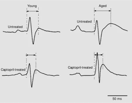
The ECG tracings were analyzed visually always by the same person, who was not aware of the protocol. The following ECG parameters were examined: 1) RR interval, defined as the interval between the apex of adjacent R waves; 2) P wave duration; 3) PR interval, defined as the interval between the apex of the P wave and the Q wave (begin-ning of the QRS complex); 4) QRS duration; 5) QT interval (defined as the interval be-tween Q wave and T wave apex), and 6) corrected QT interval [QTc, defined as the QT interval corrected for the heart rate by means of Bazett’s equation: corrected QTc = QT (in s)/RR (in s)1/2]. In small rodents, in
contrast to humans, the T wave is not well characterized and appears as a shoulder of the QRS complex (Figure 1). Accordingly, in order to measure the QT interval we used the apex of the T wave which can be deter-mined with high accuracy. ECG recordings were carried out for 20 s for each lead, and the ECG parameters described above were determined from each lead and averaged.
The 30-min ECG recordings were
care-fully examined on the screen of the com-puter to identify premature heart beats. The total number of supraventricular (SVPB) and ventricular premature beats (VPB) were counted over the 30-min period. The classic definition of arrhythmias in humans, adapt-ed to the high heart rate of the rat, has been described elsewhere (19,20) and was used to define the severity of ventricular arrhythmias. Briefly: class 0 no VPB, class 1 -infrequent isolated unifocal VPB (<30/h), class 2 - frequent unifocal VPB (>30/h), class 3 multifocal ectopic beats, class 4 -couplets of VPB, class 5 - triplets of VPB and non-sustained ventricular tachycardia (<6 ectopic beats), and class 6 - ventricular tachycardia.
Data are reported as means ± SEM. For ECG parameters and arterial pressure data, two-way ANOVA followed by Tukey’s mul-tiple comparison test was performed to evalu-ate the effects of treatment (captopril vs tap water) and age (young vs aged). The arrhyth-mia data, i.e., incidence of premature beats, were analyzed by the Kruskal-Wallis
Untreated
Untreated
Captopril-treated Captopril-treated
50 ms
Young Aged
ANOVA test. The differences were consid-ered significant when P<0.05.
Re sults
Untreated aged rats were significantly heavier than untreated young rats (512 ± 22
vs 460 ± 15 g, P<0.05). In contrast, young
rats (treated or not) and aged rats treated with captopril presented similar body weights (434 ± 26, 460 ± 24 and 460 ± 15 g, respec-tively).
The basal mean arterial pressure and heart rate of conscious rats are shown in Table 1. Arterial pressure and heart rate were signifi-cantly lower in old rats treated with captopril compared to other groups.
The pressor response to angiotensin I was 35 ± 3 mmHg in young untreated rats and 4 ± 2 mmHg in young rats treated with captopril (P<0.001). Comparison of these values indicates a blockade of ACE by cap-topril of approximately 89%. In aged rats, the pressor response to angiotensin I was 68 ± 18 mmHg in untreated rats, and 1 ± 2 mmHg in rats treated with captopril (P<0.0001), indicating an ACE blockade of approximately 98%.
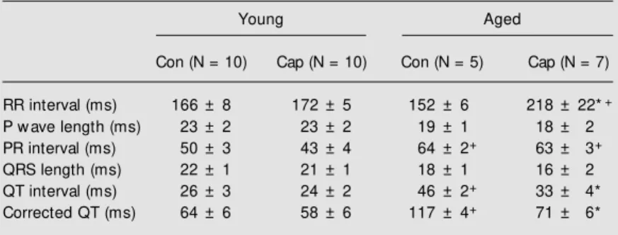
The ECG parameters are presented in Table 2. The RR interval was similar in young (treated or not) and old untreated rats, but significantly larger in aged subjects treated with captopril. P wave length did not differ among groups. The PR interval was significantly larger in old than in young rats, but was not affected by captopril in either old or young rats. QRS length did not differ among groups. The QT and QTc intervals were significantly larger in old than in young rats, but were significantly reduced by cap-topril in old, but not in young rats (Table 2). The incidence of SVPB was low in young rats, but high in old rats (Table 3). Captopril did not change the incidence of SVPB. VPB were absent in young rats (treated or not), but highly frequent in untreated old subjects (Table 3). Nevertheless, captopril signifi-cantly reduced VPB in old rats. Ventricular arrhythmias was 30 times less frequent than in untreated old rats (Table 3). According to the classification of ventricular arrhythmia adopted in the present study (20), all young rats belonged to class 0, whereas 80% of the untreated old rats belonged to class 1, 2 or 3. As a result of treatment with captopril, the Table 3. Number (mean ± SEM ) of supraventricular (SVPB) and ventricular premature
beats (VPB) in conscious male young and aged rats treated (Cap) or not (control, Con) w ith captopril.
Young Aged
Con (N = 10) Cap (N = 10) Con (N = 5) Cap (N = 7)
SVPB 0.5 ± 0.1 0.3 ± 0.1 5.2 ± 1.0+ 3.0 ± 0.5+
VPB 0.0 ± 0.0 0.0 ± 0.0 8.4 ± 3.0+ 0.3 ± 0.1*
Total 0.5 ± 0.1 0.3 ± 0.1 13.6 ± 3.3+ 3.3 ± 0.5*
* P<0.05 vs untreated aged rats (Kruskal-Wallis ANOVA test).
+P<0.05 vs young rats (Kruskal-Wallis ANOVA test).
Table 2. M ean values (± SEM ) of ECG parameters measured in anesthetized young and aged rats treated (Cap) or not (control, Con) w ith captopril.
Young Aged
Con (N = 10) Cap (N = 10) Con (N = 5) Cap (N = 7)
RR interval (ms) 166 ± 8 172 ± 5 152 ± 6 218 ± 22*+
P w ave length (ms) 23 ± 2 23 ± 2 19 ± 1 18 ± 2
PR interval (ms) 50 ± 3 43 ± 4 64 ± 2+ 63 ± 3+
QRS length (ms) 22 ± 1 21 ± 1 18 ± 1 16 ± 2
QT interval (ms) 26 ± 3 24 ± 2 46 ± 2+ 33 ± 4*
Corrected QT (ms) 64 ± 6 58 ± 6 117 ± 4+ 71 ± 6*
* P<0.05 vs untreated aged rats (Tukey’s multiple comparison test).
+P<0.05 vs young rats (Tukey’s multiple comparison test).
Table 1. Baseline values (mean ± SEM ) of mean arterial pressure (M AP) and heart rate (HR) in conscious male young and aged rats treated (Cap) or not (control, Con) w ith captopril.
Young Aged
Con (N = 10) Cap (N = 10) Con (N = 5) Cap (N = 7)
M AP (mmHg) 89 ± 2 93 ± 4 97 ± 2 85 ± 2*
HR (bpm) 381 ± 11 374 ± 15 405 ± 11 311 ± 25*
percentage of old rats in class 1 was 29%. No old rat treated with captopril was found in class 2 or 3, and no rat was found in classes 4, 5 or 6.
Relative heart weight (mg/g of body weight) was found to be similar in young rats treated (2.62 ± 0.08 mg/g) or not (2.70 ± 0.05 mg/g) with captopril. Aged rats without any treatment presented an increase in relative heart weight (3.44 ± 0.14 mg/g, P<0.05) as compared to young rats, but captopril brought the relative heart weight of aged rats (3.07 ± 0.10 mg/g, P<0.05) within the range of young rats.
D iscussio n
The remarkable attenuation of the pres-sor response produced by angiotensin I dem-onstrated the efficacy of ACE blockade by captopril.
Heart rate did not change with aging, in agreement with previous reports in the lit-erature (21-23). Berg (24) also found no heart rate changes in rats aged 219 days (~7 months) or 557 days (~18 months), whereas they detected bradycardia in older rats aged 851 days (~28 months) and 951 days (~31 months). In the present study, chronic (20 months) captopril treatment of aged rats re-duced the basal heart rate compared to age-matched control rats.
The bradycardia observed in aged rats treated with captopril may be associated with an increase in the vagal reflex controlling the heart rate caused by ACE inhibition (25), even though further studies are required to better understand this mechanism.
Concerning the other ECG parameters, the results of the present study have shown some differences in cardiac electrical activ-ity with major alterations of PR and QT interval with aging. The PR interval is an index that correlates well with atrioventricu-lar conduction, and an increase in this pa-rameter indicates an impairment in atrioven-tricular conduction. The present finding that
aging decreases electrical conduction in the atrioventricular node is in accordance with previous findings in experimental animals (24) and humans (26).
The QT interval, i.e., the time elapsed for ventricular repolarization (ventricular refrac-tory period), was also increased in aged rats, corroborating ECG (24) and electrophysi-ological data (27) obtained for aged rats. A prolonged QT interval has been associated with cardiac arrhythmia and sudden death in humans (1,2,5). At the cellular level, ven-tricular repolarization is prolonged in a num-ber of cardiac disturbances such as myocar-dial hypertrophy, ischemia or congestive heart failure (1,2,5). Changes of the QT in-terval have also been described in experi-mental models of hypertension (28). The other parameters examined in the present study were found to be unchanged by aging. SVPB and VPB were the most frequent cardiac arrhythmias found in aged rats in the present study. Despite the short period of ECG monitoring (30 min), this finding sub-stantiates previous literature reports describ-ing a higher incidence of this kind of arrhyth-mia, linked to aging, in rats submitted to 24-h Holter monitoring (20).
The impaired atrioventricular conduction (larger PR interval), the prolonged ventricu-lar repoventricu-larization (ventricu-larger QT interval), and the higher incidence of cardiac arrhythmias could be produced by degenerative lesions due to myocardial fibrosis and/or ischemia, as well as alterations in gene expression associated with the aging process (7,8,29).
direct anti-trophic effect on cardiac myocyte proliferation, preventing myocardial fibro-sis by means of the attenuation of the effect of angiotensin II, as well as inhibition of bradykinin degradation (12,13). The devel-opment of apoptosis in heart tissue is also associated with the hyperactivity of local ACE (13). ACE inhibitors also improve the coronary blood flow (12,13) which is im-paired with aging (7,8).
The prolonged PR interval observed in aged rats is probably related to the same structural and functional changes due to ag-ing (7,8). A possible explanation for the failure of captopril to normalize the PR in-terval is that ACE inhibition increases vagal control of the heart in aged rats (25), and it has been well documented that augmented vagal nerve activity increases the delay of atrioventricular conduction under physiologi-cal conditions (32). Therefore, the larger PR interval found in aged rats treated with cap-topril could be associated with a shift in the sympathovagal balance toward an increased parasympathetic activity. However, further studies are necessary to clarify this issue.
Chronic treatment with captopril almost eliminated the ventricular arrhythmias but did not affect SVPB in aged rats. This effect of ACE inhibitors seems not to be restricted to aging. In fact, a significant reduction in the incidence of ectopic beats has been ob-served in experimental (33,34) and clinical (35) arterial hypertension, acute myocardial infarction (36) and congestive heart failure (37) after chronic ACE inhibition. There are a number of hypotheses to explain this anti-arrhythmic effect. For instance, structural
changes in fibrosis and/or cardiac hypertro-phy and remodeling (12,13), functional changes in coronary blood flow (12,13), au-tonomic imbalance (25), ion channel dys-function (20) and intracellular gene expres-sion (38).
In the present study a cardiac hypertro-phy evaluated by relative cardiac weight was reported in untreated aged rats. Cardiac hy-pertrophy in aged male rats is a common feature reported by many investigators (7,8, 28,38). The heart undergoes myocardial cell enlargement associated with myocardial fi-brosis (7,8). It is well accepted that this increase in heart weight is caused by the reduced diastolic stiffness of the left ven-tricle, and by changes in the properties of large arteries during the aging process (7,8,28). At the cellular level, excitation-contraction coupling is prolonged by aging (7,8,28).
Captopril was able to prevent cardiac hypertrophy in aged rats. This effect could be ascribed to the hemodynamic effects of captopril, which reduced the mean arterial pressure of aged rats. However, a direct ef-fect of captopril inhibiting the trophic efef-fect of angiotensin II on myocardial cells or fi-brosis should be considered as well.
Rat aging is characterized by the devel-opment of ECG alterations associated with cardiac hypertrophy, typical of myocardial disorders. Chronic treatment with the ACE inhibitor, captopril, prevents some of these ECG alterations, as well as cardiac hypertro-phy in rats. Therefore, ACE inhibitors have a remarkable beneficial effect on the heart during the aging process in rats.
Re fe re nce s
1. Locati E & Schw artz PJ (1987). Prognostic value of QT interval prolongation in post myocardial infarction patients. European Heart Journal, 8 (Suppl A): 121-126. 2. Goldberg RJ, Bengt son J, Chen ZY,
Anderson KM , Locati E & Levy D (1991). Duration of the QT interval and total and cardiovascular mortality in healthy
per-sons (The Framingham Heart Study expe-rience). American Journal of Cardiology, 67: 55-58.
3. Statters DJ, M alik M , Ward DE & Camm AJ (1994). QT dispersion: problems of methodology and clinical significance. Journal of Cardiovascular Electrophysiol-ogy, 5: 672-685.
4. M ayet J, Shahi M , M cGrath K, Poulter NR, Sever PS, Foale RA & Thom SA (1996). Left ventricular hypertrophy and QT dispersion in hypertension. Hyperten-sion, 28: 791-796.
myocardial infarction: the BHAT experi-ence. The BHAT Study Group. Journal of Clinical Epidemiology,43: 167-172. 6. M oreno FL, Villanueva T, Karagounis LA &
Anderson JL (1994). Reduction in QT in-terval dispersion by successful throm-bolytic therapy in acute myocardial infarc-tion. TEAM -2 Study Investigators. Circula-tion, 90: 94-100.
7. Folkow B & Svanborg A (1993). Physiolo-gy of cardiovascular aging. Physiological Review s, 73: 725-764.
8. Lakatta EG (1993). Cardiovascular regula-tory mechanisms in advanced age. Physi-ological Review s, 73: 413-467.
9. Reardon M & M alik M (1996). QT interval change w ith age in an overtly healthy older population. Clinical Cardiology, 19: 949-952.
10. Perkiomaki JS, Sourander LB, Levomaki L, Raiha IJ, Puukka P & Huikuri HV (2001). QT dispersion and mortality in the elderly. Annals of Noninvasive Electrocardiology, 6: 183-192.
11. Fleg JL (1988). Ventricular arrhythmias in the elderly: prevalence, mechanisms, and therapeutic implications. Geriatrics, 43: 23-29.
12. Unger T, Gohlke P & Gruber M G (1990). Converting enzyme inhibitors. In:Ganten D & M ulrow PJ (Editors), Handbook of Experim ent al Pharm acology. Vol. 93. Springer-Verlag, Berlin, Germany, 377-481.
13. Brow n NJ & Vaughan DE (1998). Angio-tensin-converting enzyme inhibitors. Cir-culation, 97: 1411-1420.
14. Heudes D, M ichel O, Chevalier J, Scalbert E, Ezan E, Bariety J, Zimmerman A & Corman B (1994). Effect of chronic ANG I-converting enzyme inhibition on aging processes. I. Kidney structure and func-tion. American Journal of Physiology, 266: R1038-R1051.
15. M ichel JB, Heudes D, M ichel O, Poitevin P, Philippe M , Scalbert E, Corman B & Levy BI (1994). Effect of chronic ANG I-converting enzyme inhibition on aging processes. II. Large arteries. American Journal of Physiology, 267: R124-R135. 16. Atkinson J, Tatchum-Talom R & Corman
B (1994). Effect of chronic ANG I-convert-ing enzyme inhibition on agI-convert-ing processes. III. Endothelial function of mesenteric ar-terial bed of rat. American Journal of Phys-iology, 267: R136-R143.
17. Lartaud I, M akki T, Bray-Des-Boscs L, Niederhoffer N, Atkinson J, Corman B &
Capdeville-Atkinson C (1994). Effect of chronic ANG I-converting enzyme inhibi-tion on aging processes. IV. Cerebral blood flow . American Journal of Physiolo-gy, 267: R687-R694.
18. Sun Y & M endelsohn FA (1991). Angio-tensin converting enzyme inhibition in heart, kidney, and serum studied ex vivo after administration of zofenopril, capto-pril, and lisinopril. Journal of Cardiovascu-lar Pharmacology, 18: 478-486.
19. Low n B & Wolf M (1971). Approaches to sudden death from coronary heart dis-ease. Circulation, 46: 130-139.
20. Carré F, Lessard Y, Coumel P, Ollivier L, Besse S, Lecarpentier Y & Sw ynghedauw B (1992). Spontaneous arrhythmias in vari-ous models of cardiac hypertrophy and senescence of rats: a Holter monitoring study. Cardiovascular Research, 26: 698-705.
21. Tanabe S & Bunag RD (1989). Age-related central and baroreceptor impairment in female Sprague-Daw ley rats. American Journal of Physiology, 256: H1399-H1406. 22. Werner A, Rosa NR, Oliveira AR, Fernan-des TG, Belló AA & Irigoyen M C (1995). Changes in blood pressure control in aged rats. Brazilian Journal of M edical and Bio-logical Research, 28: 603-607.
23. Irigoyen M C, M oreira ED, Werner A, Ida F, Pires M D, Cestari IA & Krieger EM (2000). Aging and baroreflex control of RSNA and heart rate in rats. American Journal of Physiology,279: R1865-R1871. 24. Berg BN (1955). The electrocardiogram in aging rats. Journal of Gerontology, 10: 420-423.
25. Bunag R, M ellick J & Allen B (1999). Abat ed cardiovascular responses t o chronic oral lisinopril treatment in con-scious elderly rats. American Journal of Physiology, 276: R1408-R1415. 26. Fleg JL, Das DN, Wright J & Lakatta EG
(1990). Age-associated changes in the components of atrioventricular conduc-tion in apparently healthy volunteers. Journal of Gerontology, 45: M 95-M 100. 27. Walker KE, Lakatta EG & Houser SR
(1993). Age associated changes in mem-brane currents in rat ventricular myocytes. Cardiovascular Research, 27: 1968-1977. 28. Baillard C, M ansier P, Ennezat PV, M angin L, M edigue C, Sw ynghedauw B & Cheva-lier B (2000). Converting enzyme inhibi-tion normalizes QT interval in spontane-ously hypertensive rats. Hypertension, 36: 350-354.
29. Lakatta EG (1993). M yocardial adaptations in advanced age. Basic Research in Cardi-ology,88 (Suppl 2): 125-133.
30. Thollon C, Kreher P, Charlon V & Rossi A (1989). Hypertrophy induced alteration of action potential and effects of the inhibi-tion of angiotensin converting enzyme by perindopril in infarcted rat hearts. Cardio-vascular Research,23: 224-230. 31. Gonzalez-Juanatey JR, Garcia-Acuna JM ,
Pose A, Varela A, Calvo C, Cabezas-Cerrato J & de la Pena M G (1998). Reduc-tion of QT and QTc dispersion during long-term treatment of systemic hypertension w ith enalapril. American Journal of Cardi-ology, 81: 170-174.
32. Thomas Jr JX & Randall WC (1983). Auto-nomic influences on atrioventricular con-duction in conscious dogs. American Jour-nal of Physiology, 244: H102-H108. 33. Pahor M , Bernabei R, Sgadari A,
Gam-bassi G, Lo Guidice PL, Pacifici L, Ramacci M T, Lagrasta C, Olivetti G & Carbonin P (1991). Enalapril prevents cardiac fibrosis and arrhythmias in hypertensive rats. Hy-pertension, 18: 148-157.
34. Chevalier B, Heudes D, Heymes C, Bas-set A, Dakhli T, Bansard Y, Jouquey S, Hamon G, Bruneval P, Sw ynghedauw B & Carré F (1995). Trandolapril decreases prevalence of ventricular ectopic activity in middle-aged SHR. Circulation,92: 1947-1953.
35. Gonzalez-Fernandez RA, Rivera M , Rodri-guez PJ, Fernandez-M artinez J, Soltero LH, Diaz LM & Luggo JE (1993). Preva-lence of ectopic ventricular activity after ventricular mass regression. American Journal of Hypertension, 6: 308-313. 36. Singh SN, Karasik P, Hafley GE, Pieper
KS, Lee KL, Wyse DG & Buxton AE (2001). Electrophysiologic and clinical effects of angiotensin-converting enzyme inhibitors in patients w ith prior myocardial infarc-tion, nonsustained ventricular tachycardia, and depressed left ventricular function. M USTT Investigators. M ulticenter Unsus-tained Tachycardia Trial. American Jour-nal of Cardiology, 87: 716-720.
37. Hattori Y, Atsushi S, Hiroaki F & Toyama J (1997). Effects of cilazapril on ventricular arrhythmia in patients w ith congestive heart failure. Clinical Therapeutics, 19: 481-486.