Antioxidant responses of Laeonereis acuta
(Polychaeta) after exposure to hydrogen
peroxide
C.E. da Rosa, A. Bianchini and J.M. Monserrat
Departamento
de Ciências Fisiológicas, Programa de PósGraduação em Ciências Fisiológicas
-Fisiologia Animal Comparada, Fundação Universidade Federal do Rio Grande, Rio Grande, RS, Brasil
Correspondence to: J.M. Monserrat, Departamento de Ciências Fisiológicas, Programa de
Pós-graduação em Ciências Fisiológicas - Fisiologia Animal Comparada, Fundação Universidade
Federal do Rio Grande, A. Itália, km 8, Campus Carreiros, 96201-900 Rio Grande, RS, Brasil
Fax: +55-53-3233-8680. E-mail: josemmonserrat@pesquisador.cnpq.br
The effects of H2O2 were evaluated in the estuarine worm Laeonereis acuta (Polychaeta, Nereididae) collected at the Patos
Lagoon estuary (Southern Brazil) and maintained in the laboratory under controlled salinity (10 psu diluted seawater) and temperature (20°C). The worms were exposed to H2O2 (10 and 50 µM) for 4, 7, and 10 days and the following variables were
determined: oxygen consumption, catalase (CAT) and glutathione peroxidase activity in both the supernatant and pellet fractions of whole body homogenates. The concentrations of non-protein sulfhydryl and lipid peroxides (LPO) were also measured. The oxygen consumption response was biphasic, decreasing after 4 days and increasing after 7 and 10 days of exposure to 50 µM H2O2 (P < 0.05). At the same H2O2 concentration, CAT activity was lower (P < 0.05) in the pellet fraction of worms exposed for
10 days compared to control. Non-protein sulfhydryl concentration and glutathione peroxidase activity were not affected by H2O2
exposure. After 10 days, LPO levels were higher (P < 0.05) in worms exposed to 50 µM H2O2 compared to control. The reduction
in the antioxidant defense was paralleled by oxidative stress as indicated by higher LPO values (441% compared to control). The reduction of CAT activity in the pellet fraction may be related to protein oxidation. These results, taken together with previous findings, suggest that the worms were not able to cope with this H2O2 concentration.
Key words: Worms; Laeonereis acuta; Hydrogen peroxide; Oxidative stress; Antioxidant enzymes; Oxygen consumption Research supported by FINEP (No. 23.01.0719.00), CNPq (No. 300536/90-9), and PGCF-FAC. C.E. da Rosa was the recipient of a CAPES fellowship. J.M. Monserrat and A. Bianchini are recipients of CNPq research fellowships.
Received March 16, 2007. Accepted October 18, 2007
Hydrogen peroxide (H2O2) is a non-radical reactive oxygen species and the most stable intermediate in the four-electron reduction of O2 to water. In the aquatic envi-ronment, H2O2 predominantly derives from UV-driven photoactivation of dissolved organic matter (1). Since H2O2 is uncharged, it easily passes through cell membranes by diffusion, and when inside the cells it can react with transi-tion metals liberating hydroxyl radicals (HO•) (2). At high
concentrations, these radicals induce peroxidation of lip-ids and proteins, affecting cell integrity (2,3).
Aquatic organisms have to cope with a wide variety of
environmental oxidants, as well as with those pro-duced by normal aerobic metabolism, leading to the re-quirement of efficient antioxidant mechanisms. Aerobic cells have acquired a variety of antioxidant mechanisms, including enzymatic (superoxide dismutase, catalase (CAT), glutathione peroxidase (GPx)) and non-enzymatic (glutathione, carotenoids, α-tocopherol, etc.) defenses (4).
enzymes (CAT, superoxide dismutase) (2) and a reduction in oxygen consumption (2,4,5).
Since H2O2 is a conspicuous environmental pro-oxi-dant, the aim of the present study was to evaluate the effects of H2O2 exposure on oxygen consumption, on H2O2 detoxification enzymes in the pellet and supernatant frac-tions, non-protein sulfhydryl groups and oxidative damage (LPO) of the estuarine worm Laeonereis acuta (Polychaeta, Nereididae). The estuarine worm L. acuta has been widely employed in toxicological and environmental studies for analysis of their antioxidant and oxidative damage re-sponses to copper, cadmium and cyanobacterium bloom events and in biomonitoring programs (6-9).
Specimens of L. acuta (60-120 mg) were collected in a salt marsh (“Saco do Justino”) near Rio Grande city (South-ern Brazil, 32° S, 52° W). This site was reported to be unpolluted (6).
The organisms were maintained under laboratory con-ditions as previously described (10). Briefly, the worms were kept individually in glass dishes (6.0 cm in diameter) containing a thin sand layer and approximately 100 mL of 10 psu diluted seawater at pH 8.0, 20°C. The fixed photo-period was a 12-h light:12-h dark cycle. During the accli-mation period (5 days) the animals were fed frozen Artemia sp and 100% of the water was renewed every 2 days.
After the acclimation period the animals were divided into three groups: a control group exposed to diluted seawater (salinity = 10 psu; N = 60), and a second (N = 60) and a third group (N = 60) receiving diluted seawater (salinity = 10 psu) containing 10 and 50 µM hydrogen peroxide, respectively. The condition was the same as employed in the acclimation period except for the absence of sand in dishes and for water renewal, which was done daily. No mortality was recorded during the experimental period.
At the end of the exposure period of 4, 7, and 10 days, some of the animals (N = 10 per experimental group) were used for the oxygen consumption assay and the remaining ones were frozen at -70°C for later analyses.
For the determination of oxygen consumption (11) the animals were transferred to 10-mL chambers containing 10 psu diluted seawater, pH 8.0, at 20°C. Oxygen con-sumption was recorded with a manual oximeter (DIGIMED, São Paulo, SP, Brazil). Values are reported as mg O2 h-1 g wet weight-1.
For the enzymatic determinations (12), whole animals were homogenized with cold buffer (1:3, v/v) containing 0.5 M sucrose and 0.15 M NaCl in 20 mM Tris-HCl, pH 7.6. The homogenate was centrifuged at 500 g for 15 min at 4°C and the resulting supernatant was centrifuged at 12,000 g for 30 min at 4°C. The 12,000-g pellet (peroxisomal and
mitochondrial fraction) was resuspended with the homog-enization buffer in the same volume as employed for homogenization. Both extracts (supernatant of 12,000 g and the resuspended pellet) were used for the determina-tion of CAT and GPx activity since CAT activity is expected to occur only in the pellet fraction and GPx activity in the cytosolic fraction.
CAT activity was measured as the rate of enzymatic decomposition of H2O2 monitored as a decrease of ab-sorbance at 240 nm (13). Enzyme activity is reported as CAT units, with one unit being the amount of enzyme needed to hydrolyze 1 µmol H2O2 min-1 mg protein-1 at 30°C and pH 8.0. GPx-Se activity (14) was measured as NADPH oxidation measured at 340 nm in the presence of excess glutathione reductase, reduced glutathione, H2O2, and aliquots of the homogenate. The results are reported as GPx units, with one unit being the amount of enzyme necessary to oxidize 1 µmol NADPH min-1 mg protein-1 at 30°C and pH 7.2.
For the measurement of non-protein sulfhydryl groups (NP-SH) (15), tissues were homogenized (1:20) in 20 mM EDTA. NP-SH content was measured after deproteinization with 50% trichloroacetic acid. NP-SH were detected using 5,5-dithiobis(2-nitrobenzoic acid) and absorbance at 405 nm was determined in a microplate reader. The result was divided by the protein concentration of each sample prior to deproteinization and is reported as specific activity,
ηmol glutathione/mg protein.
LPO was measured by the Fox method (16) based on Fe2+ oxidation by lipid hydroperoxides (FOX reactive sub-stances) at acid pH in the presence of the Fe3+-complexing dye xylenol orange. Samples were homogenized (1:9) in 100% cold (4°C) methanol. The homogenate was centri-fuged at 1000 g for 10 min at 4°C and the supernatant was collected and used for LPO determination (580 nm). Cumene hydroperoxide was employed as standard. The results are reported as ηmol cumene hydroperoxide/g
tissue.
Significant differences between treatments were as-sessed by ANOVA in combination with the Newman-Keuls a posteriori test, with the level of significance set at 5%.
Figure 1. Figure 1.Figure 1. Figure 1.
Figure 1. Catalase (CAT) activity (in units, U) in the supernatant (A) and pellet (B) fraction of Laeonereis acuta homogenates after exposure to 0 (control), 10, or 50 µM hydrogen peroxide. Glutathione peroxidase (GPx) activity (in units, U) in the supernatant (C) and pellet (D) fraction of L. acuta homogenates after exposure to 0 (control), 10, or 50 µM hydrogen peroxide. Data are reported as means ± SD for N = 5-9. *P < 0.05 compared to control (ANOVA and Newman-Keuls test).
The presence of CAT activity in the supernatant fraction would be related to peroxisome damage during homogeni-zation (12). Concerning CAT activity, no statistical differ-ence (P > 0.05) was observed in the supernatant fraction (Figure 1A), whereas a lower CAT activity (P < 0.05) was observed in the pellet fraction of worms exposed to 50 µM H2O2 for 10 days (Figure 1B). The other enzyme that degrades H2O2, GPx, was not affected by exposure to this oxidant either in the supernatant or in the pellet fraction (P > 0.05; Figure 1C and D). The other antioxidant mechan-ism analyzed, NP-SH content, was not affected by H2O2 exposure (P > 0.05). The mean values (± SEM) after 7 days of exposure were 2.5 ± 0.5, 2.1 ± 0.7, and 2.5 ± 0.3 nmol/mg protein for the control and 10 and 50 µM H2O2 groups, respectively.
The absence of the induction of CAT and GPx activity induction does not mean that the animal is vulnerable to daily variations in H2O2 concentration in the natural envi-ronment. It has been demonstrated that this species pos-sesses an alternative mechanism to deal with environmen-tal H2O2 since it presents a conspicuous mucus secretion that protects its body against environmental H2O2 because of high CAT and GPx activities (17). However, during the experimental period the dishes containing the animals were cleaned daily, with the consequent removal of this
protection.
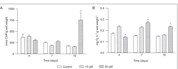
The absence of the induction of activity of the antioxi-dant enzymes involved in H2O2 degradation was paral-leled by an increase of almost 441% in LPO levels after 10 days of exposure to the higher H2O2 concentration (50 µM; P < 0.05; Figure 2A). This result agrees with the reduction of CAT activity in the pellet fraction during the same period, suggesting oxidative damage at the protein level (18).
The oxygen consumption response was biphasic, de-creasing after 4 days and inde-creasing after 7 and 10 days of exposure to 50 µM H2O2 (P < 0.05; Figure 2B). The hehavior of the first phase corroborates reports about Nereis diversicolor exposed for 6 h to 5 µM H2O2 (4), about the isolated body wall of Arenicola maritma (Polychaeta) ex-posed to 300 µM H2O2 (1) and about the shrimp Crangon crangon exposed for 5 h to 20 µM H2O2 (2). This decrease was related to damage to the membrane transporter mech-anisms and to the consequent reduction in intracellular pH (2).
After the period of exposure to both concentrations of H2O2, morphological alterations were observed, similar to these described in the aquatic oligochaeta Tubifex tubifex after 96-h exposure to copper and lead (19) and in L. acuta chronically exposed to copper (6). Worms exposed to H2O2 showed coiling and necrosis, particularly in the posterior region of their body, where the cuticle is thinner than the other parts (5), with this region being more susceptible to
Figure 2. Figure 2. Figure 2. Figure 2.
Figure 2. A, Lipid peroxide (LPO) content of Laeonereis acuta after exposure to 0 (control), 10, or 50 µM hydrogen peroxide. Units of LPO are in terms of cumene hydroperoxide (CHP) equivalents per gram wet weight (w/w). B, Oxygen consumption of L. acuta after exposure to 0 (control), 10, or 50 µM hydrogen peroxide. Units of oxygen consumption are reported as mg O2 h-1 g wet weight-1 (w/
w). Data are reported as means ± SEM for N = 4-8. *P < 0.05 compared to control (ANOVA and Newman-Keuls test).
the effects of H2O2.
The present study demonstrates that exposure to higher concentrations of H2O2 causes a significant alteration in the metabolism of L. acuta, as shown by the oxygen consumption measurements. It was also demonstrated that its antioxidant defense system was not sufficient to deal with H2O2 exposure, as evidenced by the oxidative damage and necrosis observed.
References
1. Storch D, Abele D, Portner HO. The effect of hydrogen peroxide on isolated body wall of the lugworm Arenicola marina (L.) at different extracellular pH levels. Comp Bio-chem Physiol C Toxicol Pharmacol 2001; 128: 391-399. 2. Abele-Oeschger D, Sartoris FJ, Pörtner HO. Hydrogen
per-oxide causes a decrease in aerobic metabolic rate and in intracellular pH in the shrimp Crangon crangon. Comp Bio-chem Physiol 1997; 117: 123-129.
3. Halliwel B, Gutteridge JM. Free radicals in biology and medicine. New York: Oxford University Press; 1998. 4. Buchner T, Abele-Oeschger D, Theede H. Biochemical
adaptations of Nereis diversicolor (Polychaeta) to tempo-rarily increased hydrogen peroxide levels in intertidal sandflats. Mar Ecol Prog Ser 1994; 106: 101-110. 5. da Rosa CE, Iurman MG, Abreu PC, Geracitano LA,
Monserrat JM. Antioxidant mechanisms of the Nereidid Laeonereis acuta (Anelida: Polychaeta) to cope with envi-ronmental hydrogen peroxide. Physiol Biochem Zool 2005; 78: 641-649.
6. Geracitano LA, Bocchetti R, Monserrat JM, Regoli F, Bianchini A. Oxidative stress responses in two populations of Laeonereis acuta (Polychaeta, Nereididae) after acute
and chronic exposure to copper. Mar Environ Res 2004; 58: 1-17.
7. da Rosa CE, de Souza MS, Yunes JS, Proenca LA, Nery LE, Monserrat JM. Cyanobacterial blooms in estuarine eco-systems: characteristics and effects on Laeonereis acuta (Polychaeta, Nereididae). Mar Pollut Bull 2005; 50: 956-964.
8. Sandrini JZ, Laurino J, Hatanaka T, Monserrat JM. cDNA cloning and expression analysis of the catalytic subunit of glutamate cysteine ligase gene in an annelid polychaete after cadmium exposure: a potential tool for pollution bio-monitoring. Comp Biochem Physiol C Toxicol Pharmacol 2006; 143: 410-415.
9. Ferreira-Cravo M, Piedras FR, Moraes TB, Ferreira JL, de Freitas DP, Machado MD, et al. Antioxidant responses and reactive oxygen species generation in different body re-gions of the estuarine polychaeta Laeonereis acuta (Nerei-didae). Chemosphere 2007; 66: 1367-1374.
11. Nithart M, Alliot E, Salen-Picard C. Production, respiration and ammonia excretion of two polychaete species in a north Norfolk saltmarsh. J Mar Biol Ass U K 1999; 79: 1029-1037. 12. Cavaletto M, Ghezzi A, Burlando B, Evangelisti V, Ceratto N, Viarengo A. Effect of hydrogen peroxide on antioxidant enzymes and metallothionein level in the digestive gland of Mytilus galloprovincialis. cavalett@unipmn.it. Comp Bio-chem Physiol C Toxicol Pharmacol 2002; 131: 447-455. 13. Beutler E. The preparation of red cells for assay. In: Beutler
E (Editor), Red cell metabolism: a manual of biochemical methods. New York: Grune & Straton Editor; 1975. p 8-18. 14. Arun S, Subramanian P. Antioxidant enzymes in freshwater prawn Macrobrachium malcolmsonni during embryonic de-velopment. Comp Biochem Physiol 1998; 121: 273-277. 15. Sedlak J, Lindsay RH. Estimation of total, protein-bound,
and nonprotein sulfhydryl groups in tissue with Ellman’s
reagent. Anal Biochem 1968; 25: 192-205.
16. Hermes-Lima M, Willmore WG, Storey KB. Quantification of lipid peroxidation in tissue extracts based on Fe(III)xylenol orange complex formation. Free Radic Biol Med 1995; 19: 271-280.
17. Moraes TB, Ferreira JL, da Rosa CE, Sandrini JZ, Votto AP, Trindade GS, et al. Antioxidant properties of the mucus secreted by Laeonereis acuta (Polychaeta, Nereididae): a defense against environmental pro-oxidants? Comp Bio-chem Physiol C Toxicol Pharmacol 2006; 142: 293-300. 18. Kono Y, Fridovich I. Superoxide radical inhibits catalase. J
Biol Chem 1982; 257: 5751-5754.
