Cyst infection in hospital-admitted
autosomal dominant polycystic kidney
disease patients is predominantly
multifocal and associated with kidney
and liver volume
B.E.P. Balbo
1, M.T. Sapienza
2, C.R. Ono
2, S.K. Jayanthi
3, J.B. Dettoni
4, I. Castro
1and L.F. Onuchic
1 1Divisa˜o de Nefrologia, Faculdade de Medicina, Universidade de Sa˜o Paulo, Sa˜o Paulo, SP, Brasil 2
Divisa˜o de Medicina Nuclear, Faculdade de Medicina, Universidade de Sa˜o Paulo, Sa˜o Paulo, SP, Brasil 3
Divisa˜o de Radiologia, Faculdade de Medicina, Universidade de Sa˜o Paulo, Sa˜o Paulo, SP, Brasil 4
Divisa˜o de Patologia, Faculdade de Medicina, Universidade de Sa˜o Paulo, Sa˜o Paulo, SP, Brasil
Abstract
Positron-emission tomography/computed tomography (PET/CT) has improved cyst infection (CI) management in autosomal dominant polycystic kidney disease (ADPKD). The determinants of kidney and/or liver involvement, however, remain uncertain. In this study, we evaluated clinical and imaging factors associated with CI in kidney (KCI) and liver (LCI) in ADPKD. A retrospective cohort study was performed in hospital-admitted ADPKD patients with suspected CI. Clinical, imaging and surgical data were analyzed. Features of infected cysts were evaluated by PET/CT. Total kidney (TKV) and liver (TLV) volumes were measured by CT-derived multiplanar reconstruction. CI was detected in 18 patients who experienced 24 episodes during an interval of 30 months (LCI in 12, KCI in 10 and concomitant infection in 2). Sensitivities of CT, magnetic resonance imaging and PET/CT were 25.0, 71.4, and 95.0%. Dysuria (P,0.05), positive urine culture (P,0.01), and previous hematuria (P,0.05) were associated with KCI. Weight loss (P,0.01) and increased C-reactive protein levels (P,0.05) were associated with LCI. PET/CT revealed that three or more infected cysts were present in 70% of the episodes. TKV was higher in kidney-affected than in LCI patients (AUC=0.91, P,0.05), with a cut-off of 2502 mL (72.7% sensitivity, 100.0% specificity). TLV was higher in liver-affected than in KCI patients (AUC=0.89, P,0.01) with a cut-off of 2815 mL (80.0% sensitivity, 87.5% specificity). A greater need for invasive procedures was observed in LCI (P,0.01), and the overall mortality was 20.8%. This study supports PET/CT as the most sensitive imaging method for diagnosis of cyst infection, confirms the multifocal nature of most hospital-admitted episodes, and reveals an association of kidney and liver volumes with this complication.
Key words: Autosomal dominant polycystic kidney disease; Cyst infection; Positron-emission tomography/computed tomography; Kidney volume; Liver volume; Mortality
Introduction
Autosomal dominant polycystic kidney disease (ADPKD) is the most common monogenic kidney disorder, with an estimated prevalence of 1:500-1000 (1). The disease is characterized by bilateral and progressively enlarging kidney cysts, often leading to end-stage renal disease. In addition to kidney manifestations, a variety of extrarenal abnormalities can also occur, comprising cystic and non-cystic phenotypes (2).
Kidney infection in ADPKD includes acute pyelone-phritis and/or cyst infection (CI) (3). Although CI tends to be
associated with a defined area of tenderness and to be more insidious than parenchymal infection, both are typically manifested by fever and abdominal pain. This scenario often makes the distinction between these two clinical entities challenging (4). Liver CI is also a common event in ADPKD, representing a potentially serious complication, and it can also occur in autosomal dominant polycystic liver disease (ADPLD) (5).
Conventional imaging methods have been shown to be of limited use in the diagnosis of renal or hepatic CI (4).
Correspondence: L.F. Onuchic, Divisa˜o de Nefrologia, Faculdade de Medicina, USP, Avenida Dr. Arnaldo, 455, Sala 4304, 01246-903 Sa˜o Paulo, SP, Brasil. Fax: ++55-11-3061-8361. E-mail: lonuchic@usp.br
In recent years, however, the management of CI in ADPKD has been redefined by the use of increasingly sophisticated nuclear medicine diagnostic techniques and the increased safety and effectiveness of image-guided, minimally invasive procedures that are sometimes required to drain infected foci, in addition to antibiotic therapy (ATB). The role of positron-emission tomography/computed tomogra-phy (PET/CT) has been analyzed in two retrospective series that support it as the gold standard imaging strategy for CI diagnosis (4,6). However, key questions and points remain unanswered and/or unaddressed (7), including the establishment of factors distinctly associated with kidney and liver CI, how often it affects multiple cysts and/or organs, to what extent this imaging strategy can be used in CI management, and a proposal of how and when invasive procedures should be performed. To address these issues, we carried out a retrospective cohort study in hospital-admitted ADPKD patients with clinically suspected CI.
Material and Methods
Study design, population and diagnostic criteria This retrospective cohort study was conducted at Hospital das Clı´nicas, Universidade de Sa˜o Paulo, Sa˜o Paulo, SP, Brazil. The medical files of all ADPKD patients with suspected CI who were admitted between May 2010 and November 2012 were reviewed, and relevant clinical, imaging, and invasive/surgical data were collected and categorized. ADPKD was diagnosed based on the criteria proposed by Pei et al. (8). Owing to the similar clinical presentation of CI episodes, ADPLD patients with sus-pected liver CI were also included in the medical file review. The diagnosis of ADPLD followed the criteria developed by Qian et al. (9).
The diagnosis of CI was based on the criteria proposed by Salle´e et al. (4) and was considered definite when cyst aspiration revealed microorganisms and/or neutrophil debris. We have extended these criteria by including tissue pathology analysis, from biopsy or autopsy, as an additional diagnostic tool. CI was considered likely in the presence of fever (temperature.38.56C for.3 days), abdominal pain (particularly a palpable area of kidney or liver tenderness), increased C-reactive protein (CRP,.50 mg/L), absence of intracystic bleeding on CT, and exclusion of other inflam-matory abdominal conditions. The patients were divided into two groups, definite and likely CI, and categorized according to the affected organ(s): kidney cyst(s) infection (KCI), liver cyst(s) infection (LCI), and concomitant kidney and liver cyst(s) infection (KLCI; Figure 1).
CI was considered severe in the presence of sepsis at admission, liver CI, or lack of improvement after 1 week of ATB therapy. The need for intervention was individualized. Image-guided drainage was performed in severe CI cases as soon as possible, if feasible, regardless of cyst size. Open surgery was considered in cases with no clinical response to less invasive approaches. CI resolution was
defined by disappearance of fever, CRP,5 mg/L and at least two negative blood and/or urine cultures.
Radiological analyses
Kidney or liver ultrasound was considered positive for CI based on detection of debris, thick wall and/or distal acoustic enhancement. CT scan was called positive for CI when enhanced wall thickening and/or perilesional inflam-mation were observed. Recent intracystic bleeding was excluded by CT. The same criteria for detecting CI were applied for magnetic resonance imaging (MRI), with the addition of marked reduction in diffusion on diffusion-weighted MRI (10). When possible, contrast media were used for CT or MRI. The presence of air within cysts was indicative of emphysematous CI.
Nuclear medicine analysis
Suspected cases of CI were further characterized by PET/CT, especially when ultrasound, CT and/or MRI were negative or inconclusive. A dedicated full-ring lutetium orthosilicate crystal-based scanner (Biograph 2, Siemens Medical Solutions, Germany, or Discovery 690, GE Health-care, USA) was used. Studies were carried out 60-90 min after intravenous injection of 370 MBq of18 fluorodeoxyglu-cose (18FDG). Delayed images, when necessary, were obtained approximately 3 h after furosemide administration, except in anuric patients. Two independent, experienced nuclear medicine physicians (NMPs) reviewed the images. When consensus was not achieved, a third NMP opinion was obtained.
Attenuation-corrected PET/CT images were analyzed in transverse, coronal and sagittal planes and built into a three-dimensional maximum-intensity projection. Two pat-terns were considered positive for CI. The first was increased 18FDG activity lining the cyst, in contrast to
physiologic accumulation in parenchyma, and the second was diffuse signal accumulation within the cyst after exclusion of cyst hemorrhage by CT. Distinction between infection and pelvicalyceal activity, resultant from physio-logic excretion of 18FDG, was sought by analyzing the decrease in activity following furosemide in delayed images.
Imaging features of each infectious episode, such as number of infected cyst(s) and location, were determined by PET/CT. Incidental findings, such as neoplasia or infection in other organs, were also analyzed. The18FDG level was measured by the maximum-standardized uptake value (SUVmax), determined by drawing regions of interest in the attenuation-corrected18FDG-PET images around the suspected lesion (6). A third NMP opinion was requested for SUVmaxdifferences.10%. Adequate glycemic control was checked before each examination.
Organ and cyst measurements
Total kidney (TKV) and liver (TLV) volumes were measured using CT-derived multiplanar reconstruction, with DICOM images processed by the OsiriX software (11). Evaluation of infected cyst size was also performed using CT, by measuring the largest internal diameter of each infected cyst. In cases of multiple CI, the three largest infected cysts were included in the analysis.
Statistical analysis
Bivariate analysis was performed by the Pearson chi-square test for categorical data and reported as frequency and percentage. The Kolmogorov-Smirnov test was used to determine normality for continuous data. Differences between two samples were compared using the unpaired Student t-test, with data reported as means±SD. Nonparametric data were compared by the Mann-Whitney test, with results reported as medians and percentiles. Variable discrimination was calculated using receiver operating characteristic (ROC) curves, assessing the area under the curve and asymptotic significance. Significance
level was set at 5% (P=0.05). The SPSS v.19.0 Statistical Package (IBM Corp., USA) was used for analyses.
Results
Clinical evaluation and characterization of CI events Thirty-four episodes of suspected abdominal infection were identified in 27 ADPKD and 1 ADPLD subjects. Ten ADPKD patients had a diagnosis other than CI. In this group, CI was excluded based not only on the absence of PET/CT imaging criteria but also by the establishment of alternative diagnoses supported by imaging and/or clinical findings. In those 10 cases, four patients presented kidney cyst hemorrhage, one had liver cyst rupture, and two had fever of unknown origin despite extensive work-up. Each of the following diagnoses were detected in one patient: vertebral osteomyelitis detected by PET/CT; acute cholecystitis, not initially seen on CT scan but diagnosed with PET/CT; and retroperitoneal abscess detected months after a unilateral nephrectomy because of chronic kidney pain. Twelve of the 18 remaining patients completed treatment with cure, 5 died from CI, and 1 LCI patient died intraoperatively during liver transplantation.
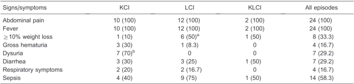
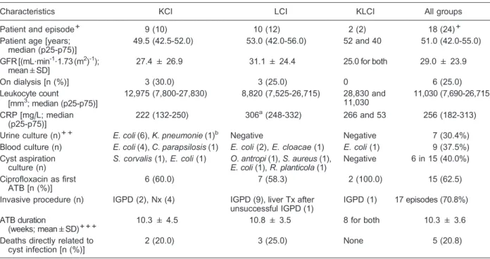
Reinfection occurred in 6 patients, a high recurrence rate. All patients had fever and abdominal pain at admission. An association between dysuria and KCI was observed (P,0.01), while a history of recent weight loss was more frequently associated with LCI (P,0.01; Table 1). CRP levels were higher in LCI than in KCI episodes (P,0.05; Table 2). No significant differences in estimated glomerular filtration rate (GFR using MDRD equation) or previous history of renal replacement therapy were observed between groups (Table 2). Positive blood cultures were obtained in KCI and LCI, but only KCI patients presented positive urine cultures (Table 2).
A high prevalence of recurrent gross hematuria and hypertension was observed in this patient population (Supplementary Table S1). The initial infectious foci,
Table 1. Clinical manifestations at hospital admission in patients with autosomal dominant polycystic kidney disease/autosomal dominant polycystic liver disease with kidney and/or liver cyst infections.
Signs/symptoms KCI LCI KLCI All episodes
Abdominal pain 10 (100) 12 (100) 2 (100) 24 (100)
Fever 10 (100) 12 (100) 2 (100) 24 (100)
§10% weight loss 1 (10) 6 (50)a 1 (50) 8 (33.3)
Gross hematuria 3 (30) 1 (8.3) 0 4 (16.7)
Dysuria 7 (70)b 0 0 7 (29.2)
Diarrhea 3 (30) 3 (25) 1 (50) 7 (29.2)
Respiratory symptoms 2 (20) 2 (16.7) 0 4 (16.7)
Sepsis 4 (40) 9 (75) 1 (50) 14 (58.3)
before CI diagnosis, were attributed to a myriad of locations (Supplementary Table S2); while urinary tract involvement occurred exclusively in KCI, the source was not identified in 16.7% of the episodes, all of which were LCI.
Management and decision making in CI events All patients with suspected CI received ATB. Intra-venous ciprofloxacin, alone or in combination, was the preferred option prior to obtaining culture results (Table 2). A decision for combination ATB was made in 75% of the episodes, particularly in severe presentations. All surviving patients received ATB for at least 4 weeks, and 66.7% required 8 or more weeks to reach cure criteria. Change to trimethoprim-sulfamethoxazole (5 cases) or chloramphen-icol (1 case), supported by an antibiogram, was made to achieve cure. An apparent higher prevalence of sepsis was observed in LCI than in KCI events.
Escherichia coliwas isolated in blood culture in 29.2% of all episodes and in urine culture in 25% (Table 2).
Salmonella corvaliswas detected by cyst drainage in one patient following a diarrheal episode, suggesting transloca-tion; the identification of a quinolone-resistant microorgan-ism prompted change to trimethoprim-sulfamethoxazole.
Image-guided percutaneous drainage (IGPD) was per-formed in 10 of 12 LCI episodes (Table 2). Need for intervention was also observed in KCI, including two IGPD and four nephrectomies. The retrospective analysis also detected one case of ADPLD that developed LCI with multiple infected cysts following liver fenestration. Cure came after effective IGPD, which revealedS. aureusand guided change to vancomycin. Moreover, in seven episodes, the diagnosis of CI was established by tissue pathology of the removed organ or at autopsy (Figure 2H and I).
Diagnostic impact of imaging methods
All patients were evaluated by CT, with an overall diagnostic sensitivity of 25% (Figure 3). Seven CT-negative cases were evaluated by MRI. The result was positive in 5 and negative in 2. While the positive MRI results were confirmed by PET/CT, this exam also revealed that the negative MRIs were false, resulting in an overall sensitivity of 71.4% (Figure 2A-C). PET/CT was performed in 20 CI episodes, with an overall sensitivity of 95% (Figure 3). Diagnostic discordance was limited to a single case that was considered positive by a third NMP and confirmed as CI by pathology following nephrectomy.
Table 2.Clinical features of cyst infection episodes in patients with autosomal dominant polycystic kidney disease/autosomal dominant polycystic liver disease.
Characteristics KCI LCI KLCI All groups
Patient and episode+ 9 (10) 10 (12) 2 (2) 18 (24)+
Patient age [years; median (p25-p75)]
49.5 (42.5-52.0) 53.0 (42.0-56.0) 52 and 40 51.0 (42.0-55.0)
GFR [(mL?min-1
?1.73 (m2)-1); mean±SD]
27.4 ± 26.9 31.1 ± 24.4 25.0 for both 29.0 ± 23.9
On dialysis [n (%)] 3 (30.0) 3 (25.0) 0 6 (25.0)
Leukocyte count
[mm3; median (p25-p75)]
12,975 (7,800-27,830) 8,820 (7,525-26,715) 28,830 and 11,030
11,030 (7,690-26,715)
CRP [mg/L; median (p25-p75)]
222 (132-250) 306a(248-332) 266 and 53 256 (182-313)
Urine culture (n)++ E. coli(6),K. pneumonie(1)b Negative Negative 7 (30.4%) Blood culture (n) E. coli(4),C. parapsilosis(1) E. coli(2),E. cloacae(1) E. coli(1) 9 (37.5%) Cyst aspiration
culture (n)
S. corvalis(1),E. coli(1) O. antropi(1),S. aureus(1), E. coli(1),R. planticola(1)
Negative 6 in 15 (40.0%)
Ciprofloxacin as first ATB [n (%)]
6 (60.0) 7 (58.3) 2 (100.0) 15 (62.5)
Invasive procedure (n) IGPD (2), Nx (4) IGPD (9), liver Tx after unsuccessful IGPD (1)
IGPD (1) 17 episodes (70.8%)
ATB duration
(weeks; mean±SD)+++
10.3 ± 4.5 10.8 ± 3.5 8 for both 10.3 ± 3.6
Deaths directly related to cyst infection [n (%)]
2 (20.0) 3 (25.0) None 5 (20.8)
KCI: kidney cyst infection; LCI: liver cyst infection; KLCI: concomitant kidney and liver cyst infection; GFR: glomerular filtration rate; CRP: C-reactive protein; ATB: antibiotic; IGPD: image-guided percutaneous drainage; Nx: nephrectomy; Tx: transplantation; p25-p75: 25 to 75 percentile.+One patient with KCI also had an episode of LCI; both KLCI patients also had an LCI episode (Figure 1).++Urine culture was available in 23 of 24 episodes (all but 1 anuric patient on dialysis).+++Includes patients who achieved cure and excludes those for whom ATB was interrupted due to death.aP
,0.05vsKCI group (Mann-Whitney test);bP
Our calculations showed, for our sample size of 24 events, a.95% power to detect CI by comparing CT with PET/CT and CT with MRI, and a .80% power by comparing MRI with PET/CT.
Cyst infection and multifocality
Cyst infection was predominantly multifocal (Figures 4 and 2D-F). A single CI occurred in only 15% of the events, but three or more infected cysts were detected in 70% of all episodes analyzed by PET/CT. Interestingly, in KCI limited to one kidney, the dominant organ was affected in the majority of the cases (Table 3). However, bilateral involvement was not uncommon in the KCI group, being detected in 30% of the cases. Another remarkable observation was the fact that the majority of the infectious episodes affected nondominant cysts. The involvement of the dominant cyst, as shown by all image data combined, was in fact limited to 40% KCI, 25% LCI, no KLCI, and 29.2% overall.
PET/CT features and incidental findings
The PET/CT analyses detected two CI imaging patterns. The first, characterized by increased 18FDG activity lining the cyst, was more prevalent and occurred in 85% of CI episodes (Figure 2C, E, and F). The second was characterized by diffuse18FDG signal accumulation within the cyst and occurred in 60% of CI episodes (Figure 2D and E). The mean SUVmax was 6.6±1.2 in KCI episodes, 7.9±3.2 in LCI episodes and 6.9±2.4 in all events combined.
Concomitant episodes of pneumonia, aortitis and subphrenic abscess were also revealed by PET/CT. Incidental neoplastic findings included gastric and thyroid cancer. In all CI events, PET/CT was performed at a median of 12 days (7.0 to 20.5 days) after initiating ATB. This method yielded a false-negative result in only one case, where LCI was confirmed at autopsy (Supplementary Table S3). MRI, in turn, had intermediate sensitivity for CI detection (Figure 3).
Cyst infection associates with kidney and liver volume Patients with KCI, isolated or combined with LCI, had higher TKVs than ADPKD cases with isolated LCI. The area under the ROC (AUC) was 0.91 [95% confidence interval (95%CI)=0.76-1.06, P=0.013; Figure 5A]. The highest accuracy point detected was for TKV=2502 mL, with 72.7% sensitivity and 100.0% specificity (Figure 5B). KCI was also associated with HtTKV (height-adjusted TKV, a potential prognostic biomarker (12); P=0.013). In this case, the highest accuracy point detected was for
HtTKV=1599 mL/m. Similarly, patients with LCI, isolated or combined with KCI, had higher TLVs compared with isolated KCI subjects (AUC=0.89, 95%CI=0.72-1.05, P=0.004; Figure 5C). The highest accuracy point detected was for TLV=2815 mL, with 80.0% sensitivity and 87.5% specificity (Figure 5D).
Most hospital-admitted episodes of CI require invasive procedures
Eleven CI episodes were associated with §5-cm Figure 3.Sensitivity of different imaging methods to detect cyst infection in autosomal dominant polycystic kidney disease/autosomal dominant polycystic liver disease. CT: computed tomogra-phy; MRI: magnetic resonance imaging; PET: positron-emission tomography; KCI: kidney cyst infection; LCI: liver cyst infection; KLCI: conco-mitant kidney and liver cyst infection.
infected cysts (6 KCI, 4 LCI, and 1 KLCI). Seven of them were successfully treated by invasive procedures and 3 achieved cure with only ATB. One was a late referral, evolving to death before invasive intervention was per-formed. In contrast, 13 CI episodes were associated with
,5-cm infected cysts (4 KCI, 8 LCI, and 1 KLCI). Ten were treated by invasive interventions, 7 of which were success-ful; 1 episode was cured with ATB only, and 2 patients died before the invasive procedure. One was a late referral and one was a postmortem diagnosis. In these cases, therefore, the vast majority of hospital-admitted patients required invasive intervention, including,5-cm infected cysts. The details of patients who died are shown in Supplementary Table S3.
Carbohydrate antigen 19-9 (CA19-9) and liver CI CA19-9 was evaluated in 1 KLCI, 4 KCI and 11 LCI episodes. Using the proposed cut-off of 106 U/mL (13), CA19-9 failed to support the diagnosis of LCI in 6 episodes (54.5%).
PET/CT for control of treatment efficacy
A second PET/CT analysis was performed in four patients as a control evaluation. Notably, it changed decision-making in two of them. In a patient with previous KLCI and gastric cancer, the absence of CI signs allowed for chemotherapy administration. In another case with multiple infected cysts and preclusion of IGPD due to technical difficulties, a positive PET/CT supported extend-ing ATB therapy for seven additional weeks despite the relative decrease in CRP levels that occurred before the second imaging examination. In the other two cases, it confirmed CI cure in patients with borderline clinical status but did not change overall decision-making.
Discussion
The diagnosis of CI in ADPKD is often challenging (4,6,7). In this scenario, attempts to define diagnostic criteria based on imaging techniques have been proposed (14-18). Conventional imaging, however, and even gallium or indium scanning, have low sensitivity (3,19). Newer imaging strategies, together with the increasing safety and effectiveness of minimally invasive procedures, have
Table 3. Organ volume and anatomical analysis in autosomal dominant polycystic kidney disease (ADPKD)/autosomal dominant polycystic liver disease (ADPLD) patients with cyst infection.
Group KCI LCI Total
TKV per patient+(mL) 4210 (1972-5803)a 1814 (962-2773) 2502 (1579-4556)
TLV per patient++(mL) 1842 (1608-2472) 4470 (2824-9081)b 2816 (1842-5832)
Cyst diameter per episode+++(cm) 5.2 (3.4-5.4) 4.2 (3.2-6.5) 5.2 (3.4-7.7)
Infection in dominant cyst/episode 4/10 (40.0%) 3/12 (25.0%) 7/24 (29.2%)
Cyst infection in dominant kidney/episode 5/7 (71.4%) Not applicable 7/9 (77.8%) Concomitant cyst infection in both kidneys/episode 3/10 (30.0%) Not applicable 3/12 (25%)
Data are reported as median with p25-p75 in parentheses or as number with percent in parentheses. TKV: total kidney volume; TLV: total liver volume; KCI: kidney cyst infection; LCI: liver cyst infection; p25-p75: 25 to 75 percentile.+An ADPKD patient with previous unilateral nephrectomy and 1 ADPLD subject were excluded from the TKV analysis.++The ADPLD patient was included in the TLV analysis. +++The largest internal diameter of each infected cyst was measured by computed tomography. In each episode, when multiple cysts
were infected, the three largest cysts were measured.aP
,0.05vsLCI group;bP
,0.01vsKCI group (Mann-Whitney test).
redesigned the management of such a potentially devas-tating complication (20). Recent reports suggest that PET/ CT is the best tool for CI detection (4,6), a finding that we have confirmed in our series. Despite PET/CT availability in our center, this retrospective cohort reflected an effort toward accurate diagnosis balanced against cost-effec-tiveness. For that reason, CT was used as the first imaging approach in all suspected CI episodes. Compared with MRI and PET/CT, CT offers the shortest waiting time, relatively low cost, and high accuracy to exclude nephrolithiasis and cyst hemorrhage. On the other hand, MRI with diffusion-weight acquisition, emerges as a potentially reliable tool, apparently with higher sensitivity for CI detection than CT. This observation is, however, limited by the low number of patients with MRI results, and merits further study.
Like others, we have been confronted with the clinical dilemma imposed by difficulties to accurately determine CI. The confirmation of such a diagnosis was achieved following the criteria proposed by Salle´e et al. (4), by searching for neutrophil debris and/or microorganisms, a procedure only feasible when drainage was performed. To improve sensitivity, we have broadened the definite diagnosis criteria to include CI episodes documented by pathology evaluation, when available.
Our patients had multiple comorbid conditions, includ-ing risk factors for infection. In the current series, the numbers of LCI and KCI episodes were similar. Interestingly, in these two groups, patient age and need for renal replacement therapy did not differ.
Fever and abdominal pain were universal at admis-sion. The search for attributable infectious foci was based on the assumption that a primary infection precedes CI eclosion. Indeed, in the present study, the primary site was detected in 83.3% of all episodes. The observation that urinary tract manifestations were common in KCI and associated with a high prevalence of Gram-negative bacteria supports the concept that the ascending route was the predominant source of infection in this group. The hematogenous spread from endocarditis and a probable intestinal translocation of Salmonella corvalis, support alternative sources for kidney CI. Moreover, the high reinfection rate suggests a prominent role of anatomical abnormalities in kidney and liver for determining CI (21).
We have performed quantitative evaluation of the number of infected cysts, as well as volumetric analyses of kidneys and livers, an original approach not performed in previously published series (4,6). Our data showed that CI was predominantly multifocal in both kidney and liver. These results were supported not only by PET/CT imaging but also by drainage and histopathologic data confirming multiple affected cysts. Hematogenous spread, low oxygen tension within the cyst wall, and anatomical abnormalities in ADPKD/ADPLD could contribute to the creation of niches for infection development. Our study indicates, therefore, that diagnosis of CI in one kidney does not preclude its occurrence in the contralateral kidney or even
in the liver.
Remarkably, the dominant cyst was infected in only 29.2% of CI episodes, indicating that a search focused on it can often be misleading. In addition, the size of infected cysts in the kidney and liver did not differ. A key finding of this series, given the overall CI severity, was the need for invasive intervention in the vast majority of the patients, even in those with,5-cm infected cysts.
Since the design of a control group to best address the roles of TKV and TLV in CI development would be very difficult, requiring patients with similar additional risk factors for CI but different kidney and/or liver volumes, we approached this issue in the best possible way. We compared TKV and HtTKV in the infected and noninfected kidney groups, comprised of patients with supposedly higher risk for CI than the general ADPKD population. This strategy provided a relatively similar background between these two groups, except for the potential differences offered by organ volume. In this context, our data revealed that KCI was associated with higher TKV and HtTKV, suggesting kidney volume as a risk factor for KCI. Interestingly, history of urinary tract infection has been regarded as an independent prognostic factor for GFR decline in ADPKD patients (22). Notably, the organ-volume approach has also been used to estimate risk for nephrolithiasis in ADPKD (23). Our results showed, in addition, that LCI was also associated with higher TLV, supporting liver volume as a potential risk factor for LCI. Possible explanations for such findings likely include: architectural changes in the renal parenchyma induced by the progressive increase in cystic burden, resulting in areas of outflow obstruction and ectasia that would favor bacterial growth, areas of focal ischemia, changes in the microenvironment driven by local inflammation associated with release of cytokines and chemokines, and increased generation of reactive oxygen species. Taken together, such abnormalities would favor the formation of niches particularly susceptible to infection.
PET/CT is not available, we favor an approach using MRI to investigate CI, particularly when CT is negative. However, when CT and/or MRI remain inconclusive in a patient without clinical improvement, we recommend rapid referral to a polycystic kidney disease center.
CRP levels are useful in the clinical follow-up of CI in ADPKD. In this study, all CRP levels were measured at admission in all patients; however, due to its retrospective nature, the sequential monitoring of CRP was not standardized, limiting the interpretation of the results. On the other hand, it should be mentioned that in the small group in which a second PET/CT was performed, the CRP levels decreased in one patient preceding improvement on PET/CT, as previously reported (7). Further studies should address the role of CRP monitoring in CI, particularly when evaluating criteria of cure.
Ciprofloxacin was our first-line ATB (24); however, we favored a switch to trimethoprim-sulfamethoxazole in outpatients to avoid quinolone-induced resistance (25,26). We balanced the approach to empirically treat with antibiotics all hospital-admitted ADPKD patients with abdominal pain and fever against the risk of unnecessary treatment in cases of noninfectious complications, such as cyst hemorrhage. Indeed, of 5 patients with cyst hemor-rhage, 3 received empirical antibiotics, which were stopped after PET/CT excluded infection. Considering these data, one could consider the restricted use of antibiotics until the release of the initial laboratory and/or imaging results to be a better strategy. This tailored strategy, however, should not be applied to those with sepsis at admission, in whom empirical antibiotics are definitely beneficial. The high severity and mortality associated with CI at our institution, should therefore justify empiric antibiotic administration in
patients with clinical signs of potentially severe infection and in those whose initial laboratory and imaging work-up are suggestive of such an etiology.
Despite robust care and follow-up, the mortality rate reached 20.8%. This outcome reflects the severity of CI cases referred to our tertiary-care center and suggests some key considerations. First, a surgical approach should be considered early in emphysematous CI (27,28); second, cyst drainage should be considered in CI events with partial clinical improvement; and third, PET/CT is an outstanding diagnostic tool, but when performed late after ATB initiation, can lead to false-negative results.
Our findings suggest, moreover, that repeating PET/ CT after CI diagnosis may improve management in some situations, such as to confirm cure in patients with CI when surgical drainage was indicated, but not feasible/ performed due to technical difficulties. Repeat PET/CI may also benefit ADPKD patients with CI in whom documentation of cure is essential for other reasons, such as to initiate chemotherapy or to undergo a surgical procedure. The negative predictive value of PET/CT to exclude CI, however, needs to be assessed in future studies.
Supplementary Material
Click here to view [pdf].
Acknowledgments
We thank Rui T. Barros and Viktoria Woronik for clinical support to the patients included in the present study.
References
1. Bastos AP, Onuchic LF. Molecular and cellular pathogen-esis of autosomal dominant polycystic kidney disease.Braz J Med Biol Res 2011; 44: 606-617, doi: 10.1590/S0100-879X2011007500068.
2. Pirson Y. Extrarenal manifestations of autosomal dominant polycystic kidney disease. Adv Chronic Kidney Dis 2010; 17: 173-180, doi: 10.1053/j.ackd.2010.01.003.
3. Schwab SJ, Bander SJ, Klahr S. Renal infection in autosomal dominant polycystic kidney disease.Am J Med 1987; 82: 714-718, doi: 10.1016/0002-9343(87)90005-2. 4. Salle´e M, Rafat C, Zahar JR, Paulmier B, Grunfeld JP,
Knebelmann B, et al. Cyst infections in patients with autosomal dominant polycystic kidney disease.Clin J Am Soc Nephrol 2009; 4: 1183-1189, doi: 10.2215/CJN.01870309.
5. Schnelldorfer T, Torres VE, Zakaria S, Rosen CB, Nagorney DM. Polycystic liver disease: a critical appraisal of hepatic resection, cyst fenestration, and liver transplantation. Ann Surg2009; 250: 112-118, doi: 10.1097/SLA.0b013e3181ad 83dc.
6. Jouret F, Lhommel R, Beguin C, Devuyst O, Pirson Y, Hassoun Z, et al. Positron-emission computed tomography in
cyst infection diagnosis in patients with autosomal dominant polycystic kidney disease.Clin J Am Soc Nephrol2011; 6: 1644-1650, doi: 10.2215/CJN.06900810.
7. Jouret F, Lhommel R, Devuyst O, Annet L, Pirson Y, Hassoun Z, et al. Diagnosis of cyst infection in patients with autosomal dominant polycystic kidney disease: attributes and limitations of the current modalities. Nephrol Dial Transplant2012; 27: 3746-3751, doi: 10.1093/ndt/gfs352. 8. Pei Y, Obaji J, Dupuis A, Paterson AD, Magistroni R, Dicks E,
et al. Unified criteria for ultrasonographic diagnosis of ADPKD. J Am Soc Nephrol 2009; 20: 205-212, doi: 10.1681/ASN. 2008050507.
9. Qian Q, Li A, King BF, Kamath PS, Lager DJ, Huston J III, et al. Clinical profile of autosomal dominant polycystic liver disease.Hepatology2003; 37: 164-171, doi: 10.1053/jhep. 2003.50006.
software for navigating in multidimensional DICOM images. J Digit Imaging 2004; 17: 205-216, doi: 10.1007/s10278-004-1014-6.
12. Chapman AB, Bost JE, Torres VE, Guay-Woodford L, Bae KT, Landsittel D, et al. Kidney volume and functional outcomes in autosomal dominant polycystic kidney disease.Clin J Am Soc Nephrol2012; 7: 479-486, doi: 10.2215/CJN.09500911. 13. Kanaan N, Goffin E, Pirson Y, Devuyst O, Hassoun Z.
Carbohydrate antigen 19-9 as a diagnostic marker for hepatic cyst infection in autosomal dominant polycystic kidney disease.Am J Kidney Dis2010; 55: 916-922, doi: 10.1053/ j.ajkd.2009.12.023.
14. Sklar AH, Caruana RJ, Lammers JE, Strauser GD. Renal infections in autosomal dominant polycystic kidney disease. Am J Kidney Dis1987; 10: 81-88.
15. Gibson P, Watson ML. Cyst infection in polycystic kidney disease: a clinical challenge.Nephrol Dial Transplant1998; 13: 2455-2457, doi: 10.1093/ndt/13.10.2455.
16. Migali G, Annet L, Lonneux M, Devuyst O. Renal cyst infection in autosomal dominant polycystic kidney disease.Nephrol Dial Transplant2008; 23: 404-405, doi: 10.1093/ndt/gfm665. 17. Telenti A, Torres VE, Gross JB Jr, Van Scoy RE, Brown ML,
Hattery RR. Hepatic cyst infection in autosomal dominant polycystic kidney disease.Mayo Clin Proc1990; 65: 933-942, doi: 10.1016/S0025-6196(12)65154-4.
18. Christophe JL, van Ypersele de Strihou C, Pirson Y. Complications of autosomal dominant polycystic kidney disease in 50 haemodialysed patients. A case-control study. The U.C.L. Collaborative Group. Nephrol Dial Transplant 1996; 11: 1271-1276, doi: 10.1093/ndt/11.7.1271. 19. Gilbert BR, Cerqueira MD, Eary JF, Simmons MC, Nabi HA,
Nelp WB. Indium-111 white blood cell scan for infectious complications of polycystic renal disease.J Nucl Med1985; 26: 1283-1286.
20. Kaim AH, Burger C, Ganter CC, Goerres GW, Kamel E, Weishaupt D, et al. PET-CT-guided percutaneous puncture of an infected cyst in autosomal dominant polycystic kidney
disease: case report.Radiology2001; 221: 818-821, doi: 10.1148/radiol.2213010445.
21. Hsu CT, Chang HR, Lee JK, Weng JH, Kao PF. FDG PET/ CT repeatedly demonstrated hepatic cyst infection in a patient with autosomal dominant polycystic kidney disease. Clin Nucl Med 2013; 38: e188-e190, doi: 10.1097/ RLU.0b013e318266d056.
22. Rule AD, Torres VE, Chapman AB, Grantham JJ, Guay-Woodford LM, Bae KT, et al. Comparison of methods for determining renal function decline in early autosomal domi-nant polycystic kidney disease: the consortium of radiologic imaging studies of polycystic kidney disease cohort.J Am Soc Nephrol2006; 17: 854-862, doi: 10.1681/ASN.2005070697. 23. Nishiura JL, Neves RF, Eloi SR, Cintra SM, Ajzen SA,
Heilberg IP. Evaluation of nephrolithiasis in autosomal dominant polycystic kidney disease patients. Clin J Am Soc Nephrol2009; 4: 838-844, doi: 10.2215/CJN.031006 08.
24. Elzinga LW, Golper TA, Rashad AL, Carr ME, Bennett WM. Ciprofloxacin activity in cyst fluid from polycystic kidneys. Antimicrob Agents Chemother 1988; 32: 844-847, doi: 10.1128/AAC.32.6.844.
25. Schwab SJ, Weaver ME. Penetration of trimethoprim and sulfamethoxazole into cysts in a patient with autosomal-dominant polycystic kidney disease.Am J Kidney Dis1986; 7: 434-438.
26. Elzinga LW, Golper TA, Rashad AL, Carr ME, Bennett WM. Trimethoprim-sulfamethoxazole in cyst fluid from autosomal dominant polycystic kidneys.Kidney Int1987; 32: 884-888, doi: 10.1038/ki.1987.290.
27. Sooraj YS, Nainan GK, Joseph F, Thara P. Emphysematous polycystic renal infection.Indian J Nephrol2010; 20: 205-206, doi: 10.4103/0971-4065.73457.


![Figure 5. Association between organ volume and cyst infection. A, Patients admitted to the hospital with KCI/KLCI presented higher TKV in comparison with those with isolated LCI [3491 mL (2136-5167; p25-p75) vs 1021 mL (745-2157; p25-p75)] (*P,0.05, Mann](https://thumb-eu.123doks.com/thumbv2/123dok_br/15819388.653201/7.918.108.816.208.342/association-infection-patients-admitted-hospital-presented-comparison-isolated.webp)
