Effect of age on upper and lower
eyelid saccades
1Departamento de Oftalmologia, Otorrinolaringologia,
Cirurgia de Cabeça e Pescoço, Faculdade de Medicina de Ribeirão Preto, Universidade de São Paulo, Ribeirão Preto, SP, Brasil
2Departamento de Física Geral, Instituto de Física,
Universidade Federal da Bahia, Salvador, BA, Brasil L.V.O. Leite1,A.A.V. Cruz1,
A. Messias1
and J.M.C. Malbouisson2
Abstract
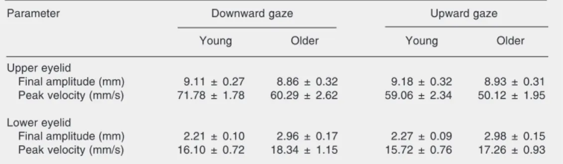
To study the effect of age on the metrics of upper and lower eyelid saccades, eyelid movement of two groups of 30 subjects each were measured using computed image analysis. The patients were divided on the basis of age into a younger group (20-30 years) and an older group (60-91 years). Eyelid saccade functions were fitted by the damped harmonic oscillator model. Amplitude and peak velocity were used to compare the effect of age on the saccades of the upper and lower eyelid. There was no statistically significant difference in saccade amplitude between groups for the upper eyelid (mean ± SEM; upward, young = 9.18 ± 0.32 mm, older = 8.93 ± 0.31 mm, t = 0.56, P = 0.58; downward, young = 9.11 ± 0.27 mm, older = 8.86 ± 0.32 mm, t = 0.58, P = 0.56) However, there was a clear decline in the peak velocity of the upper eyelid saccades of older subjects (upward, young = 59.06 ± 2.34 mm/s, older = 50.12 ± 1.95 mm/s, t = 2.93, P = 0.005; downward, young = 71.78 ± 1.78 mm/s, older = 60.29 ± 2.62 mm/s, t = 3.63, P = 0.0006). In contrast, for the lower eyelid there was a clear increase of saccade amplitude in the elderly group (upward, young = 2.27 ± 0.09 mm, older = 2.98 ± 0.15 mm, t = 4.33, P < 0.0001; downward, young = 2.21 ± 0.10 mm, older = 2.96 ± 0.17 mm, t = 3.85, P < 0.001). These data suggest that the aging process affects the metrics of the lid saccades in a different manner according to the eyelid. In the upper eyelid the lower tension exerted by a weak aponeurosis is reflected only on the peak velocity of the saccades. In the lower eyelid, age is accompanied by an increase in saccade amplitude which indicates that the force transmission to the lid is not affected in the elderly.
Correspondence
A.A.V. Cruz
Departamento de Oftalmologia, Otorrinolaringologia, Cirurgia de Cabeça e Pescoço HF, FMRP, USP Av. Bandeirantes, 3900 14049-900 Ribeirão Preto, SP Brasil
E-mail: [email protected]
Research supported by CNPq.
Publication supported by FAPESP.
Received March 6, 2006 Accepted September 11, 2006
Key words
•Upper eyelid saccade •Lower eyelid saccade •Aging
•Digital image processing
Introduction
When the gaze shifts from the primary position to up- or downgaze the eyelids also move accompanying the direction of the eye rotation. These eyelid movements are named eyelid saccades because their metrics closely
match the metrics of the vertical eye sac-cades (1).
The eyelid mechanics are also affected by aging. In the upper eyelid, aponeurotic pto-sis is a common finding (3) and in the lower eyelid horizontal laxity due to stretching of the canthal tendons can be demonstrated in the majority of elderly patients (4).
Very few studies have described the ef-fect of aging on eyelid saccades. We were able to find only one article with data for upper eyelid saccades in young and older subjects (5). The effect of aging on the lower eyelid saccades has not been well docu-mented. The few data on the amplitude of lower eyelid saccades have been measured with rulers (6), a procedure that does not allow a precise description of the effect of age on the metrics of these movements.
We have recently demonstrated that both upper and lower eyelid saccades can be ac-curately measured with a commercial video camera and mathematically analyzed with a model extensively used in engineering to describe elastic movements (damped har-monic oscillator) (7).
In the present study, we measured the upper and lower eyelid saccades of young and old normal subjects to determine the effect of aging.
Subjects, Material and Methods
Subjects
Sixty healthy subjects without history of eye or eyelid surgery were included in the study. They were divided into two groups of 30 each according to their age. Group A consisted of subjects (10 males and 20 fe-males) aged 20 to 30 years (mean ± SD = 26.33 ± 3.11) and group B consisted of older subjects (13 males and 17 females) aged 60 to 91 years (mean ± SD = 70.63 ± 8.28).
None of the subjects showed any sign of ophthalmic or neurological abnormality ex-cept for a moderate degree of cataract in the older group.
Written informed consent was obtained
from all subjects and the study was approved by the institutional Ethics Committee.
Procedure
First, the palpebral fissure of all subjects was photographed in the primary position of gaze with a digital camera. The images were then transferred to a microcomputer and the Image J software (available at http://rsb.info. nih.gov/ij/) was used to measure the midpupil-eyelid distance of the upper and lower midpupil-eyelids. The saccades of the upper and lower eyelids were then measured. Only one eye randomly chosen was used. The methods employed for this measurement have been described in detail elsewhere (7). Briefly, with their heads stabilized on a chin rest, the subjects were instructed to look down and up between two targets 20º above and below a central light that was aligned with the subject’s visual axis in the primary position of gaze (40º of eye rotation across the upper and lower hemifields).
Eyelid motion during downward and up-ward eyelid saccades was recorded with a coupled-charged device (CCD) camera con-nected to a personal computer by a frame grabber. The temporal resolution of the cam-era was the standard NTSC (30 Hz or 30 frames per second). Movement was ana-lyzed with a software which calculated in real time the center of a blue spot in each frame. In order to provide the signal for the software a small piece of blue paper was attached to the eyelashes of the central por-tion of the upper eyelid. A similar spot was made on the skin of the lower eyelid margin with blue make-up.
Results
SD; upper eyelid: young = 3.77 ± 0.73, older = 2.97 ± 0.95 mm, t = 3.68, P = 0.0005; lower eyelid: young = 6.45 ± 0.69, older = 5.05 ± 1.10 mm, P < 0.00001) indicating a clear tendency for the eyelids to rest close to the pupil center in the elderly population.
The mean values of the saccade param-eters for the downward and upward saccades of the two groups of subjects are shown in Table 1 (upper eyelid and lower eyelid). For the upper eyelid there was no difference in amplitude between the saccades of the older and young subjects (independent t-test,
up-ward: t = 0.56, P = 0.58; downup-ward: t = 0.58, P = 0.56). However, the saccades of the older subjects were slower than the saccades of the young group (upward: t = 2.93, P = 0.005; downward: t = 3.63, P = 0.0006). The decrease in velocity demonstrated in the older subjects was not accompanied by any change in the functions relating amplitude and ve-locity to time (Figure 2). The linear relation-ship between amplitude and velocity shown in young subjects was also observed in older subjects (Figure 3).
Aging had a completely different effect on the dynamics of the lower eyelid. The saccade amplitude of the older group was significantly greater than the amplitude of the young (independent t-test, upward: t =
4.33, P < 0.0001; downward: t = 3.85, P < 0.001). Similarly to the upper eyelid, the relationships between amplitude and velocity and time were normal (Figure 4). A linear
Figure 1. Mid-pupil eyelid dis-tances for the upper and lower eyelid in young and older sub-jects.
relationship was found between amplitude and velocity (Figure 5). When the velocity data were grouped (downward + upward, Figure 6) the mean velocity was also higher in the older group (young = 15.87, old = 17.85 mm/s, t = 2.20, P = 0.03). When velocity was compared according to the di-rection of gaze (downward vs downward
and upward vs upward) the difference was
not statistically significant.
Discussion
Aging induces a process of deterioration of collagen and other tissues resulting in the attenuation of the levator muscle aponeuro-sis in the upper eyelid (8). This process is the underlying cause of the aponeurotic ptosis. The decline in the mean mid-pupil eyelid distance verified here in the older group is consistent with this concept. From a me-chanical perspective, weak aponeurosis
Table 1. Parameters of upper and lower eyelid saccades.
Parameter Downward gaze Upward gaze
Young Older Young Older
Upper eyelid
Final amplitude (mm) 9.11 ± 0.27 8.86 ± 0.32 9.18 ± 0.32 8.93 ± 0.31 Peak velocity (mm/s) 71.78 ± 1.78 60.29 ± 2.62 59.06 ± 2.34 50.12 ± 1.95
Lower eyelid
Final amplitude (mm) 2.21 ± 0.10 2.96 ± 0.17 2.27 ± 0.09 2.98 ± 0.15 Peak velocity (mm/s) 16.10 ± 0.72 18.34 ± 1.15 15.72 ± 0.76 17.26 ± 0.93
Figure 3. Linear relationships between upper eyelid saccade amplitude and velocity for young (left, r = 0.71) and older subjects (right, r = 0.75).
Figure 2. Upper eyelid saccade amplitude: A, down; C, up. The evolution of velocity with time (B, down; D, up) is illustrated on the right. Note that, although the saccades of the older subjects are slower, they have essentially the same amplitude as the sac-cades of the young subjects.
means less tension on the tarso-ligament system of the upper eyelid. It is interesting to note that the effect of this decrease in tension is reflected only in the velocity of the upper eyelid saccades. In elderly subjects, the func-tions relating upper eyelid position to time were shifted to the right but otherwise were essentially normal. The decline of the peak velocity of the upper eyelid saccades with
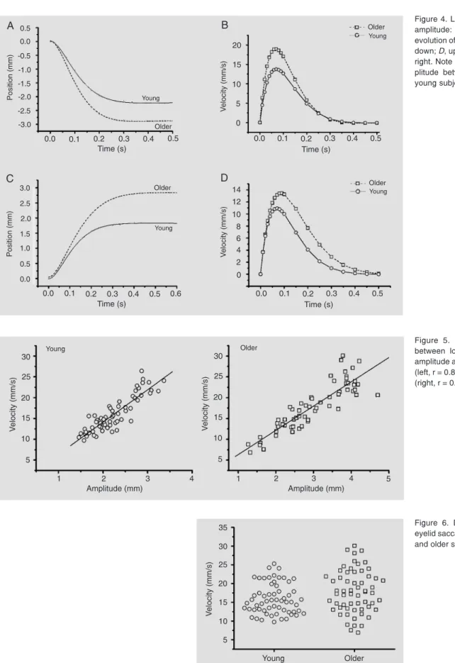
Figure 4. Lower eyelid saccade amplitude: A, down; C, up. The evolution of velocity with time (B, down; D, up) is illustrated on the right. Note the difference in am-plitude between the older and young subjects.
Figure 5. Linear relationships between lower eyelid saccade amplitude and velocity for young (left, r = 0.88) and older subjects (right, r = 0.89).
References
1. Becker W, Fuchs AF. Lid-eye coordination during vertical gaze changes in man and monkey. J Neurophysiol 1988; 60: 1227-1252. 2. Castanares S. Classification of baggy eyelids deformity. Plast
Reconstr Surg 1977; 59: 629-633.
3. Frueh BR. The mechanistic classification of ptosis. Ophthalmology
1980; 87: 1019-1021.
4. Dalgleish R, Smith JL. Mechanics and histology of senile entropion.
Br J Ophthalmol 1966; 50: 79-91.
5. Wouters RJ, van den Bosch WA, Mulder PG, Lemij HG. Upper eyelid motility in blepharoptosis and in the aging eyelid. Invest
Ophthalmol Vis Sci 2001; 42: 620-625.
6. Shore JW. Changes in lower eyelid resting position, movement, and tone with age. Am J Ophthalmol 1985; 99: 415-423.
7. Malbouisson JM, E Cruz AA, Messias A, Leite LV, Rios GD. Upper and lower eyelid saccades describe a harmonic oscillator function.
Invest Ophthalmol Vis Sci 2005; 46: 857-862.
8. Dortzbach RK, Sutula FC. Involutional blepharoptosis. A histopatho-logical study. Arch Ophthalmol 1980; 98: 2045-2049.
9. Jones LT. The anatomy of the lower eyelid and its relation to the cause and cure of entropion. Am J Ophthalmol 1960; 49: 29-36.
and extends onto both the anterior and poste-rior surfaces of the tarsus (10).
Aging is also believed to induce attenua-tion of the lower eyelid retractors. The so-called reverse lower eyelid ptosis is charac-terized by a reduction of the distance from the lower eyelid margin to the pupil center (11). This tendency of age to induce lower eyelid ptosis was seen in our older subjects who presented a smaller mean mid-pupil eyelid distance than the young subjects.
The exact mechanism of transmission of active forces to the lower eyelids during downgaze is still obscure. In one study using dynamic high-resolution magnetic resonance imaging it was found that the length of the anterior capsulopalpebral fascia between the tarsus and inferior oblique muscle remained constant in downgaze (12). The authors con-cluded that the source of the tension of the lower eyelid was posterior to the capsulo-palpebral fascia.
The forces involved in the upward dis-placement of the lower eyelid have not been investigated. Jones (9) suggested that the pull of the upper eyelid structures at the lateral canthus and the contraction of the lower preseptal muscle were the factors that explained the lower eyelid movement up-wards. The lateral pull during upward eye saccades is easily demonstrated. In fact, when the direction of gaze shifts from down- to upgaze, the lateral canthus also moves up, increasing the palpebral fissure angle (13). However, contraction of the orbicularis
muscle is unlikely to occur, because it has been shown that the orbicularis does not play a role during upper eyelid saccades (14).
10. Hawes MJ, Dortzbach RK. The microscopic anatomy of the lower eyelid retractors. Arch Ophthalmol 1982; 100: 1313-1318. 11. Bartley GB, Frueh BR, Holds JB, Linberg JV, Patel BC, Hawes MJ.
Lower eyelid reverse ptosis repair. Ophthal Plast Reconstr Surg
2002; 18: 79-83.
12. Goldberg RA, Lufkin R, Farahani K, Wu JC, Jesmanowicz A, Hyde JS. Physiology of the lower eyelid retractors: tight linkage of the anterior capsulopalpebral fascia demonstrated using dynamic ultrafine surface coil MRI. Ophthal Plast Reconstr Surg 1994; 10:
87-91.
13. Alves LA, Cruz AAV, Souza ADA, Ruiz ES. Palpebral fissure changes during upgaze and downgaze. Invest Ophthalmol Vis Sci
1998; 39: 1011 (Abstract).
14. Evinger C, Manning KA, Sibony PA. Eyelid movements. Mechan-isms and normal data. Invest Ophthalmol Vis Sci 1991; 32: 387-400. 15. Danks JJ, Rose GE. Involutional lower lid entropion: to shorten or