Iodine-131 treatment of thyroid cancer cells leads to
suppression of cell proliferation followed by induction
of cell apoptosis and cell cycle arrest by regulation of
B-cell translocation gene 2-mediated JNK/NF-
k
B
pathways
L.M. Zhao
1and A.X. Pang
2 1Department of Nuclear Medicine, Linyi People’s Hospital, Linyi, China 2Department of Urology, Linyi People
’s Hospital, Linyi, China
Abstract
Iodine-131 (131I) is widely used for the treatment of thyroid-related diseases. This study aimed to investigate the expression of p53andBTG2genes following131I therapy in thyroid cancer cell line SW579 and the possible underlying mechanism. SW579
human thyroid squamous carcinoma cells were cultured and treated with131I. They were then assessed for131I uptake, cell viability, apoptosis, cell cycle arrest, p53 expression, andBTG2gene expression. SW579 cells were transfected with BTG2 siRNA, p53 siRNA and siNC and were then examined for the same aforementioned parameters. When treated with a JNK inhibitor of SP600125 and131I or with a NF-kB inhibitor of BMS-345541 and131I, non-transfected SW579 cells were assessed in JNK/NFkB pathways. It was observed that131I significantly inhibited cell proliferation, promoted cell apoptosis and cell cycle arrest. Both BTG2 and p53 expression were enhanced in a dose-dependent manner. An increase in cell viability by up-regulation in Bcl2gene, a decrease in apoptosis by enhancedCDK2gene expression and a decrease in cell cycle arrest at G0/G1phase
were also observed in SW579 cell lines transfected with silencedBTG2gene. When treated with SP600125 and131I, the non-transfected SW579 cell lines significantly inhibited JNK pathway, NF-kB pathway and the expression of BTG2. However, when treated with BMS-345541 and131I, only the NF-kB pathway was suppressed.131I suppressed cell proliferation, induced cell
apoptosis, and promoted cell cycle arrest of thyroid cancer cells by up-regulating B-cell translocation gene 2-mediated activation of JNK/NF-kB pathways.
Key words: Iodine-131; P53; BTG2; SW579; Thyroid cancer; JNK/NF-kB pathways
Introduction
The history of radionuclide therapy for the treatment of various diseases dates back to early 1900’s. A parameter considered while choosing a particular radionuclide for therapy is the effective half-life, which is the net half-life considering both physical and biological half-life within the patient’s body or organs. The biological half-life of a radio-nuclide depends on parameters like radiotracer delivery, uptake, metabolism, clearance, and excretion within the patient’s body. The ionizing radiation leads to DNA damage, which is primarily caused by both direct or indirect inter-action of radiation leading to molecular damage such as single strand break, double-strand breaks, base damage and DNA-protein cross links (1–4). It is established that cancer cells are more prone to damage following exposure
to ionizing radiation than normal cells, which leads to the death of cancerous cells (5). The most widely used ther-apeutic radionuclide for the treatment of thyroid-related diseases such as differentiated thyroid cancer, Grave’s disease, solitary hyper-functioning nodule, and toxic multi-nodular goiter is iodine-131 (131I).131I, an isotope of127I, is commonly used as a beta emitter in radiation therapy, causing mutation and cell death. It is known that 10% of the energy and radiation dose is via gamma radiation. In a study by Eriksson et al. (6), radio-immunotherapy triggered apoptosis in tumor cells.
Expression of p53 at post-translational level is enhanced due to DNA damage by radiation (7), subsequently lead-ing to the arrest of cell growth at G1 and/or G2 phase,
Correspondence: L.M. Zhao:<[email protected]>
DNA repair, senescence or apoptosis (7–10). B-cell trans-location gene 2 (BTG2) acts as a tumor suppressor gene for a number of cancers and it is stimulated by a p53-dependent pathway, which subsequently leads to the DNA damage.BTG2gene belongs to an anti-proliferative family protein which has highly conserved domains of BTG-Box A (Y50–N71) and BTG-Box B (L97–E115) (11–14). It has been reported that amongst the numerous molecules that are involved in diverse anti- or pro-apoptotic signaling pathways, NF-kB is one of the key factors controlling anti-apoptotic responses. The anti-anti-apoptotic effect is thought to be mediated through not only transcriptional activation of dependent genes but also by cross talking with the JNK pathway (15). In the present study, we have assessed the effects of 131I in thyroid cancer cell line SW579 with special emphasis on cell proliferation, apoptosis, and cell cycle arrest, and also explored the possible underlying mechanisms in JNK/NF-kB pathways.
Material and Methods
Cell culture
SW579 human thyroid squamous cell carcinoma cells were obtained from American Type Culture Collection (USA), and cultured in L-15 medium (GE Healthcare Life Sciences, USA) supplemented with 10% fetal calf serum (Gibco, USA), 2 mM glutamine (Gibco), penicillin (100 U/mL; Sigma-Aldrich, USA) and streptomycin (100 mg/mL; Amresco, USA), and maintained at 37°C without CO2in a humidified atmosphere. SP600125 (10mM) and BMS-345541 (10mM) were used as JNK and NF-kB inhibitors to treat SW579 for 3 days, respectively (16).
131I uptake assay
The cells were seeded at 1105/well on 6-well plates for 24 h. Subsequently, the cells were cultured for 24 h with 2 mL culture medium per well containing 7.4, 14.8, 29.4 MBq/mL131I (9).
CCK-8 assay
SW579 cells were seeded on 96-well plate with 5000 cells/well, and cell proliferation was assessed by the Cell Counting Kit-8 (CCK-8, Dojindo Molecular Technologies, USA). Briefly, after stimulation, the CCK-8 solution was added to the culture medium, and the cultures were incubated for 1 h at 37°C in humidified 95% air and 5% CO2. The absorbance was measured at 450 nm using a Microplate Reader (Bio-Rad, USA).
Apoptosis assay
Cell apoptosis analysis was performed using propi-dium iodide (PI) and fluorescein isothiocynate (FITC)-conjugated Annexin V staining. Briefly, cells were washed in phosphate-buffered saline (PBS) and fixed in 70% ethanol. Fixed cells were then washed twice in PBS and stained in PI/FITC-Annexin V in the presence of 50mg/mL
RNase A (Sigma-Aldrich), and then incubated for 1 h at room temperature in the dark. Flow cytometry analysis was done by using a FACScan (Beckman Coulter, USA). Data were analyzed with FlowJo software.
Cell cycle assay
For analysis of cell cycle, cells with different treatments were trypsinized, washed twice in PBS, and fixed over-night at –20°C in 300mL PBS and 700mL ethanol. The fixed cells were spun down gently in 200 mL extraction buffer (0.1% Triton X-100, 45 mM Na2HPO4and 2.5 mM sodium citrate) at 37°C for 20 min and then stained with PI (BD Biosciences, USA) (50 mg/mL) containing 50 mg/mL RNase A for 30 min at 37°C in the dark, and subsequently analyzed by FACScan. The experiment was repeated at least three times, and the data were analyzed using Cell-Quest and ModFit softwares (Verity Software House, USA).
qRT-PCR
Total RNA was extracted with TRIzol reagent accord-ing to the manufacturer’s protocol (Sigma) and 2mg were reverse-transcribed with the Omniscript RT kit (Qiagen, Italy) using random primers (1 mM) at 37°C for 1 h. Real time PCR was performed in triplicate in 20 mL reaction volumes using the Power SYBER Green PCR Master Mix (Applied Biosystems, USA). All primers were purchased from Invitrogen Life Technologies (USA). Real time PCR reactions were carried out in a MJ MiniTM Personal Thermal Cycler apparatus (Bio-Rad Laboratories, USA). Melting curves were obtained by increasing the tempera-ture from 60 to 95°C with a temperatempera-ture transition rate of 0.5°C/s. The comparative threshold cycle number (CT) method was used to assess the relative quantification of gene expression. The fold change of the target gene was calculated as 2-DDCT.
siRNAs transfection
BTG2 siRNA, p53 siRNA, and siNC were designed and synthesized by GenePharma (China). Cell transfection was performed using Lipofectamine 3000 (Invitrogen Life Tech-nologies) according to the manufacturer’s instructions.
Statistical analysis
All experiments were repeated three times. The results of multiple experiments are reported as means±SD.
Statistical analyses were performed using SPSS 19.0 statistical software. Differences were compared using a one-way analysis of variance (ANOVA). A P-value of
o0.05 was considered to be statistically significant.
Results
131
I inhibited cell proliferation, promoted cell apoptosis, and induced cell cycle arrest
131I was found to inhibit cell proliferation when admin-istered to SW579 human thyroid squamous cell carcinoma
cell lines. Cell viability was lesser than 0.5% at 14.8 (Po0.05) and 29.4 MBq/mL (significantly lower than cell
viability at 7.4 MBq/mL; Figure 1A). A significant increase in apoptosis was observed when SW579 cells was treated with131I at 29.6 and 14.8 MBq/mL (Po0.05; Figure 1B).
Furthermore, expression of Bcl-2 was suppressed by 0.5 fold, andBax andcleaved-Cas 3genes were enhanced by 1.5 and 1.5 folds, respectively, at 14.8 MBq/mL com-pared to GAPDH expression used as endogenous control. 131
I induced cell cycle arrest significantly by more than 60% at Go/G1at the concentration of 14.8 MBq/mL com-pared to the arrest at G1/S and S/G2, by suppressing the expression of cyclin-dependent kinases 2 (CDK2) and cyclin E by 0.5 and 0.4 folds, respectively, at 14.8 MBq/mL compared to GAPDH expression (Po0.05). Furthermore,
the expressions ofp27andp21genes were enhanced by 1.5 and 2 folds, respectively, at 14.8 MBq/mL compared to GAPDH expression (Figure 1C).
131
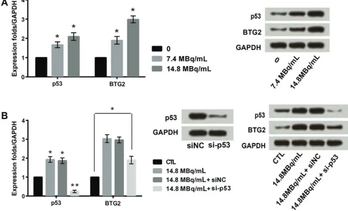
I induced the expressions of p53 and BTG2 As shown in Figure 2A,131I increased the expressions of p53 and BTG2 in a concentration-depended manner. The expression of BTG2 was raised even after silencing of p53, thereby indicating that the higher expression of BTG2 was only partly dependent on p53 expression (Po0.05)
(Figure 2B).
Silencing of BTG2 reversed the effects of131I on cell proliferation, cell apoptosis, and cell cycle arrest
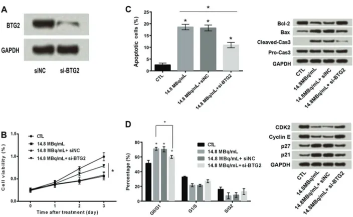
SW579 cells transfected with silenced BTG2 gene (Figure 3A) and treated with131I, presented an increase in cell viability (more than 0.5%) at 14.8 MBq/mL (Figure 3B), unlike in non-transfected cells, shown in Figure 1. Similarly, a significant decrease in apoptosis (approximately 10%) was found in cells transfected with silenced BTG2gene compared to non-transfected cells, where apoptosis was approximately 20%, when treated with 131I (Figure 3C). A down-regulation in Bcl2 and an up-regulation in Bax by 2.0 and 1.2 folds, respectively, were observed in cell proliferation pathway. A significant decrease in cell cycle arrest (less than 60%) was also observed at Go/G1stage in cells transfected with silencedBTG2gene compared to non-transfected cells. Assessment of the molecular path-way revealed that there was an up-regulation in CDK2, followed by down-regulation in cyclin E and p27 genes and down-regulation inp21gene (Figure 3D).
131
I up-regulated BTG2 expression by activation of JNK/NF-kB pathways
As shown in Figure 4, non-transfected SW579 cells treated with SP600125, a JNK inhibitor, and131I at 14.8 MBq/ mL not only had a significant inhibition of JNK pathway but also of NF-kB pathway. The expression of BTG2 was
also down-regulated. Furthermore, non-transfected SW579 cells treated with BMS-345541, a NF-kB inhibitor, and 131I at 14.8 MBq/mL had only the expression of
NF-kB pathway affected but not of the JNK pathway. The expres-sion of BTG2 was down-regulated, thus indicating that 131
I up-regulated BTG2 expression by activation of JNK/NF-kB pathways.
Discussion
It is well known that 131I destroys residual thyroid cancer tissue after surgical resection of differentiated thyroid carcinoma. The degree to which DNA is damaged by ionizing radiation depends on factors like type and dose of radiation (17,18). In the present study, we eval-uated the role of131I in cell proliferation, apoptosis and cell cycle arrest in a thyroid cancer cell line, together with the exploration of the possible underlying mechanism (increased expression of BTG2gene-mediated activation of the JNK/NF-kB pathways).131I significantly inhibited cell proliferation as assessed in terms of cell-viability, enhanced cell apoptosis by down-regulatingBcl2gene, and promoted cell cycle arrest at G0/G1phase by down-regulatingCDK2 gene. Cell apoptosis is largely regulated by protein-protein
interactions between members of the Bcl-2 protein family. It is known that members ofBcl-2family genes have con-served domains called Bcl-2 homology domains, which are differentially modulated in various cancers (19,20).
Furthermore,131I increased both BTG2 and p53 expres-sion in a dose-dependent manner. It is mportant to men-tion that 131I enhanced the expression of BTG2, after silencing p53 gene in SW579 cells, suggesting that the expression of BTG2 was partly dependent on the p53
gene. An increase in cell viability by up-regulation inBcl2
gene, a decrease in apoptosis by enhancedCDK2gene expression and a decrease in cell cycle arrest at G0/G1 phase were also observed in SW579 cells transfected with silencedBTG2gene. Moreover, it was observed that not only the JNK pathway in the non-transfected SW579 cells, treated with SP600125, a JNK inhibitor, and131I at 14.8 MBq/mL, was significantly inhibited but also the NF-kB pathway was inhibited along with the down-regulation of the BTG2 expression. Again, when treated with BMS-345541, a NF-kB inhibitor, and131I, SW579 cells revealed only suppression of the NF-kB pathway but not that of the JNK pathway. Considering the aforementioned effects of 131I, we can conclude that 131I up-regulated BTG2 expression by activation of JNK/NF-kB pathways.
Figure 2.Effects of different concentrations of iodine-131 (131I) on expression of p53 and BTG2 (A).B, Expression of BTG2 was raised even with silencing of p53 (si-p53). Data are reported as means±SD. *Po0.05. **Po0.01 compared with control (CTL–GAPDH) (ANOVA).
Figure 3.A, Transfection efficiency of BTG2. Silencing of BTG2 increased iodine-131 (131I)-induced cell proliferation (B),131I-induced cell apoptosis (C), and down-regulated131I-induced cell cycle arrest (
D). Data are reported as means±SD. *Po0.05 compared with control (CTL–GAPDH) (ANOVA).
References
1. Oleinick NL, Chiu SM, Ramakrishnan N, Xue LY. The formation, identification, and significance of DNA-protein cross-links in mammalian cells.Br J Cancer Suppl1987; 8: 135–140. 2. Nikjoo H, O’Neill P, Goodhead DT, Terrissol M.
Computa-tional modelling of low-energy electron-induced DNA damage by early physical and chemical events.Int J Radiat Biol1997; 71: 467–483, doi: 10.1080/095530097143798.
3. van Gent DC, Hoeijmakers JH, Kanaar R. Chromosomal stability and the DNA double-stranded break connection.
Nat Rev Genet2001; 2: 196–206, doi: 10.1038/35056049. 4. Walters K. Modelling the probability distribution of the number
of DNA double-strand breaks due to sporadic alkylation of nucleotide bases. J Theor Biol 2007; 245: 161–168, doi: 10.1016/j.jtbi.2006.09.028.
5. Neshasteh-Riz A, Koosha F, Mohsenifar A, Mahdavi SR. DNA Damage Induced in Glioblastoma Cells by I-131: A Comparison between Experimental Data and Monte Carlo Simulation.Cell J2012; 14: 25–30.
6. Eriksson D, Blomberg J, Lindgren T, Lofroth PO, Johansson L, Riklund K, et al. Iodine-131 induces mitotic catastrophes and activates apoptotic pathways in HeLa Hep2 cells.Cancer Biother Radiopharm 2008; 23: 541–549, doi: 10.1089/cbr. 2008.0471.
7. Reinhardt HC, Schumacher B. The p53 network: cellular and systemic DNA damage responses in aging and cancer.
Trends Genet2012; 28: 128–136, doi: 10.1016/j.tig.2011. 12.002.
8. Jin S, Levine AJ. The p53 functional circuit.J Cell Sci2001; 114: 4139–4140.
9. Feng Z, Levine AJ. The regulation of energy metabolism and the IGF-1/mTOR pathways by the p53 protein.Trends Cell Biol 2010; 20: 427–434, doi: 10.1016/j.tcb.2010. 03.004.
10. Zhang W, Gao R, Yu Y, Guo K, Hou P, Yu M, et al. Iodine-131 induces apoptosis in HTori-3 human thyrocyte cell line and G2/M phase arrest in a p53-independent pathway.Mol Med Rep2015; 11: 3148–3154.
11. Chiang KC, Tsui KH, Chung LC, Yeh CN, Feng TH, Chen WT, et al. Cisplatin modulates B-cell translocation gene 2 to attenuate cell proliferation of prostate carcinoma cells in
both p53-dependent and p53-independent pathways. Sci Rep2014; 4: 5511, doi: 10.1038/srep05511.
12. Lim IK. TIS21 (/BTG2/PC3) as a link between ageing and cancer: cell cycle regulator and endogenous cell death molecule.J Cancer Res Clin Oncol2006; 132: 417–426, doi: 10.1007/s00432-006-0080-1.
13. Cortes U, Moyret-Lalle C, Falette N, Duriez C, Ghissassi FE, Barnas C, et al. BTG gene expression in the p53-dependent and -independent cellular response to DNA damage.Mol Carcinog 2000; 27: 57–64, doi: 10.1002/(SICI)1098-2744 (200002)27:2o57::AID-MC143.0.CO;2-I.
14. Rouault JP, Falette N, Guehenneux F, Guillot C, Rimokh R, Wang Q, et al. Identification of BTG2, an antiproliferative p53-dependent component of the DNA damage cellular response pathway. Nat Genet 1996; 14: 482–486, doi: 10.1038/ng1296-482.
15. Namba H, Saenko V, Yamashita S. Nuclear factor-kB in thyroid carcinogenesis and progression: a novel therapeutic target for advanced thyroid cancer. Arq Bras Endocrinol Metabol 2007; 51: 843–851, doi: 10.1590/S0004-273020 07000500023.
16. Yu J, Ren P, Zhong T, Wang Y, Yan M, Xue B, et al. Pseudolaric acid B inhibits proliferation in SW579 human thyroid squamous cell carcinoma.Mol Med Rep2015; 12: 7195–7202.
17. Reisz JA, Bansal N, Qian J, Zhao W, Furdui CM. Effects of ionizing radiation on biological molecules -mechanisms of damage and emerging methods of detection.Antioxid Redox Signal2014; 21: 260–292, doi: 10.1089/ars.2013.5489. 18. Moore S, Stanley FK, Goodarzi AA. The repair of
environ-mentally relevant DNA double strand breaks caused by high linear energy transfer irradiation - no simple task.DNA Repair
2014; 17: 64–73, doi: 10.1016/j.dnarep.2014.01.014. 19. Thomadaki H, Scorilas A. BCL2 family of apoptosis-related
genes: functions and clinical implications in cancer. Crit Rev Clin Lab Sci2006; 43: 1–67, doi: 10.1080/1040836050 0295626.
20. van Delft MF, Huang DC. How the Bcl-2 family of proteins interact to regulate apoptosis.Cell Res2006; 16: 203–213, doi: 10.1038/sj.cr.7310028.