NOP14 inhibits melanoma proliferation and metastasis
by regulating Wnt/
b
-catenin signaling pathway
Jingrong Li
1,2, Ruihua Fang
2, Jianqin Wang
3and Liehua Deng
11Department of Dermatology, The First Af
filiated Hospital of Jinan University, Guangzhou, China 2Department of Dermatology, Guangzhou First People
’s Hospital, School of Medicine, South China University of Technology, Guangzhou, China 3Department of Dermatology, Guangzhou Institute of Dermatology, Guangzhou, China
Abstract
Malignant melanoma is an aggressive skin cancer with a high mortality rate. Nucleolar protein 14 (NOP14) has been implicated in cancer development. However, the role of NOP14 in malignant melanoma progression remains largely unclear. In this study, we observed that malignant melanoma tissue showed NOP14 down-regulation compared to melanocytic nevi tissues. Moreover, we observed that NOP14 expression was significantly associated with melanoma tumor thickness and lymph node metastasis. NOP14 overexpression in melanoma cells suppressed proliferation, caused G1 phase arrest, promoted apoptosis, and inhibited melanoma cell migration and invasion. Further investigations revealed that NOP14 overexpression reduced the expression levels of Wnt3a,b-catenin, and GSK-3bof the Wnt/b-catenin pathway. In summary, we demonstrated that NOP14 inhibited melanoma cell proliferation and metastasis by regulating the Wnt/b-catenin signaling pathway.
Key words: Malignant melanoma; Metastasis; NOP14; Proliferation; Wnt/b-catenin pathway
Introduction
The incidence of malignant melanoma, an aggressive cancer of the skin with a high mortality rate, has continued to increase in the past two decades (1). Malignant melanoma ranks as the fifth and seventh most common malignant cancer for males and females, respectively (2). Exposure to ultraviolet radiation, fair skin, dysplastic nevi syndrome, age, and family history are the most common risk factors for the development of malignant melanoma (3). Early diagnosis of melanoma is associated with good prognosis; however, the median survival of patients with metastatic malignant melanoma is 6–9 months (4,5). The current therapeutic interventions for metastatic melanoma, including surgery, radiation therapy, and chemotherapy, are not sufficient, and only negligible improvement in survival has been achieved overall (6). Therefore, under-standing the biology of malignant melanoma initiation and progression is imperative.
Nucleolar protein 14 (NOP14) is a stress-response gene that encodes a nucleolar protein required for the maturation of 18S rRNA and production of 40S ribosome (7). Studies show that ribosomes are involved in DNA repair and regulation of cell development and differentiation, and perturbations in ribosome function may result in tumor formation (8). An increasing body of evidence indicates that
NOP14 is involved in cancer development. For example, Zhou et al. (9) observed that NOP14 overexpression pro-moted pancreatic cancer cell proliferation and metastasis bothin vitroandin vivo. Lei et al. (10) reported that NOP14 suppressed the tumorigenesis and metastasis of breast cancer by inhibiting the NRIP1/Wnt/b-catenin pathway. However, the role of NOP14 in melanoma progression remains largely unclear.
In this study, we investigated the roles of NOP14 in malignant melanoma development. We determined the expression levels of NOP14 in malignant melanoma tissues, and its association with clinicopathological features. We also studied the functional role of NOP14 overexpression in regulating melanoma growth, cell cycle, apoptosis, migra-tion, and invasion. Furthermore, the underlying mechanism via which NOP14 suppresses melanoma cell proliferation and metastasis was investigated.
Material and Methods
Sample collection
Forty malignant melanoma tissues were collected from patients with malignant melanoma at the Department of Dermatology, Guangzhou First People’s Hospital.
Correspondence: Liehua Deng:<[email protected]>
Received July 9, 2018 | Accepted September 27, 2018
The diagnosis of malignant melanoma was confirmed pathologically. None of these patients had received radiation therapy or chemotherapy before surgical resection. Twenty melanocytic nevi tissue samples were collected as negative control. Written informed consents were obtained from patients involved in this study. The tissue samples were immediately snap-frozen in liquid nitrogen after surgical resection and stored at–80°C for further use.
NOP14 immunohistochemistry (IHC)
IHC was performed to determine NOP14 expression in tissue samples. Four-micrometer sections cut from formalin-fixed, paraffin-embedded blocks were placed on charged slides, which were then dried and melted. After antigen retrieval in citrate buffer, IHC streptavidin-biotin complex was formed and 3,30diaminobenzidine staining was performed. Results of IHC analysis were reviewed independently by two senior pathologists who were blinded to the outcome of the study. Semi-quantitative assessment of target proteins was performed by con-sensus, which involved determination of the staining intensity (negative, 0; light yellow, 1; brown, 2; tan, 3) of each cell and the extent of staining (ratio of the number of positive cells to the number of counted cells, being a ratio of 1, 25%; ratio of 2, 26–50%; ratio of 3, 51–75%; ratio of 4, 475%) in each random field. Scores for the intensity and extent of staining were multiplied to obtain weighted scores for each patient (maximum possible score was 12). For statistical analysis, the weighted scores were grouped into four categories, with a score of 0 con-sidered negative, 1–4 (+) considered as weakly positive, and 5–8 (++), 9–12 (+++), and (++)–(+++) consid-ered highly positive.
Cell culture and transfection
Human melanoma cell lines A375 and SK-ML110 were purchased from the Cell Bank of the Chinese Academy of Sciences (China). All cell lines were cultured in Hyclone Dulbecco’s modified Eagle’s medium (DMEM) (Thermo Fisher Scientific, USA) containing 10% fetal bovine serum (FBS) and 100 U/mL each of penicillin and streptomycin (Gibco, USA) at 37°C in a humidified atmosphere with 5% CO2. For NOP14 overexpression, full-length human
NOP14 cDNA was amplified by PCR and inserted into the pcDNA3.1 vector (Realgene, China) according to the manufacturer’s instructions. The forward and reverse primer sequences were F: 50-CGGGGTACCGCCAC CATGGCGAAGGCGAAGAAGGTCGGGGC-30, and R: 50-CTAGTCTAGATTATTTTTTGAACTTTTTCCTCTTC-30. Cells (1105cells/well) were seeded in 24-well plates, and the NOP14 overexpression and empty vectors were transfected into cells using the FuGENEsHD transfection reagent (Roche Applied Science, USA), according to the manufacturer’s instructions. The cells were then cultured at 37°C in a 5% CO2incubator. After 48 h of transfection,
the cells were harvested for quantitative reverse transcrip-tion-polymerase chain reaction (qRT-PCR) and western blot analyses.
qRT-PCR
Total RNA was extracted from cultured cells using the TRIzol reagent (Invitrogen, USA) according to the manufacturer’s instructions. Total RNA concentration was determined using the NanoDrop ND-1000 spectro-photometer (Agilent Technologies, USA). Total RNA (1mg) was then reverse-transcribed to cDNA using Superscript III reverse transcriptase (Invitrogen). qRT-PCR was per-formed with SYBR Green (Takara, China) and 7500 real-time PCR system (Applied Biosystems, USA). The primers were synthesized by Takara, and their sequences were: forward (F): ATCACTGGGCTGCTATTTCC, NOP14-reverse (R): CTCTGGGACAAAGCCACATA; Wnt3a-F CC CAAGAGCCCAAAAGAG, Wnt3a-R CAGTGGATATAGC AGCATCAG;b-catenin-F: TCTTGGCCATCCTTCTGTGT, b-catenin-R GGGCTTTTATGTGGGTTCTG; GSK-3b: FC TGCACCTTCTTTCCAGTGA, GSK-3b-R: GCATTGGTG CAGACAAGATG; 18s-F: CCTGGATACCGCAGCTAGGA, 18s-R: GCGGCGCAATACGAATGCCCC. The 18s rRNA was used as an internal control. Relative expression was calculated using the 2-DDCtmethod. All experiments were performed in triplicate.
Western blotting
Cells were lysed using ice-cold mammalian radio-immunoprecipitation assay (RIPA) buffer (Beyotime, China), containing a protease inhibitor cocktail (Invitrogen) and phenyl methanesulfonate (PMSF) (Invitrogen). Proteins were quantified by the bicinchoninic acid (BCA) protein assay (Thermo Fisher Scientific, USA). Equal amounts of protein samples were separated by sodium dodecyl sulfate-polyacrylamide gel electrophoresis (SDS-PAGE) and transferred onto a polyvinylidene difluoride (PVDF) membrane (Thermo Fisher Scientific), followed by incu-bation with 10% nonfat milk overnight at 4°C. After washing thrice with phosphate buffered saline (PBS) containing Tween 20 (PBST), the membrane was incubated with primary antibodies for 1 h at room temperature with the following primary antibodies: NOP14 (1:500), Wnt3a (1:800),b-catenin (1:1000), GSK-3b(1:500), and GAPDH (1:2000). After washing thrice with PBST, the membrane was incubated with 1:10,000 dilution of horseradish peroxidase (HRP)-conjugated goat anti-rabbit IgG H & L secondary antibodies (Southern Biotech, USA). The membrane was rinsed, and protein bands were visua-lized using an enhanced chemiluminescence detection kit (Thermo Scientific).
Cell proliferation assay
96-well plate, where each well contained 100 mL fresh serum-free medium. After culturing for 0, 24, 48, 72, and 96 h, the cells were treated with 10mL MTT and incubated at 37°C for 4 h. One hundred microliters of formazan solvent was added to dissolve the formazan crystals. The absorbance was read at 570 nm using a microplate reader (Thermo Fisher Scientific, USA). All assays were performed in triplicate.
Migration and invasion assays
Cell migration and invasion assays were performed using transwell chambers (Corning Co., USA) with or without Matrigel (BD Biosciences, USA). After 48 h of transfection, cells (2105) were seeded in the upper wells with or without 10mg/mL Matrigel in DMEM, whereas the lower well contained the same medium with 10% FBS. After 48 h of incubation at 37°C in a humid atmosphere containing 5% CO2, non-migrating cells on the upper side of thefilter were removed by wiping with a cotton swab, whereas cells that migrated through the membranes were fixed with 70% cold ethanol, stained with 0.1% crystal violet, and counted under 200 magnification of the micro-scope (Olympus, Japan). The experiment was performed in triplicate.
Cell cycle analysis
One million cells were harvested 48 h after transfection and washed in cold PBS, followed by fixing in 90% ice-cold ethanol for 1 h at room temperature. Before cell cycle analysis, the cells were washed thrice in cold PBS, followed by incubation with propidium iodide (PI, 50mg/ mL) and RNase A (2 mg/mL; Sigma, USA) for 20 min at 37°C in the dark. Cell cycle analysis was performed using flow cytometry (BD Biosciences). Populations in the G1, S, and G2 phases are shown as percentages of total gated cells. Each experiment was repeated thrice.
Analysis of apoptosis byflow cytometry
Apoptosis was assessed using an annexin V-FITC-PI dual staining kit (Biolegend, USA) followed by flow cytometry analysis per manufacturer’s instructions. Briefly, the cell pellet (5103cells) was resuspended in 500mL binding buffer. Next, 5mL each of annexin V-FITC and PI were added to the cell suspension, and the cells were cultured in the dark for 15 min at room temperature, followed by flow cytometry analysis (BD Biosciences). Each experiment was repeated thrice.
Statistical analysis
Statistical analysis was performed using the SPSS 19.0 software (IBM, USA). Data are reported as means±SD. Data were analyzed using a Student’st-test for two-group comparison and one-way analysis of variance for multiple-group comparison. The chi-squared test was used to analyze the correlation between NOP14 protein levels and clinicopathological characteristics of patients with melanoma. A P-valueo0.05 was considered statistically significant.
Results
NOP14 expression and clinicopathological characteristics
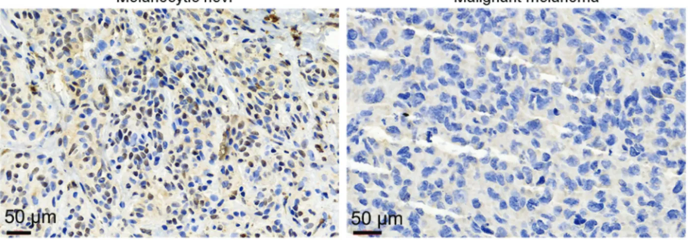
To investigate the relationship of NOP14 and human malignant melanoma development, wefirst analyzed NOP14 expression levels in 40 paired malignant melanoma tissues and melanocytic nevi tissues by IHC. As shown in Figure 1, NOP14 expression was significantly lower in malignant melanoma tissues than in melanocytic nevi. This indicated that abnormal NOP14 expression levels might be related to malignant melanoma pathogenesis. Moreover, the associa-tion between the NOP14 expression and the clinicopatho-logical features of malignant melanoma were analyzed. As shown in Table 1, no significant association between NOP14
Table 1. Correlation between nucleolar protein 14 (NOP14) protein levels and clinicopathological characteristics of patients with melanoma.
Characteristic n NOP14 protein levels P-value
Low expression (–,+) High expression (++,+++)
Age (years) 0.427
o60 21 8 13
X60 19 5 14
Gender 0.919
Male 18 6 12
Female 22 7 15
Tumor thickness (mm) 0.002
o1 14 9 5
X1 26 4 22
Site 0.427
Sun-exposed 21 8 13
Sun-protected 19 5 14
Lymph node metastasis 0.010
No 11 7 4
Yes 29 6 23
Statistical analyses were carried out with the chi-squared test. Bold type indicates statistical significance (Po0.05).
Figure 2.Effect of nucleolar protein 14 (NOP14) overexpression on melanoma cell proliferation. NOP14 mRNA levels (A) and protein levels (B) in melanoma cell lines transfected with NOP14 overexpression and empty vectors.CandD, Cell proliferation analysis of melanoma cells after transfection of NOP14 overexpression and empty vectors. Data are reported as means±SD. **Po0.01vsempty
expression and age, gender, and lesion site were observed. Instead, NOP14 expression was significantly associated with an increase in tumor thickness and lymph node metastasis.
NOP14 overexpression suppressed melanoma cell proliferation
To assess the biological function of NOP14 in mela-noma cell lines A375 and SK-ML110, NOP14 overexpres-sion and empty vectors were transiently transfected into
cells. As shown in Figure 2A and B, the mRNA and protein levels of NOP14 were significantly increased in melanoma cell lines harboring the NOP14 overexpression vector compared to those containing the empty vector (Po0.01).
Next, we investigated the role of NOP14 in the regula-tion of cell proliferaregula-tion. As shown in Figure 2C and D, the NOP14 overexpression vector significantly suppressed cell proliferation after 72 h of transfection compared to cells transfected with the empty vector. These results
suggested that NOP14 may be involved in the regula-tion of melanoma cell proliferaregula-tion.
NOP14 overexpression promoted apoptosis and induced cell cycle arrest
To investigate the underlying mechanism that NOP14 overexpression suppressed melanoma cell proliferation, we assessed the effect NOP14 overexpression on apoptosis in A375 and SK-ML110 cell lines. As shown in Figure 3A and B, NOP14 overexpression significantly promoted apoptosis in both melanoma cell lines. In addition, results of flow cytometry showed that the proportion of cells in
the G1 phase increased, whereas those in the G2 phase decreased after overexpression of NOP14 in A375 and SK-ML110, indicating that NOP14 induced G1 arrest (Figure 3C and D).
NOP14 overexpression inhibited migration and invasion of melanoma cells
We further examined the effects of NOP14 over-expression on melanoma cell migration and invasiveness. The transwell assay revealed that NOP14 overexpression remarkably reduced the number of A375 and SK-ML110 cells that passed through the transwell membrane compared
to the control group (Figure 4). These results suggested that NOP14 overexpression significantly inhibited the migratory ability and invasiveness of melanoma cells (Po0.01).
NOP14 overexpression suppressed the Wnt/b-catenin pathway in melanoma cells
The Wnt/b-catenin signaling pathway plays an impor-tant role in tumor progression, including in melanoma. Hence, we assessed the changes in mRNA and protein levels of genes encoding various components of the Wnt/ b-catenin pathway, such as Wnt3a,b-catenin, and GSK-3b, by overexpressing NOP14 in melanoma cell lines. qRT-PCR showed that compared to the empty vector control, the mRNA levels of Wnt3a,b-catenin, and GSK-3b were decreased by NOP14 overexpression in both melanoma cell lines (Figure 5A to C). Moreover, western blot analysis showed that the levels of Wnt3a, b-catenin, and GSK-3bwere reduced by overexpressing NOP14 in both melanoma cell lines (Figure 5D). These results indicated that NOP14 inhibited the Wnt/b-catenin pathway.
Discussion
NOP14 is highly conserved in eukaryotes (7), and its down-regulation inhibits ribosome biogenesis after DNA damage (11). In recent years, increasing evidence shows
that NOP14 participates in cancer progression, cellular proliferation, metastasis, and apoptosis (12–15). However, the role of NOP14 in melanoma was unknown. In this study, we showed that compared to melanocytic nevi, malignant melanoma tissues showed down-regulation of NOP14 expression. Moreover, we observed that NOP14 expression was significantly associated with melanoma tumor thickness and lymph node metastasis. These results indicated that abnormal NOP14 expression might be related to malignant melanoma pathogenesis in human. Upon further assessment of the biological function of NOP14 in melanoma cell lines A375 and SK-ML110, we observed that NOP14 overexpression inhibited mela-noma cell proliferation. Cell proliferation is association with protein synthesis, cell cycle progression, and apoptosis (16). Analysis of cell cycle distribution and apoptosis in melanoma cells overexpressing NOP14 showed that NOP14 overexpression induced cell cycle arrest at the G1 phase and promoted apoptosis in both melanoma cell lines. Malignant cancer cells are characterized by their ability to migrate and invade tissues into blood vessels, where they initiate metastasis (17). Lei et al. (10) showed that NOP14 suppressed tumorigenesis and metastasis of breast cancer cellsin vivoandin vitro. The transwell assay in this study revealed that NOP14 overexpression reduced the migratory ability and invasiveness of A375
and SK-ML110 cells. These results suggested that NOP14 might be an important regulatory molecule for melanoma formation and development and a potential predictor of melanoma.
Tumor progression is driven by molecular changes that confer proliferative advantage and promote invasive and metastatic phenotypes. Reports show that the Wnt/b-catenin pathway is important for embryonic development and adult tissue homeostasis, including cell migration, proliferation, hematopoiesis, and wound repair (18). Deregulation or mutations in the Wnt/b-catenin pathway are implicated in both tumor formation and progression of various types of cancer. b-catenin accumulates in the nucleus, binds to T-cell factor/lymphoid enhancer binding factor (TCF/LEF), and activates its target genes such as cyclin D1 (CCND1) and the cellular myelo-cytomatosis (MYC) oncogene (19). b-catenin levels are subjected to tight regulation, particularly through the GSK3b-dependent phosphorylation of exon3, which plays a key role in controlling proteasomal degradation (20). Moreover, the Wnt/b-catenin pathway has been reported not only to be a predisposing factor for melanoma, but also to contribute to the progression and deterioration of melanoma (21). In the present study, we observed that NOP14 overexpression suppressed the Wnt/b-catenin pathway.
These results indicated that NOP14 exerted its func-tions on melanoma cells through the Wnt/b-catenin signaling pathway, and suggested that targeting of NOP14 may constitute a novel therapeutic approach for treating patients with melanoma or abnormally activated Wnt/b-catenin signaling pathway.
In conclusion, we showed that NOP14 was down-regulated in malignant melanoma tissue. NOP14 over-expression suppressed melanoma cell proliferation, arrested the cell cycle at G1 phase, promoted apoptosis, and inhibited cell migration and invasion. Additionally, overexpression of NOP14 decreased Wnt3a,b-catenin, and GSK-3b expression. Our findings show that over-expression of NOP14 reduced melanoma cell proliferation and metastasis by regulating the Wnt/b-catenin signaling pathway. These observations may provide new insights into the development of targeted therapeutic agents for melanoma.
Acknowledgments
This work was supported by Guangzhou General Science and Technology Project of Health and Family Planning (No. 20181A010005) and Guangdong Medical Science and Research Foundation (No. A2018556).
References
1. Chen W, Zheng R, Baade PD, Zhang S, Zeng H, Bray F, et al. Cancer statistics in China, 2015.CA Cancer J Clin 2016; 66: 115–132, doi: 10.3322/caac.21338.
2. Trotter SC, Sroa N, Winkelmann RR, Olencki T, Bechtel M. A global review of melanoma follow-up guidelines.J Clin Aesthet Dermatol2013; 6: 18–26.
3. Erdmann F, Lortet-Tieulent J, Schuz J, Zeeb H, Greinert R, Breitbart EW, et al. International trends in the incidence of malignant melanoma 1953-2008--are recent generations at higher or lower risk?Int J Cancer2013, 132: 385–400, doi: 10.1002/ijc.27616.
4. Gogas HJ, Kirkwood JM, Sondak VK. Chemotherapy for metastatic melanoma: time for a change?Cancer2007; 109: 455–464, doi: 10.1002/cncr.22427.
5. Chapman PB, Hauschild A, Robert C, Haanen JB, Ascierto P, Larkin J, et al. Improved survival with vemur-afenib in melanoma with BRAF V600E mutation.N Engl J Med 2011; 364: 2507–2516, doi: 10.1056/NEJMoa11 03782.
6. Berrocal A, Cabanas L, Espinosa E, Fernandez-de-Misa R, Martin-Algarra S, Martinez-Cedres JC, et al. Melanoma: diagnosis, staging, and treatment. Consensus group recom-mendations.Adv Ther 2014; 31: 945–960, doi: 10.1007/ s12325-014-0148-2.
7. Liu PC, Thiele DJ. Novel stress-responsive genes EMG1 and NOP14 encode conserved, interacting proteins required for 40S ribosome biogenesis.Mol Biol Cell2001; 12: 3644– 3657, doi: 10.1091/mbc.12.11.3644.
8. Ruggero D, Grisendi S, Piazza F, Rego E, Mari F, Rao PH, et al. Dyskeratosis congenita and cancer in mice deficient in
ribosomal RNA modification.Science2003; 299: 259–262, doi: 10.1126/science.1079447.
9. Zhou B, Wu Q, Chen G, Zhang TP, Zhao YP. NOP14 promotes proliferation and metastasis of pancreatic cancer cells. Cancer Lett 2012; 322: 195–203, doi: 10.1016/ j.canlet.2012.03.010.
10. Lei JJ, Peng RJ, Kuang BH, Yuan ZY, Qin T, Liu WS, et al. NOP14 suppresses breast cancer progression by inhibiting NRIP1/Wnt/beta-catenin pathway.Oncotarget2015; 6: 25701– 25714.
11. Calkins AS, Iglehart JD, Lazaro JB. DNA damage-induced inhibition of rRNA synthesis by DNA-PK and PARP-1. Nucleic Acids Res2013; 41: 7378–7386, doi: 10.1093/nar/ gkt502.
12. Kuhn H, Hierlmeier T, Merl J, Jakob S, Aguissa-Toure AH, Milkereit P, et al. The Noc-domain containing C-terminus of Noc4p mediates both formation of the Noc4p-Nop14p submodule and its incorporation into the SSU processome. PLoS One2009; 4: e8370, doi: 10.1371/journal.pone.0008370. 13. Zhou B, Irwanto A, Guo YM, Bei JX, Wu Q, Chen G, et al. Exome sequencing and digital PCR analyses reveal novel mutated genes related to the metastasis of pancreatic ductal adenocarcinoma.Cancer Biol Ther2012; 13: 871–879, doi: 10.4161/cbt.20839.
14. Cao Q, Wang X, Zhao M, Yang R, Malik R, Qiao Y, et al. The central role of EED in the orchestration of polycomb group complexes. Nat Commun 2014; 5: 3127, doi: 10.1038/ ncomms4127.
domain-mediated protein interactions.Sci Signal2012; 5: rs6, doi: 10.1126/scisignal.2002255.
16. Merrick KA, Wohlbold L, Zhang C, Allen JJ, Horiuchi D, Huskey NE, et al. Switching Cdk2 on or off with small molecules to reveal requirements in human cell prolifera-tion. Mol Cell 2011; 42: 624–636, doi: 10.1016/j.molcel. 2011.03.031.
17. Alderman C, Sehlaoui A, Xiao Z, Yang Y. MicroRNA-15a inhibits the growth and invasiveness of malignant melanoma and directly targets on CDCA4 gene.Tumour Biol2016; 37: 13941–13950, doi: 10.1007/s13277-016-5271-z.
18. Clevers H, Nusse R. Wnt/beta-catenin signaling and disease. Cell2012, 149: 1192–1205, doi: 10.1016/j.cell.2012.05.012.
19. Nakamoto M, Hisaoka M. Clinicopathological implications of Wingless/int1 (WNT) signaling pathway in pancreatic ductal adenocarcinoma.J Uoeh2016; 38: 1–8, doi: 10.7888/ juoeh.38.1.
20. Benoit YD, Guezguez B, Boyd AL, Bhatia M. Molecular pathways: epigenetic modulation of Wnt-glycogen synthase kinase-3 signaling to target human cancer stem cells.Clin Cancer Res2014; 20: 5372–5378, doi: 10.1158/1078-0432. CCR-13-2491.