ORIGINAL
ARTICLE
Candida
species isolated from the vaginal mucosa
of HIV-infected women in Salvador, Bahia, Brazil
Authors
Paula Matos Oliveira1 Rita Elizabeth Mascarenhas2 Claire Lacroix3 Suzana Ramos Ferrer2 Rone Peterson C Oliveira4 Elaine Andrade Cravo5 André P Ribeiro Alves5 Maria Fernanda Rios Grassi6
1Obstetrics and Gynecology
Specialist; PhD student of Postgraduate Program on Medicine and Human Health, Escola Bahiana de Medicina e Saúde Pública (EBMSP), Salvador, Bahia, Brazil
2PhD; Professor of
Microbiology, EBMSP, Salvador, Bahia, Brazil
3PhD; Laboratoire de
Mycologie-Parasitologie, Paris, France
4MSc; Gynecology Assitant
Professor, EBMSP, Salvador, Bahia, Brazil
5Scientific initiation
student, EBMSP, Salvador, Bahia, Brazil
6PhD; Chief of Laboratório
Avançado de Saúde Pública, FIOCRUZ, Bahia, Brazil
Submitted on: 11/17/2010 Approved on: 02/17/2011
Correspondence to: Maria Fernanda Rios Grassi
Rua Waldemar Falcão, 121 Candeal - Salvador (BA) Brazil 40296-710 Phone: +55 71 3176-2200 Fax: +55 71 3176-2327 grassi@bahia.fiocruz.br
We declare no conflict of interest.
ABSTRACT
Background: Vulvovaginal candidiasis (VVC) is the second most common vaginal infec-tion. HIV-infection is a risk factor for this infecinfec-tion. Objective: To determine the frequency of VVC and to describe the main Candida species isolated and their susceptibility to antifun-gal drugs in HIV-infected patients, compared to HIV-uninfected women in Salvador, Brazil. Methods: Cross-sectional study including a group of 64 HIV-infected women and 76 uninfect-ed women, followuninfect-ed up at the AIDS reference center and at the Gynecological Clinic of Escola Bahiana de Medicina e Saúde Pública (Salvador, Bahia, Brazil). Results: Frequency of Candida spp. was higher in HIV-infected women (29.7%) than in HIV-uninfected controls (14.5%) (p = 0.02). The odds ratio value for vulvovaginal candidiasis in HIV-infected patients was 2.6 (95% CI: 1.07 - 6.32 p = 0.03). Candida albicans was the most commonly isolated species in both HIV-infected (52.3%) and uninfected women (85.7%), followed by C. parapsolis in 17.6% and 14.3%, respectively. In HIV-infected women, C. glabrata, C. parapsilosis, and a coinfection of C. albicans and C. glabrata were also identified. There was no significant difference between Candida species isolated from the vaginal mucosa of women with VVC and colonization of the vaginal mucosa of HIV-infected and HIV-uninfected women. One C. glabrata isolate from an HIV-infected pa-tient was resistant to fluconazole and other two isolates exhibited a dose-dependent susceptibility. Conclusion: Our results confirm a higher frequency of Candida spp. isolated from the vaginal mu-cosa of HIV-infected women and a broader spectrum of species involved. Only Candida glabrata isolates showed decreased susceptibility to fluconazole.
Keywords: HIV; Candida; candidiasis, vulvovaginal.
[Braz J Infect Dis 2011;15(3):239-244]©Elsevier Editora Ltda.
INTRODUCTION
Vulvovaginal candidiasis (VVC) is one of the most frequent vaginal infections in the world.1,2 It is estimated that 75% of women
will have VVC at least once in their life-time.3,4 Clinical signs and symptoms of
this infection include itching, white dis-charge, edema, and erythema of the vulva.4-6
Candida albicans, a saprophyte present in the vaginal mucosa, is isolated in 80% of VVC cases.7,8 Factors which predispose to
VVC include: pregnancy, uncontrolled dia-betes mellitus, corticosteroids or systemic/ vaginal antibiotic therapy, HIV infection, and Candida vaginal colonization (CVC).9,10
The frequency of VVC caused by other Can-dida species, such as Candida tropicalis, Can-dida glabrata, and CanCan-dida krusei is increasing,
especially in HIV-infected women.5,8,11 C.
albi-cans and C. glabrata are responsible for the majority of VVC cases in HIV-infected wom-en.10,12 Although resistance to azoles
antifun-gal therapy is rare in C. albicans isolates (1%), it is becoming commonplace among C. glabra-ta isolates (up to 15%) as well as among other non-albicans species.5,10,13,14
Few studies in Brazil have assessed the prevalence of VVC in HIV-infected women undergoing highly active antiretroviral ther-apy (HAART) treatment.15 The aim of this
MATERIAL AND METHODS
Study population and procedure
A group of sixty-four HIV-infected women receiving care at the AIDS reference center of Bahia (Centro Especiali-zado em Diagnóstico, Assistência e Pesquisa, CEDAP) and 76 HIV-uninfected women from the Gynecology Clinic of Escola Bahiana de Medicina e Saúde Pública (Salvador, Bahia, Brazil) were included in this study, between May 2006 and May 2007.
Patients were sequentially enrolled at the moment of the medical appointment. Inclusion criteria were wom-en aged more than 18 years old, sexually active, posi-tive serology for HIV (HIV group) or negaposi-tive (control group). Exclusion criteria were: pregnancy or post-par-tum, intra-vaginal treatment thirty days prior to vagi-nal collection, vagivagi-nal bleeding, sexual intercourse or vaginal douching within the 48 hours preceding vaginal sample collection.
The study was approved by the Institutional Research Boarding of the Fundação Oswaldo Cruz Bahia (Fiocruz - Bahia). All patients signed an informed consent form prior to admission.
Specimen collection
Demographic and clinical data were obtained by specific standardized data collection forms. CD4+ T lymphocytes count and HIV viral load values were obtained from medi-cal records. Vaginal samples were collected with sterile swabs, during gynecological examination. Samples were cul-tured on Sabouraud’s agar (BD-Difco), incubated at 35oC for 48 h, and a direct examination was performed. Yeasts isolates were sub-cultured on CHROMagar Candida (Beckton Dick-inson) in order to identify C. albicans and mixtures of yeasts. All C. albicans isolates were screened with Bichro-Dubli® (Fu-mouze) in order to identify C. dubliniensis. Other Candida
species were identified with ID32CAux® (BioMérieux). Mini-mal inhibitory concentrations (MICs) were determined by the E-Test® method with C. parapsilosis ATCC 22019 as con-trol. After 24 hours of incubation, an elliptical zone of inhibi-tion was produced and the point at which the ellipse meets the strip gives a reading for the MIC of the drug.
Candida vaginal colonization was defined as culture isolation of yeasts from asymptomatic women. Women with a positive culture were considered to have VVC if they reported symptoms (vaginal itching, burning, and/or dis-charge) at the time of the medical appointment.
Statistical analysis
Frequencies of Candida spp. of HIV-infected women were compared to those of the control group by using
t tests for continuous variables and Chi-square tests or Fisher’s exact test for categorical variables. A
mul-tiple logistic regression model was used to evaluate the prediction capacity of each independent variable in the occurrence of the expected condition. Unadjusted
odds ratios (ORs) were initially calculated to screen for inclusion in the multivariate model; variables that ex-hibited at least moderate association (p = 0.10) with the outcome in the presence of these design variables were considered for inclusion in the final models. Statistical analysis was done with the SPSS (Statistical Package for the Social Sciences) 13.0.
RESULTS
The median age of HIV-infected patients was similar to that of the uninfected control group. Age at time of first sexual intercourse was lower in HIV-infected women (16.2 ± 4.0 years) compared to the control group (17.5 ± 3.9 years) [p = 0.05]. The number of lifetime sexual partners and the frequency of reported sexually transmitted diseases were significantly higher in the HIV-infected group, compared to uninfected patients. Thirty-four HIV-infected women (56.3%) were treated with HAART. The mean CD4+ T lym-phocytes count was 644 ± 551 cells/mm3 and mean HIV viral load was 3.9 ± 4.3 log10 copies/mL.
Candida spp. isolation from the vaginal mucosa was more frequent in HIV-infected patients (29.7%), com-pared to uninfected women (14.5%) (p = 0.02). However, the frequency of VVC and Candida spp. colonization were similar in both HIV-infected and uninfected women (p = 0.95) (Table 1). The odds ratio value for the presence of Candida spp. in vaginal mucosa of HIV-infected pa-tients was 2.6 (95% CI: 1.07 – 6.32, p = 0.03), after adjust-ing for variables (Table 2).
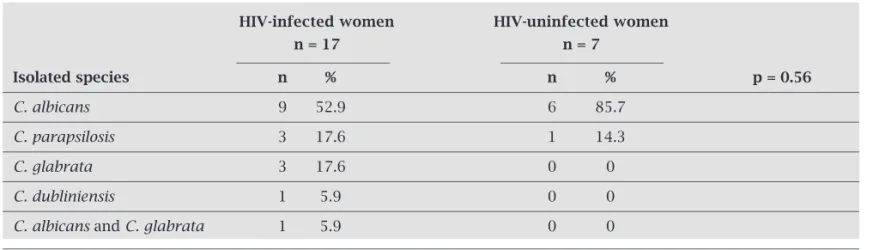
Candida spp. identification was possible in 24 vagi-nal samples (17 from HIV-infected patients and 7 from uninfected women) (Table 3). C. albicans was the most frequent species isolated in both HIV-infected (52.9%) and uninfected women (85.7%). In HIV-infected women were also identified the following species: C. glabrata, C. parapsilosis, C. dubliniensis and a coinfection of C. albi-cans and C. glabrata. In the HIV-uninfected group, the only species isolated were C. albicans and C. parapsilosis.
In both HIV-infected and uninfected women, there was no significant difference observed in the frequency of a particular species of Candida isolated from the vaginal mucosa and the presence of VVC, nor was there a differ-ence in the frequency of a particular species of Candida
colonizing the vaginal mucosa (Table 4).
Only one out of 24 yeast isolates, an isolate of
Table 1. Demographic characteristics and frequency of Candida spp. in HIV-infected and HIV-uninfected patients
HIV-infected group (n = 64) HIV-uninfected group (n = 76)
n n p
Age (years)# 64 30.4 ± 6 76 28.7 ± 6 0.07
Marital status (n, %)
Married/cohabiting 34 53.1 47 61.8 0.29
Unmarried/non-cohabiting 30 46.9 29 38.2 0.29
Education (n, %)
< 8 years 40 62.5 26 34.2 0.001
> 8 years 24 37.5 50 65.8 0.001
Family income (n, %)
< $ 240/month 48 75 34 44.7 < 0.001
≥ $ 240/month 16 25 42 55.3 < 0.001
Sexual and obstetric history
Age of first intercourse# 64 16.2 ± 4.0 76 17.5 ± 3.9 0.05
No. of partners 64 8.3 ± 13.4 76 3.6 ± 4.3 < 0.01
STD history (n, %) 26 40.6 % 10 13.2 < 0.01
No. of deliveries 64 1.7 ± 1.2 76 1.3 ± 1.2 0.02
Users of HAART 38 59.4% - - NA
T CD4+ count 64 644 ± 551 - - NA
Viral load 64 3.9 ± 4.3 - - NA
Vaginal Candida spp. (n) 19 29.7 11 14.5 0.02
Candida spp. colonization 5 26.3 3 27.3 0.95
VVC 14 73.7 8 72.7 0.95
SD, standard deviation; NA, not applicable. STD, sexually transmitted disease. HAART, highly active antiretroviral therapy; CD4: cells/mm³; viral load: log10 copies/mL. p < 0.05, χ2 test, Student’s t test.
# mean ± SD
Table 2. Adjusted odds ratio for Candida spp. presence in HIV infected women
Variables Adjusted OR IC 95% p
HIV infection 2.61 1.07–6.32 0.03
Age 1.00 0.92–1.08 0.98
Years of schooling 0.89 0.78–1.02 0.1 Family income 0.97 0.7–1.33 0.86 First sexual 1.02 0.91–1.14 0.73 intercourse
Condom use 0.82 0.27–2.43 0.72 Number of sexual 0.99 0.95–1.04 0.9 partners
STD history 1.00 0.36–2.75 0.99
Adjusted variables: HIV infection, age, years of schooling, family income, first sexual intercourse, condom use, number of sexual partners, STD history. OR, odds ratio. p < 0.05. Multiple logistical regression.
DISCUSSION
Table 3. Frequency of Candida species isolated from the vaginal mucosa of HIV-infected and uninfected women
HIV-infected women HIV-uninfected women
n = 17 n = 7
Isolated species n % n % p = 0.56
C. albicans 9 52.9 6 85.7
C. parapsilosis 3 17.6 1 14.3
C. glabrata 3 17.6 0 0
C. dubliniensis 1 5.9 0 0
C. albicans and C. glabrata 1 5.9 0 0
p < 0.05, χ2 test.
Table 4. Frequency of vulvovaginal candidiasis and colonization with Candida species from the vaginal mucosa of HIV-infected and uninfected women
HIV-infected HIV-uninfected
n = 17 n = 7
Candida species VVC Colonization VVC Colonization
n n n n
C. albicans 7 2 5 1
C. parapsilosis 2 1 1 0
C. glabrata 1 2 0 0
C. dubliniensis 1 0 0 0
C.albicans + C. glabrata 1 0 0 0
VVC, vulvovaginal candidiasis. p < 0.05, χ2 test, Fisher’s exact test.
Table 5. Minimal inhibitory concentrations (mg/L) of Candida spp.
Candida species MIC median, mg/L, (min-max) n = 25
Amphotericin B Flucytosine Fluconazole Voriconazole Caspofungin
C. albicans 0.22 0.5 0.25 0.008 0.11 (n = 16) (0.094-0.38) (0.023- > 32) (0.094-0.5) (0.004-0.032) (0.064-0.5)
C. dubliniensis 0.064 0.004 0.094 0.006 0.19 (n = 1)
C. glabrata 0.5 0.007 36 0.285 0.19 (n = 4) (0.38-0.5) (0.006-0.008) (8- > 256) (0.064-1) (0.19-0.38)
uninfected-control group.15 The prevalence of Candida spp.
isolated from the vaginal mucosa is variable. In Italy, 16.8% of HIV-infected women have Candida spp. in vaginal muco-sa,10 while in the United States 35% of HIV-infected women had vaginal positive culture for Candida spp. (EUA).18 Such variation could be explained by differences in the immune status of patients involved or by the presence of additional risk factors to Candida infection, such as a decreased num-ber of CD4+ T-cells or concomitant antibiotic therapy.18
There is scant information about the frequency of non- al-bicans species in vaginal mucosa of HIV-infected women in Brazil. In the present study, C. albicans was the most frequent species isolated from the vaginal mucosa in both HIV-infect-ed and uninfectHIV-infect-ed women. In other countries such as Italy and the United States, the frequency was higher ranging from 81% to 92%, compared to that found in our study (52.9%).10,11,18 In HIV-uninfected patients, our data was similar to other studies performed in Brazil, which describe the prevalence of C. albicans isolated in vaginal samples ranging from 60% to 80%19,20 and in other countries where the prevalence of
C. albicans in vaginal samples ranges between 80% to 90%.1,11,21,22 A higher diversity of non-albicans species was found in HIV-infected patients compared to uninfected con-trols. C. glabrata was the most frequent non-albicans species isolated in HIV-infected patients, followed by C. parapsilosis.
In accordance with other studies, there was no relation-ship found between symptoms and isolated yeast species.23 No correlation could be established between the level of CD4+ T-cell counts, the amount of HIV viral load, and the frequency of VVC in HIV-infected patients. These results were expected because of the observational nature of our study and the small sample size.
Regarding the susceptibility of Candida species to antifun-gal drugs, isolated yeasts were susceptible to Amphotericin B, Flucytosine, Voriconazole, and Caspofungin. Only three iso-lates had dose-dependent susceptibility or resistance to flucon-azole, all of these were C. glabrata present in the vaginal mu-cosa of HIV-1-infected patients. These data are in agreement with other studies23-25 that described azole refractory vaginitis caused by non-albicansCandida, particularly by C. glabrata.23 Non-albicansCandida infections frequently require the use of non-azole therapy, including topical therapy with boric acid, Flucytosine, or Amphotericin B.24,26
In summary, our study found higher prevalence of
Candida spp. in vaginal mucosa of HIV-infected patients than uninfected women. Thus, these patients could ben-efit from a periodic gynecological examination and VVC screening, even when undergoing HAART treatment. Although the species of C. albicans is the most frequent, it is evident the emergence of non-albicansCandida spe-cies, including C. glabrata with intrinsic resistance to azoles. Therefore, alternative agents to treat VCC caused by C. glabrata should be considered.
REFERENCES
1. Sobel JD, Faro S, Force RW et al. Vulvovaginal candidiasis: epi-demiologic, diagnostic and therapeutic considerations. Am J Obstet Gynecol. 1998; 178:203-11.
2. Foxman B, Barlow R, D’Arcy H, Gillespie B, Sobel JD. Can-dida vaginitis: self-reported incidence and associated costs. Sex Transm Dis. 2000; 27(4):230-5.
3. Mardh PA, Rodrigues AG, Genc M et al. Facts and myths on recurrent vulvovaginal candidosis-a review on epidemiology, clinical manifestations, diagnosis, pathogenesis and therapy. Int J STD AIDS 2002; 13(8):522-39.
4. Schaller M. Candida albicans-interactions with the mucosa and the immune system. J Dtsch Dermatol Ges. 2006: 4(4):328-36. 5. Mendling W, Seebacher C. Guideline vulvovaginal candidosis:
guideline of the German Dermatological Society, the German Speaking Mycological Society and the Working Group for In-fections and Infectimmunology of the German Society for Gy-necology and Obstetrics. Mycoses 2003; 46(9-10):365-9. 6. Linhares LM, Witkin SS, Miranda SD et al. Differentiation
be-tween women with vulvovaginal symptoms who are positive or negative for Candida species by culture. Infect Dis Obstet Gynecol. 2001; 9(4):221-5.
7. Rosa MI, Rumel D. Fatores Associados à Candidíase Vulvo-vaginal: Estudo Exploratório. Rev Bras Ginecol Obstet. 2004; 26(1):65-70.
8. Barrenetxea Z. Vulvovaginitis candidiásica. Rev Iberoam Mi-col. 2002; 19(1):22-4.
9. Sebitloane M.H. HIV and gynecological infections. Best Pract Res Clin Obstet Gynaecol. 2005; 19(2):231-41.
10. Beltrame A, Matteelli A, Carvalho AC et al. Vaginal colo-nization with Candida spp. in human immunodeficiency virus-infected women: a cohort study. Int J STD AIDS 2006; 17(4):260-6.
11. Sobel JD, Ohmit SE, Schuman P et al. The evolution of Can-dida spp. and fluconazole susceptibility among oral and vagi-nal isolates recovered from human immunodeficiency virus (HIV)-seropositive and at-risk HIV-seronegative women. J Infect Dis 2001; 183:286-93
12. Spinillo A, Zara F, Gardella B et al. The effect of vaginal can-didiasis on the shedding of human immunodeficiency virus in cervicovaginal secretions. Am J Obstet Gynecol. 2005; 192(3):774-9.
13. Shifrin E, Matityahu D, Feldman J, Minkoff H. Determinants of incident vulvovaginal candidiasis in human immunodefi-ciency virus-positive women. Infect Dis Obstet Gynecol. 2000; 8(3-4):176-80.
14. Gygax SE, Vermitsky JP, Chadwick SG et al. Antifungal re-sistance of Candida glabrata vaginal isolates and develop-ment of a quantitative reverse transcription-PCR-based azole susceptibility assay. Antimicrob Agents Chemother. 2008; 52(9):3424-6.
15. Grinsztejn B, Bastos FI, Veloso VG et al. Assessing sexu-ally transmitted infections in a cohort of women living with HIV/AIDS, in Rio de Janeiro, Brazil. Int J STD AIDS 2006; 17(7):473-8.
16. Duerr A, Heilig CM, Meikle SF et al. Incident and persistent vulvovaginal candidiasis among human immunodeficiency virus-infected women: Risk factors and severity. Obstet Gy-necol. 2003; 101(3):548-56.
18. Williams A, Andrews S, Tashima K, Mezger J, Yu C. Factors as-sociated with vaginal yeast infections in HIV-positive women. J Assoc Nurses AIDS Care 1998; 9(5):47-52.
19. Holanda A, Fernandes A, Bezerra C et al. Candidíase vulvo-vaginal: sintomatologia, fatores de risco e colonização anal concomitante. Rev Bras Ginecol Obstet. 2007; 29(1):3-9. 20. Lopes Consolaro ME, Aline Albertoni T, Shizue Yoshida C et
al. Correlation of Candida species and symptoms among pa-tients with vulvovaginal candidiasis in Maringa, Parana, Bra-zil. Rev Iberoam Micol. 2004; 21(4):202-5
21. Novikova N, Rodrigues A, Mardh PA. Can the diagnosis of re-current vulvovaginal candidosis be improved by use of vaginal lavage samples and cultures on chromogenic agar? Infect Dis Obstet Gynecol. 2002; 10(2):89-92.
22. Corsello S, Spinillo A, Osnengo G et al. An epidemiological survey of vulvovaginal candidiasis in Italy. Eur J Obstet Gy-necol Reprod Biol. 2003; 110(1):66-72.
23. Ringdahl E. Treatment of recurrent vulvovaginal candidiasis. Fam Physician. 2000; 61(11) 3306-12, 17.
24. Sobel J. Vulvovaginitis due to Candida glabrata. An emerging problem. Mycoses 1998; 41(Suppl 2):18-22.
25. Richter SS, Galask RP, Messer SA et al. Antifungal suscep-tibilities of Candida species causing vulvovaginitis and epidemiology of recurrent cases. J Clin Microbiol 2005; 43(5):2155-62.