Abstract
Objectives: To evaluate sleep architecture in children and adolescents with both cystic ibrosis (CF) and a clinical suspicion of sleep-disordered breathing (SDB), and to identify the respiratory polysomnographic proile of these patients.
Methods: Parents or guardians of children with CF illed out a questionnaire designed to assess their clinical and sleep conditions. Children who were identiied as having behaviors associated with SDB underwent polysomnography. After polysomnography, patients were grouped according to the obstructive apnea index (AI) obtained (either < 1 or ≥ 1), and a multiple correspondence factor analysis was used to analyze and identify the polysomnographic proile of patients.
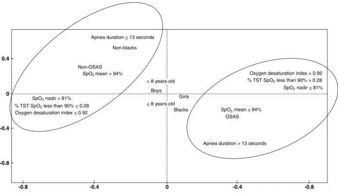
Results: Of the 74 patients who met inclusion criteria for this study, 67 underwent polysomnography, and 38 (56.7%) of the 67 patients showed an AI ≥ 1. Median age was 8 years. The group of patients with an AI ≥ 1 was characterized by total sleep time (TST) during stage 4 and rapid eye movement (REM) stage of sleep < 21 and 13%, respectively, REM sleep latency > 144 minutes, percentage of TST with pulse oxyhemoglobin saturation (SpO2) < 90% higher than 0.28 seconds, and an oxygen desaturation index higher than 0.92.
Conclusion: Results suggest that clinically stable pediatric patients with CF have a high prevalence of SDB and present frequent sleep complaints, signiicant changes in sleep architecture, and episodes of oxygen desaturation during sleep.
J Pediatr (Rio J). 2011;87(1):63-69: Cystic ibrosis, children, adolescents, polysomnography, obstructive sleep apnea.
O
RiginAlA
RtiCleCopyright © 2011 by Sociedade Brasileira de Pediatria
63
introduction
Cystic ibrosis (CF) is a recessive autosomal genetic disease with multi-systemic involvement that primarily affects the respiratory tract.1,2 Compromised lung function is responsible for the majority of deaths in CF patients, followed
by respiratory insuficiency, pulmonary hypertension and cor pulmonale.3,4
Several factors can lead to disturbed sleep and sleep fragmentations in these patients, including nocturnal hypoxia,
Sleep architecture and
polysomnographic respiratory proile
of children and adolescents with cystic ibrosis
Regina terse trindade Ramos,1 Cristina Salles,2 Carla Hilário da Cunha Daltro,3
Maria Angélica Santana,4 Paloma Baiardi gregório,5 Angelina Xavier Acosta6
1. MD, MSc, PhD. Adjunct professor, Department of Pediatrics, School of Medicine, Universidade Federal da Bahia (UFBA), Salvador, BA, Brazil. Graduate Department, Escola Bahiana de Medicina e Saúde Pública, Fundação Bahiana para Desenvolvimento das Ciências, Salvador, BA, Brazil.
2. MD, MSc. Graduate Program in Medicine and Health, School of Medicine, UFBA, Salvador, BA, Brazil. 3. MD, MSc, PhD. Graduate Program in Medicine and Health, School of Medicine, UFBA, Salvador, BA, Brazil. 4. MD, PhD. Centro de Referência em Fibrose Cística, Hospital Especializado Octávio Mangabeira, Salvador, BA, Brazil. 5. MD. Centro Especializado em Pneumologia e Sono, Salvador, BA, Brazil.
6. MD, PhD. Adjunct professor, Department of Pediatrics, School of Medicine, UFBA, Salvador, BA, Brazil.
No conflicts of interest declared concerning the publication of this article.
Financial support: Fundação de Apoio à Pesquisa do Estado da Bahia (FAPESB).
Suggested citation: Ramos RT, Salles C, Daltro CH, Santana MA, Gregório PB, Acosta AX. Sleep architecture and polysomnographic respiratory profile of children and adolescents with cystic fibrosis. J Pediatr (Rio J). 2011;87(1):63-69.
hypoventilation, chronic cough, chronic inlammation, and drug effects.3 Sleep-disordered breathing (SDB), especially obstructive sleep apnea syndrome (OSAS), can also lead to sleep disturbances, subsequently causing changes in ventilation and gas exchange in CF patients, which may be worsened by the presence of advanced pulmonary disease. These changes can result in oxyhemoglobin desaturation and hypercapnia, particularly during the rapid eye movement (REM) stage of sleep.3
Previous data have demonstrated that upper airway obstruction caused by chronic infection and nasal polyposis may contribute to the presence and severity of OSAS in CF patients.3,5,6 However, OSAS is not believed to be an important cause of sleep disorders in CF, nor is it thought to be part of the etiology of the oxyhemoglobin desaturation observed in these patients.3 Few studies have evaluated sleep architecture or the prevalence of OSAS in this population.
The aims of the present study were: 1) to evaluate sleep architecture using polysomnography in a cohort of clinically stable children and adolescents with CF and clinical signs of SDB; and 2) to identify the polysomnographic respiratory proile of patients with OSAS and compare it to that of patients without OSAS.
Materials and methods
Study population
Study participants were recruited from the Cystic Fibrosis Reference Center at the Hospital Especializado Octávio Mangabeira (HEOM) between November 2006 and April 2008, using a non-probability, sequential sampling technique. For the sample size calculation, the Pepi-Sample software (Sagebush Press, Salt Lake City, USA) was used, and the following parameters were set: a 95% conidence interval, a 5% prevalence of SDB in children and adolescents, and a 5% acceptable difference in the prevalence of SDB. The population from which the sample was recruited was comprised of approximately 200 children and adolescents with CF registered at the reference center. The initial sample included 54 patients. However, in order to achieve the study’s objectives and to include the possibility of a 10% loss, the inal sample size was increased to 59.
Inclusion criteria were as follows: 1) between 2 and 14 years of age; and 2) diagnosis of CF and attendance at routine clinical evaluations during the study period. The following exclusion criteria were also taken into consideration: 1) use of an orogastric or nasogastric tube; 2) presence of pulmonary exacerbation before enrollment in the study or during the course of the study; 3) use of home oxygen therapy; 4) previously diagnosed SDB, neuromuscular diseases, or craniofacial abnormalities; and 5) prior lung transplantation. The sleep portion of the study was conducted overnight in a specialized laboratory, and the onset of sleep was spontaneous. Race was self-reported according to the
oficial terms used in the demographic census. Using skin color as a parameter, the reported categories were white, black, and mixed race.7 All CF diagnoses met the standard Cystic Fibrosis Foundation criteria.2
This study was approved by the Research Ethics Committee at Fundação Oswaldo Cruz (CPqGM-FIOCRUZ, decision no. 119/2007). Informed consent was obtained from all parents or guardians of the children, or from the children who were mature enough to comprehend the study goals.
Sleep disturbance questionnaire
Parents/guardians completed the sleep disturbance questionnaire administered by team members of the study including demographic data and issues related to the history of the child and about sleep. Clinical suspicion of SDB was performed using a previously validated sleep-disturbance questionnaire8 that included the following key questions: “Does your child snore?,” “Does your child have pauses in breathing (or assisted apnea)?,” “Does your child have dificulty in breathing during sleep?,” and “Does your child have a disturbed sleep pattern?” Possible responses were: “never,” “only during colds,” “less than once a week,” “one to three times a week,” and “more than three times a week,” or “yes,” “no,” and “I don’t know.”
Clinical score and nutrition
The Shwachman-Kulczycki (S-K) score was calculated for each patient, and the child’s nutritional status was evaluated. The S-K score9 covers four domains, each one with ive possible scores, according to the degree of impairment: general activity, physical examination, nutrition, and radiological indings. The four domains were totaled to obtain the inal score, and the condition of the patient was categorized as excellent (86-100), good (71-85), average (56-70), poor (41-55), or severe (≤ 40). The heights and weights of all patients were measured, compared to the National Center for Health Statistics (NCHS) growth charts, and converted into weight/age (W/A) and height/age (H/A) z scores based on age and gender, using the Epi-Info program (Centers for Disease Control and Prevention, Atlanta, USA), version 3.4.1.
Polysomnography
as an additional method of monitoring airlow. Snoring was detected by a microphone. Pulse oxyhemoglobin saturation (SpO2) was measured using a inger probe (Onyx® II 9650 Bluetooth®, Nonim Medical, Plymouth, USA).
Sleep stages were scored in 30-s epochs, according to the standard criteria of Rechtschaffen & Kales.10 The proportion of time spent in each sleep stage was expressed as a percentage of the total sleep time (TST). The following parameters were calculated: 1) sleep eficiency, deined as the TST divided by the total recording time and expressed as a percentage; 2) irst-stage sleep latency, deined as the time elapsed between turning off the lights and the onset of sleep; and 3) REM sleep latency, deined as the time elapsed between the onset of sleep and the irst REM sleep period. Number of EEG arousals per hour (according to the arousal index) and periodic leg movement per hour of sleep (PLM index) were also calculated.11-14 Obstructive apnea was deined as the presence of abdominal and thoracic wall movement and absence of oronasal airlow for a minimum time of two respiratory cycles. Hypopnea was deined as a 50% or greater reduction in the airlow signal amplitude, and it was quantiied if an episode lasted more than two respiratory cycles with a 3% or greater reduction in SpO2 and/or EEG arousals. Mixed apneas were also recorded. The obstructive apnea index (AI) was deined as the total number of obstructive apneas and mixed apneas divided by the TST, and the apnea-hypopnea index was deined as the total number of obstructive apneas and hypopneas added to the number of mixed apneas per hour of sleep.13,14 OSAS was diagnosed when the AI was greater than or equal to one event/hour of sleep; OSAS was considered mild when the AI was between one and ive events/hour of sleep, moderate when the AI was greater than 5 and up to 10 events/hour of sleep, and severe when the AI > 10 events/hour of sleep.14,15 The mean duration of apnea was calculated for each patient. Desaturation was deined as a 3% or greater reduction in oxygen saturation13; the number
of desaturations per hour was calculated and reported as the desaturation index. The mean value and SpO2 nadir were measured. TST spent with SpO2 below 90% was also calculated. Nocturnal hypoxia in CF was deined as SpO2 < 90% for more than 5% of the TST.16
Statistical analysis
This is a descriptive, explanatory, cross-sectional study. Quantitative variables were compared using Student’s t test or Mann-Whitney test. To analyze
respiratory polysomnographic proiles, we used a multiple correspondence factor analysis (MCFA),17 with patients divided into two groups: patients without OSAS (AI < 1) and patients with OSAS (AI ≥ 1). This analysis allows to assess the most important relationships in a large pool of categorical and continuous variables. MCFA analysis uses graphs that are also known as factorial planes. Each
variable is represented by a point included in the plane. Relationships among variables can be assessed graphically by the proximity between the points on the plane, and the cumulative contribution (expressed as a percentage) of each variable in the irst two factorial axes can be calculated. The purpose of this analysis was to assess the strength of relationships between presence of OSAS and each one of the polysomnographic variables, so as to identify the proiles of CF children that have OSAS. Each group of data was analyzed separately. MCFA is a powerful tool for inspecting large amounts of data and selecting the most important variables for a more detailed, subsequent analysis.
All statistical tests were two-tailed, and signiicance was set at p ≤ 0.05. The software used for database management and statistical analysis was the Statistical Package for the Social Sciences for Windows, version 12.0 (SPSS Inc., Chicago, USA), and the R 2.0.2 software (R Development Core Team, Vienna, Austria). The Système Pour Analyse de Données (SPAD) software, version 3.5 (Cisia-Ceresta, Montreuil, France), was used for the correspondence analysis.
Results
The parents of 120 of the 200 pediatric patients registered at the CF reference center agreed to participate in the study and completed the sleep disturbance questionnaire; 85 of these 120 children met clinical criteria for sleep disturbance. Of the 85 children with CF and a clinical suspicion of SDB, 74 met inclusion criteria for this study, and 67 agreed to undergo polysomnography. Their demographic data are shown in Table 1, and their sleep and everyday performance complaints are shown in Figure 1.
Mean and standard deviation of TST (minutes) and sleep eficiency (%) of the patients studied were 379±60 and 81±11, respectively, and 38 (56.7%) of the 67 patients with CF showed an AI ≥ 1 event/hour of sleep. Of these 38 patients, seven (18%) were identiied as having moderate OSAS (5 < AI ≤ 10 events/hour of sleep), and two (5%) were identiied as having severe OSAS (AI > 10 events/hour of sleep); the remaining 29 (43.3%) patients were diagnosed as non-OSAS cases. Four patients had SpO2 < 90% in over 5% of the TST. The mean and standard deviation of mean SpO2 level and SpO2 nadir during sleep were 94±2 and 81±6%, respectively. The polysomnographic proile of patients is detailed in Table 2.
Proiles of CF patients in the OSAS group and in the non-OSAS group
Characteristics Results
Male (%) 56.7 (38/67)
Age (years)* 8 (5:10)
Skin color: black and mixed race (%) 80.6 (54/67)
Weight/age z score* -0.54 (-1.3:0.2)
Height/age z score* -0.50 (-1:0.5)
BMI percentile* 34 (11:64)
Total S-K† 85.6±9.1
FEV1* 78.5 (67:92.8)
BMI = body mass index; CF = cystic fibrosis; FEV1 = forced expiratory volume in 1 second; Total S-K = total Shwachman-Kulczycki score.
* Median and interquartile range. † Mean ± standard deviation.
Figure 1 - Frequency of complaints related to sleep and everyday performance in the 67 cystic ibrosis children assessed from November 2006 to April 2008
general population Patients with OSAS Patients without OSAS
Polysomnographic data (n = 67) (n = 38) (n = 29) p
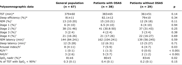
TST (min)* 379±60 383±65 361±51 0.14
Sleep eficiency (%)* 81±11 82.1±12 79±10 0.34
REM (%)† 13 (10:20) 15 (10:21) 12 (9:18) 0.11
Stage 1 (%)† 6 (4:10) 6.5 (4:10) 6 (4:10) 0.94
Stage 2 (%)† 38 (31:46) 41 (30.8:50) 37 (31:43) 0.25
Stage 3 (%)† 3 (2:4) 4 (2:4) 3 (3:4) 0.38
Stage 4 (%)† 21 (16:26) 21 (17:26) 22 (16:27) 0.69
REM latency (min)† 144 (84:241) 162 (104:241) 139 (56:245) 0.59
Sleep latency (min)† 12 (5:28) 12 (6:31) 13 (5:27) 0.71
Arousal index/h† 8 (4:11) 7 (5:9) 6 (4:7) 0.03
AI/h†‡ 1 (0:1) 1 (1:3) 0 (0:0) < 0.001
AHI/h† 3 (2:6) 5 (3:7) 2 (1:2) < 0.001
SpO2 nadir (%)* 81±6 80±5 83±6 0.02
% of TST with SpO2 < 90%† 0.3 (0:1) 0.4 (0:1) 0.2 (0:1) 0.15
table 2 - Comparison of polysomnographic data of the 67 CF patients, with and without OSAS, studied from November 2006 to April 2008
AHI = apnea-hypopnea index; AI = apnea index; CF = cystic fibrosis; OSAS = obstructive sleep apnea syndrome; REM = rapid eye movement; SpO2 = pulse oxyhemoglobin saturation; TST = total sleep time.
Results expressed as mean ± standard deviation or median with interquartile range. * Student’s t test.
† Mann-Whitney test. ‡ AI cut-off point = 1.
table 1 - Demographic and clinical data of the 67 CF patients
studied from November 2006 to April 2008 periods (> 12 minutes), higher percentages of time spent in stages 1 and 2 (> 6 and > 38%, respectively), greater
Figure 2 - Representation of polysomnographic respiratory variables of 67 cystic ibrosis patients, shown in a factorial plane graph using the irst two axes
OSAS = obstructive sleep apnea syndrome; SpO2 = pulse oxyhemoglobin saturation; TST = total sleep time. Discussion
Children and adolescents with CF and a clinical suspicion of SDB evaluated in our study had frequent sleep complaints and signiicant changes in sleep architecture. This study also found a high prevalence of OSAS in this group of patients. There is limited information available on sleep architecture from sleep studies in patients with CF, as well as on the prevalence of SDB in CF patients, particularly OSAS in children.5 The majority of studies published so far on SDB and CF include older populations, namely patients in late adolescence and young adults.18 The present study assessed a younger sample of patients (range of 5 to 10 years and median age of 8 years).
Naqviet al.19 evaluated sleep architecture in children and adolescents (14.2±3.8 years) with CF, and noticed that they presented frequent sleep complaints and changes in sleep architecture. In addition, those authors observed that the magnitude of sleep disruption was associated with the severity of lung disease, but was not directly correlated with the degree of nocturnal hypoxemia or hypoventilation. Furthermore, the majority of patients did not have signiicant OSAS. Conversely, in our study, nine patients (23.7%) had moderate and severe OSAS, and we did not observe any correlation between sleep disruption and the severity of lung disease. A possible explanation for our results is that we studied a younger, clinically stable population with shorter disease duration.
Nocturnal polysomnography13 is the standard diagnostic test to conirm OSAS. However, the reference values for this test vary between pediatric population-based studies. We adopted an AI ≥ 1 as a polysomnographic diagnostic criterion,14,15 as recommended by Sociedade Brasileira de Sono14. In this study, only one polysomnography was performed, but data from the literature support the concept that a single nocturnal exam is suficient for diagnosing OSAS, even in CF patients.5,20-22
In the present study, children with OSAS were predominantly below 8 years of age and female. OSAS occurs in children of all ages, but is more common from 3 to 6 years of age.23 Adenotonsillar hypertrophy may explain the occurrence of OSAS in this population and age group (< 8 years). Furthermore, upper airway obstruction caused by chronic infection and nasal polyposis is another possible cause of OSAS in CF patients and possibly contributes to the severity of OSAS.3,24 Ramos et al.,25 studying this same population, noted that the presence of chronic rhinosinusitis (with or without nasal polyposis) may serve as causal or aggravating factor for SDB in this population.
References
1. Mattar AC, Gomes EN, Adde FV, Leone C, Rodrigues JC. Comparison between classic Gibson and Cooke technique and sweat conductivity test in patients with and without cystic ibrosis.J Pediatr (Rio J). 2010;86:109-14.
2. Farrell PM, Rosenstein BJ, White TB, Accurso FJ, Castellani C, Cutting GR, et al. Guidelines for diagnosis of cystic ibrosis in newborns through older adults: Cystic Fibrosis Foundation consensus report. J Pediatr. 2008;153:S4-S14.
3. Milross MA, Piper AJ, Dobbin CJ, Bye PT, Grunstein RR.
Sleep disordered breathing in cystic ibrosis. Sleep Med Rev. 2004;8:295-308.
4. Frangolias DD, Wilcox PG. Predictability of oxygen desaturation during sleep in patients with cystic ibrosis: clinical, spirometric, and exercise parameters. Chest. 2001;119:434-41.
5. Milross MA, Piper AJ, Norman M, Willson GN, Grunstein RR, Sullivan CE, et al. Night-to-night variability in sleep in cystic ibrosis. Sleep Med. 2002;3:213-19.
6. Woodson BT, Franco R. Physiology of sleep disordered breathing. Otolaryngol Clin North Am. 2007;40:691-711.
7. Lessa I, Magalhães L, Araújo MJ, de Almeida Filho N, Aquino E, Oliveira MM. Arterial hypertension in the adult population of Salvador (BA) - Brazil. Arq Bras Cardiol. 2006;87:747-56. 8. Kaditis AG, Alexopoulos EI, Kalampouka E, Kostadima E, Germenis
A, Zintzaras E, et al. Morning levels of C-reactive protein in children
with obstructive sleep-disordered breathing. Am J Respir Crit Care
Med. 2005;171:282-6.
9. Shwachman H, Kulczycki LL. Long-term study of one hundred ive patients with cystic ibrosis; studies made over a ive- to
fourteen-year period. AMA J Dis Child. 1958;96:6-15.
10. Rechtschaffen A, Kales A. A manual of standardized terminology, techniques and scoring system for sleep stages of human subjects. Publication no. 204. Washington, DC: National Institutes of Health; 1968.
11. Carroll JL. Obstructive sleep-disordered breathing in
children: new controversies, new directions. Clin Chest Med. 2003;24:261-82.
12. EEG arousals: scoring rules and examples: a preliminary report from the Sleep Disorders Atlas Task Force of the American Sleep Disorders Association.Sleep. 1992;15:173-84.
OSAS group had longer REM sleep latency periods and a lower percentage of TST in stage 4 and the REM stage of sleep, and we did not ind a signiicant correlation between the degree of sleep fragmentation and the severity of lung disease.
We used the Rechtschaffen & Kales’ criteria to score the sleep architecture parameters instead of the new American Academy of Sleep Medicine rules which could have disclosed some additional differences, such as a higher percentage of stage 1 and a higher number of stage shifts in children with OSAS. Coughing occurred during the initial stages of sleep, which may have contributed to delays in the progression from these stages of sleep to slow wave stages and REM sleep. Another possible explanation is that the so-called irst-night effect interfered with sleep architecture.5,28
The respiratory events that made the greatest contribution to distinguishing between the OSAS and non-OSAS groups were desaturation index, apnea duration, percentage of the TST with SpO2 < 90%, and SpO2 nadir. Oxyhemoglobin desaturation during sleep has been documented as being more prevalent in patients with CF and severe lung disease. However, limited information describing nocturnal oxygen saturation in patients with CF and mild lung disease is available.4 Villa et al.29 studied young children with CF (mean age, 13.1 months; range, 3-36 months) and observed the presence of desaturation during sleep. We studied patients from 2 to 14 years of age and frequently noticed intermittent drops in SpO2; this was even observed in younger patients, patients with normal lung function, and patients with minor changes in lung function. Of the patients studied, 56 (84%) experienced desaturation during sleep. Nocturnal hypoxemia, deined as SpO2 < 90% for more than 5% of the patient’s TST, was observed in four patients in our study. Although OSAS has not been identiied in the medical literature as a mechanism responsible for progressive oxygen desaturation during sleep in populations of patients with CF, the OSAS group presented strong associations with a higher TST percentage with SpO2 < 90%, a higher number of desaturation events, and more prolonged apnea events. Even though there is a signiicant difference in some of these parameters in the OSAS and non-OSAS group, it is not possible to conirm that these parameters exert inluence on CF clinical progression. The coexistence of a chronic lung disease (such as CF) and OSAS might possibly contribute to a higher number of desaturation and hypoxemia episodes compared to patients with CF alone.
We needed to be careful about adopting the AI as our diagnostic criterion, since hypopneas are frequently described in CF patients,30 so that the prevalence of OSAS in our cohort of CF is not overrepresented. The strength of the present study is the wide age group of children and adolescents with CF evaluated; however, several limitations also deserve to be mentioned: 1) the study design is
cross-sectional, which does not allow us to establish a causal relationship between sleep apnea and CF-related factors; 2) capnography was not used, which limited the observation of hypoventilation; 3) sleep architecture parameters were scored using the Rechtschaffen & Kales’ criteria instead of the new American Academy of Sleep Medicine guidelines.
It is concluded that children and adolescents with CF had frequent sleep complaints, signiicant changes in their sleep architecture, and a high prevalence of SDB. Further studies should be conducted to explore the nature of the interaction between lung disease in CF and sleep, and to determine whether intervention in SDB in CF patients will modify the clinical course of the disease.
Acknowledgments
13. Standards and indications for cardiopulmonary sleep studies in children. American Thoracic Society. Am J Respir Crit Care Med. 1996;153:866-78.
14. Bittencourt LR. Síndrome da Apnéia Obstrutiva do Sono em Crianças e Adolescentes. In: Bittencourt LR. Diagnóstico e Tratamento da Síndrome da Apnéia Obstrutiva do Sono (SAOS): guia prático. São Paulo: Livraria Médica Paulista Editora; 2008. p. 81-93 15. Katz ES, Marcus CL. Diagnosis of Obstructive Sleep Apnea
Syndrome in Infants and Children. In: Sheldon SH, Ferber R, Kryger MH, editors. Principles and Practice of Pediatric Sleep Medicine. Philadelphia: Elsevier Saunders; 2005. p. 197-210 16. Uyan ZS, Ozdemir N, Ersu R, Akpinar I, Keskin S, Cakir E, et al.
Factors that correlate with sleep oxygenation in children with cystic ibrosis. Pediatr Pulmonol. 2007;42:716-22.
17. Greenacre MJ. Practical correspondence analyses. In: Casella G, Fienberg S, Olkin J, editors. Looking at multivariate data. New York: John Wiley and Sons; 1981. p. 119-46.
18. Dancey DR, Tullis ED, Heslegrave R, Thornley K, Hanly PJ. Sleep quality and daytime function in adults with cystic ibrosis and
severe lung disease. Eur Respir J. 2002;19:504-10.
19. Naqvi KS, Sotelo C, Murry L, Simakajornboon N. Sleep architecture in children and adolescents with cystic ibrosis and the association
with severity of lung disease. Sleep Breath. 2008;12:73-83.
20. Brietzke SE, Katz ES, Roberson DW. Can history and physical examination reliably diagnose pediatric obstructive sleep apnea/ hypopnea syndrome? A systematic review of the literature. Otolaryngol Head Neck Surg. 2004;131:827-32.
21. Scholle S, Scholle HC, Kemper A, Glaser S, Rieger B, Kemper G, et al. First night effect in children and adolescents undergoing
polysomnography for sleep-disordered breathing. Clin Neurophysiol. 2003;114:2138-45.
22. Verhulst SL, Schrauwen N, De Backer WA, Desager KN. First night effect for polysomnographic data in children and adolescents
with suspected sleep disordered breathing. Arch Dis Child.
2006;91:233-7.
23. Section on Pediatric Pulmonology, Subcommittee on Obstructive Sleep Apnea Syndrome. American Academy of Pediatrics. Clinical practice guideline: diagnosis and management of childhood obstructive sleep apnea syndrome. Pediatrics. 2002;109:704-12.
24. Cardiorespiratory sleep studies in children: establishment of normative data and polysomnographic predictors of morbidity. American Thoracic Society. Am J Respir Crit Care Med. 1999;160:1381-7.
25. Ramos RT, Salles C, Gregório PB, Barros AT, Santana A, Araújo-Filho JB, et al. Evaluation of the upper airway in children and adolescents with cystic ibrosis and obstructive sleep apnea syndrome. Int J Pediatr Otorhinolaryngol. 2009;73:1780-5. 26. Jankelowitz L, Reid KJ, Wolfe L, Cullina J, Zee PC, Jain M. Cystic
ibrosis patients have poor sleep quality despite normal sleep latency and eficiency. Chest. 2005;127:1593-9.
27. Amin R, Bean J, Burklow K, Jeffries J. The relationship between
sleep disturbance and pulmonary function in stable pediatric cystic ibrosis patients. Chest. 2005;128:1357-63.
28. Gee L, Abbott J, Conway SP, Etherington C, Webb AK. Quality of life
in cystic ibrosis: the impact of gender, general health perceptions
and disease severity. J Cyst Fibrosis. 2003;2:206-13.
29. Villa MP, Pagani J, Lucidi V, Palamides S, Ronchetti R. Nocturnal oximetry in infants with cystic fibrosis. Arch Dis Child. 2001;84:50-4.
30. Piper AJ, Bye PT, Grunstein RR. Sleep and breathing in cystic ibrosis. Sleep Med Clin. 2007;2:87-97.
Correspondence:
Regina Terse Trindade Ramos
Av. Reitor Miguel Calmon, s/nº – Vale do Canela CEP 40110-100 – Salvador, BA – Brazil Tel.: +55 (71) 3332.6182