Effect of mitochondrial potassium channel on the renal
protection mediated by sodium thiosulfate against ethylene
glycol induced nephrolithiasis in rat model
_______________________________________________
N. Baldev
1,2,3, R. Sriram
1,2,3, P.C. Prabu
1,2,3, A. Kurian Gino
1,2,31 School of Chemical and Biotechnology, SASTRA University, Thanjavur, Tamil Nadu, India; 2 Vascular Biology Lab, SASTRA University, Thanjavur, Tamil Nadu, India; 3 Central Animal Facility, SASTRA University, Thanjavur, Tamil Nadu, India
ABSTRACT
ARTICLE
INFO
______________________________________________________________ ______________________
Purpose: Sodium thiosulfate (STS) is clinically reported to be a promising drug in preventing nephrolithiasis. However, its mechanism of action remains unclear. In the present study, we investigated the role of mitochondrial KATP channel in the renal protection mediated by STS.
Materials and Methods: Nephrolithiasis was induced in Wistar rats by administrating 0.4% ethylene glycol (EG) along with 1% ammonium chloride for one week in drinking water followed by only 0.75% EG for two weeks. Treatment groups received STS, mi-tochondrial KATP channel opener and closer exclusively or in combination with STS for two weeks.
Results: Animals treated with STS showed normal renal tissue architecture, suppor-ted by near normal serum creatinine, urea and ALP activity. Diazoxide (mitochondria KATP channel opening) treatment to the animal also showed normal renal tissue histo-logy and improved serum chemistry. However, an opposite result was shown by gliben-clamide (mitochondria KATP channel closer) treated rats. STS administered along with diazoxide negated the renal protection rendered by diazoxide alone, while it imparted protection to the glibenclamide treated rats, formulating a mitochondria modulated STS action.
Conclusion: The present study confirmed that STS render renal protection not only through chelation and antioxidant effect but also by modulating the mitochondrial KATP channel for preventing urolithiasis.
Key words:
Diazoxide; glibenclamide receptor [Supplementary Concept]; Kidney Calculi; Calcium Oxalate; Pathology
Int Braz J Urol. 2015; 41: 1116-25
_____________________
Submitted for publication: November 13, 2014 _____________________
Accepted after revision: June 10, 2015
INTRODUCTION
Nephrolithiasis is a complex disease of kidney where crystals or foreign bodies can act as nidi, upon which ions from the supersatura-ted urine form microscopic crystalline structures (1). The formation of these crystals in the tubular fluid, followed by crystal retention and accumu-lation in the kidney are the pre-requisite for the
development of renal stone. Free particle model, fixed particle model and Randall’s plaque hypo-thesis are some well-established hypohypo-thesis for stone formation and growth (2). However, mole-cular understanding of all these theories underli-nes the importance of oxidative stress in the renal stone formation.
have antioxidant and chelation property (3). So-dium thiosulfate, FDA approved drug is used to lessen the side effects of cisplatin (4) and widely used in the emergency treatment of cyanide poi-soning (5). Due to the availability of the sulfur group for the donations and free radical scaven-ging potential, STS can essentially act as an anti--urolithiatic agent (6). Few studies by Asplin and his co-workers and Yatzidis showed that thiosulfa-te can prevent calcium phosphathiosulfa-te nephrolithiasis. However, LaGrange et al., 2009 reported negative results for thiosulfate in the prevention of calcium stone disease (7). In fact, in both studies, the me-chanism by which STS affects calcium deposition was not clearly mentioned.
Potassium channels in nephrons have varied functions ranging from maintaining io-nic equilibrium to regulating the volume during hypotonic stress environments. Their activation depends on location across the nephrons, as they can be activated either by altering pH, calcium, sodium, chloride or ATP levels. Thus, effects of K+
channels are very complex to study (8). The pre-sent study was designed to understand the speci-fic role of ATP sensitive potassium channel using diazoxide (channel opener) and glibenclamide (channel closer) in sodium thiosulfate mediated renal protection from ethylene glycol induced ne-phrolithiasis in a rat model. The calcium chelating potential of STS was evaluated in vitro using gel diffusion method.
MATERIALS AND METHODS
Chemicals
Diazoxide was purchased from Sigma-Al-drich. All other commercial reagents used were of analytical grade.
Animals
All animal experiments were conducted in accordance with the CPCSEA (Committee for the purpose of conduct and supervision of expe-riments on animals) guidelines, approved by the institutional animal ethical committee (IAEC No. 214/SASTRA/IAEC), Central Animal Facility, SAS-TRA University. To demonstrate the anti-urolithia-sis property and mechanism of action of sodium
thiosulfate we used male albino Wistar rats aged 7 to 8 weeks (180-200g). Animals were kept in polycarbonate cages at a controlled temperature of 25±3ºC and 60±10% relative humidity with a 12 h each of dark and light cycle. Rats were ac-climatized for one week with standard laboratory diet and tap drinking water before the start of ex-periment.
Study design
Forty two male Wistar rats were assigned randomly into seven equal groups. All the doses were selected based on previous studies (6, 9).
Group-1 (Normal control): received water ad libitum for 21 days.
Group-2 (Induction control): received 0.4% ethylene glycol (EG) along with 1% ammo-nium chloride for one week followed by only 0.75% EG in drinking water for two subsequent weeks.
Groups-3 to 7 received the same treatment as group 2 along with the following drug treatments:
Group-3 (STS): received sodium thiosulfate (400mg/Kg b.wt.) intraperitoneally for 21 days.
Group-4 (Diazoxide): received diazoxide (mito KATP channel opener; 5mg/Kg b.wt.) intraperitoneally for 21 days.
Group-5 (Glibenclamide): received gliben-clamide (mito KATP channel closer; 10mg/ Kg b.wt.) intraperitoneally for 21 days Group-6 (STS+Diazoxide): received diazo-xide 30min. before administration of STS for 21 days.
Group-7 (STS+Glibenclamide): received glibenclamide 30min. before administra-tion of STS for 21 days.
Biochemical Parameters
tripli-cates for calcium, creatinine, urea, ALP using the respective diagnostic kits purchased from Agappe diagnostics Ltd. & Span diagnostics Ltd. (India).
Antioxidant assays
After necropsy left kidney was cut into four equal sections. Each section was weighed separately, crushed and homogenized in 3mL ice cold Tris buffer (pH=7.4) for performing various assays. Total protein content was measured by Lowry et al., (1956) (10) and used for further cal-culation. The remaining sample was used for the estimation of various antioxidant levels in kidney homogenate such as TBARS, SOD, GPX and ca-talase by previously described standard methods (11) while the level of ALP was measured using commercial kit.
Histopathology
After 21 days of treatment, rats were eu-thanized by carbon dioxide inhalation followed by cervical dislocation. Immediate laparotomy was performed to collect both the kidneys. Iso-lated kidneys were cleaned off the extraneous tissue, weighed and rinsed with ice-cold normal saline. A section from both kidneys was fixed with 10% v/v neutral formalin and processed through graded alcohol series and xylene, embedded in pa-raffin, sectioned at 5µm, and stained with hemato-xylin and eosin for histopathological examination under a light microscope. Three kidney tissues per group were analyzed for nephropathy, obstruction and stone deposition.
In vitro gel diffusion model
To find the inhibitory effect of STS on calcium oxalate stone formation the gel diffu-sion assay was performed according to Li et al. with minor modifications (12). A microscope slide was uniformly coated with 3mL of 1% agar. After the agar solidified, two pairs of equidistant wells were made perpendicularly. Sodium oxalate and calcium chloride each 20µl was placed in vertical wells. The horizontal wells were filled either with 20µl distilled water as standard or 20µl of STS at varying concentrations. Then the slide was left in a moist chamber for 24h at room temperature. The calcium and oxalate ions diffuse through the gel
and form crystals of calcium oxalate, visible as a cloudy streak in the center. The intensity of crystal formation and size of the crystals was dependent on the molarity of the crystal forming solutions employed. Depending on concentration, the inhi-bitory substances would modify the density and width of the crystal streak. This was carried out in triplicates with different concentrations (200mM, 100mM, 50mM, and 25mM) of STS. The slides were photographed using Gel Documentation System ‘BioRad Chemidoc XRS’. The images were analyzed using Image J software and densitometry plots were obtained. Relative density of the sam-ple with respect to the control was obtained, and the percentage inhibition was calculated by the following formula: inhibition=1-(relative density of the sample/relative density of the control)*100.
Statistical Analysis
Data was expressed as mean±SD. The com-parison between groups, at various time points during the experiment was conducted using ANO-VA followed by multiple comparison tests, parti-cularly Dunnett’s test using GraphPad Prism sof-tware version 5.0.
RESULTS
Preliminary observations of the rats indi-cate that ethylene glycol consumption reduced the body weight while the urine output was elevated at the end of 21 days.
Urine and Serum chemistry
Table-1 shows the levels of urea, creati-nine and calcium in the urine, and their corres-ponding serum concentration is depicted in Ta-ble-2. Induction group rats showed a significant decrease in the concentration of urea, creatinine in urine, while its serum concentration was sig-nificantly higher as compared to normal control rats. Administration of rats with STS, diazoxide, glibenclamide+STS exhibits near normal levels of urea, creatinine and calcium in both urine and se-rum as compared to normal control rats.
its levels in the blood can be used as an index to assess the effectiveness of the treatment. Adminis-tration of STS, diazoxide and glibenclamide+STS to rats reduced the significant elevation of serum ALP activity (Table-2) shown in induction control groups as compared to normal control rats.
Antioxidant status
Ethylene glycol administration to the rat was reported to alter the oxidant and antioxidant balance in the kidney and thereby induces ne-phrolithiasis in two phases. Initially, it causes the production of free radicals and in the later stage, it initiates infiltration of leukocytes (14). Ethylene glycol treatment significantly (P<0.001) increased the TBARS levels, decreased superoxide dismu-tase and glutathione peroxidase in the induction control group compared to normal rats. The treat-ment with STS (400mg/kg) to the rats significantly
(P<0.05) reduced the TBARS levels and improved the antioxidant enzymes activities compared to group 2 (Table-3).
Histopathology
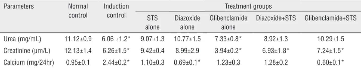
Histopathological analysis of renal tissues in the control group showed no calcium oxalate deposits or other abnormalities in different seg-ments of the nephrons (Figure-1A). But in the uro-lithiasis induction group, a substantial amount of calcium oxalate deposition was observed, and this was present in whole parts of three major areas of the kidney (Figure-1B). Renal tubular dilations with tubular basophilic and epithelial damage were also observed on pathological examination. In sodium thiosulfate treated group the number of calcium oxalate deposits was significantly lower than that in the disease control group with only mild nephropathy in one of the animals in that
Table 1 - Urine Chemistry.
Parameters Normal
control
Induction control
Treatment groups
STS alone
Diazoxide alone
Glibenclamide alone
Diazoxide+STS Glibenclamide+STS
Urea (mg/mL) 11.12±0.9 6.06 ±1.2* 9.07±1.3 10.77±1.5 7.33±0.8* 8.92±1.3 10.29±1.5
Creatinine (µm/L) 12.13±1.4 6.26±1.5* 9.42±0.4 8.99±2.9 3.94±0.2* 6.93±1.8* 7.24±1.5*
Calcium (mg/24hr) 0.95±0.1 2.44±0.2* 1.10±0.3 0.69±0.1* 1.23±0.3 1.28±0.2 0.60±0.1*
Group-1 = served as normal control; Group-2 = as a stone induction control; Group-3 = was given STS; Group-4 andGroup-5 = were administered diazoxide and
glibenclamide respectively and Groups 6 and 7 = were pretreated with diazoxide and glibenclamide respectively half an hour before administration of STS. Data of all results
are presented as mean±SD (*) p<0.05, statistically different from normal controls.
Table 2 - Serum Chemistry.
Parameters Normal
control
Induction control
Treatment groups
STS alone Diazoxide alone
Glibenclamide alone
Diazoxide +STS
Glibenclamide +STS
Urea (mg/dL) 16.50±0.9 41.94±1.1* 18.76±1.2 19.55±1.3 29.93±1.2* 16.42±1.6 13.74±1.3
Creatinine (mg/ dL)
0.35±0.02 1.03±0.07* 0.38±0.02 0.35±0.06 0.58±0.02* 0.38±0.04 0.35±0.06
ALP (U/L) 57.03±2.3 115.96±2.9* 49.25±2.4 50.73±3.2 76.18±2.4* 70.14±4.2* 41.39±2.1
Calcium (mg/dL) 6.21±0.5 5.14±0.9 4.46±0.7* 3.34±0.7* 1.57±0.2* 4.31±0.7* 4.10±0.9*
Group-1 = served as normal control; Group-2 = as a stone induction control; Group-3 = was given STS; Group-4 and Group-5 = were administered diazoxide and
glibenclamide respectively and Groups 6 and 7 were pretreated with diazoxide and glibenclamide respectively half an hour before administration of STS. Data of all results
Table 3 - Lipid peroxidation and antioxidant levels.
Parameters Normal
control
Induction control
Treatment groups
STS alone Diazoxide
alone
Glibenclamide alone
Diazoxide +STS
Glibenclamide +STS
TBARS (mM/100g tissue)
1.78±0.1 4.21±0.5* 2.10±0.4 0.84±0.09* 0.74±0.05* 0.71±0.03* 0.72±0.08*
Superoxide Dismutase (Units/mg protein)
34.7±4.5 17.64±1.2* 31.24±2.4 26.47±1.2* 25.63±1.3* 29.91±2.3 32.30±2.6
Glutathione peroxidase (µg of GSH utilized/min/ mg protein)
22±1.2 12.13±1.1* 19.14±1.6 15.54±1.3* 16.66±1.5* 19.82±1.1 19.57±1.4
Group-1 = served as normal control; Group-2 = as a stone induction control; Group-3 = was given STS; Group-4 and Group-5 = were administered diazoxide and
glibenclamide respectively and Groups 6 and 7 = were pretreated with diazoxide and glibenclamide respectively half an hour before administration of STS. Data of all results
are presented as mean±SD (*) p<0.05, statistically different from normal controls.
group (Figure-1C). Rats treated with diazoxide alone showed less obstructive damage (Figure-1D) while glibenclamide treated rats showed severe damage and obstruction (Figure-1E). Apparently more renal damage, inflammation and hemolysis were observed in rats co-administered with STS and diazoxide (Figure-1F) while STS administra-tion along with glibenclamide showed preserved renal tissue with mild obstruction (Figure-1G).
In vitro gel analysis to study chelation effect
In order to re-confirm the inhibitory effect of STS on calcium oxalate crystal, we performed an in-vitro analysis. The calcium oxalate crystals that have been produced in this study were similar to the crystals in the urine of patients with calcium oxalate crystals. The crystals were predominantly of monohydrate type, confirmed by FTIR. Accor-ding to Figure-2, STS showed a dose-dependent inhibition of calcium oxalate stone formation. Apparently, only 18% direct inhibition was shown with maximum STS concentration of 200mM, in-dicating an additional tissue based mechanism for its renal protective action.
DISCUSSION
Urinary lithiasis is a multifactorial urologi-cal disorder that generally occurs as a result of an imbalance between inhibitors and promoters for renal stone formation (15). The human kidney
sto-nes predominantly comprised of calcium oxalate, and few studies have examined the effect of the sodium thiosulfate on calcium oxalate crystalliza-tion (16) as well. However, the conclusions from those studies were not consistent as few studies claim the beneficial effect of STS and while others, its negative result (6, 7). In the present study, we investigated the effect of sodium thiosulfate on renal stone formation in both in vivo and in vi-tro models and evaluated its mechanism of action. Our study results were in agreement with previous reports that suggested anti-urolithiatic property of STS but provides a new direction for its mode of action where STS may modulate mitochondrial KATP channel in rendering renal protection.
Figure 1 - Light microscopic architecture of kidney showing (A) Renal tissue of control (group 1) rats showing no sign of crystal deposition. (B) Renal tissue of urolithiatic rats (group 2) showing crystals deposition and severe obstructive nephropathy (C) Renal tissue of (group 3) STS treated rats showing mild crystal deposition with mild nephropathy. (D) Renal tissue of (group 4) diazoxide treated rats showing mild crystal deposition with moderate obstructive nephropathy (E) Renal tissue of (group 5) glibenclamide treated rats showing prominent crystal deposition with severe obstructive nephropathy (F) Renal tissue of (group 6) diazoxide +STS treated rats showing crystal deposition with severe obstructive nephropathy (G) Renal tissue of (group 7) glibenclamide + STS pretreated rats low crystal deposition with mild nephropathy (H) Crystal's deposition as observed under 40X zoom.
A
E
G C
B
F
H
serum ALP activity may be derived from the in-jury to the brush border membrane of the renal tubular cells (19). The near-normal activity of ALP in the present study was in agreement with the other group that showed a significant decline of ALP activity after STS administration in uremic rats (20). Further evidence for STS protection was confirmed through histological results where pa-pillary crystalline deposits and calcium parenchy-ma deposits were absent. The direct interaction of STS on calcium oxalate formation was confirmed by using in vitro gel technique and found around 18% inhibition in calcium oxalate formation with 200mM STS, suggesting indirect action of STS in preventing renal stone formation.
A direct co-relation between renal mito-chondrial dysfunction and ethylene glycol indu-ced calcium oxalate formation was reported, where the exposure of the proximal tubule with calcium oxalate crystals resulted in rapid and progressive osmotic swelling and dissipation of transmembra-ne potential of mitochondria resulted in its dama-ge (21). The peroxidation of protein had greater influence on the nucleation and aggregation pro-perty of calcium oxalate crystal growth and that
predominantly occur in mitochondria (22). Mi-tochondrial permeability transition pore (mPTP) opening is a terminal event leading to mitochon-drial dysfunction and cell death under conditions of oxidative stress. In fact, the vulnerability of the renal tissue towards oxidative stress depends on the functional cross talks between mPTP and mi-tochondrial KATP channel (23). In the present stu-dy, we evaluated the specific role of mitochondrial potassium ATP channel in the sodium thiosulfate mediated renal protection.
Acccording to previous study results, it is believed that renal protection mediated by sodium thiosulfate is mainly attributed to its chelation and antioxidant potential (6, 24). However, thiosulfa-tes are metabolized in mitochondria, and thus we anticipated a mitochondria based mechanism for its renal protection. In this connection, we used a mitochondrial potassium channel blocker gliben-clamide (binds to sulphonylurea receptor subtypes of KATP channel) and channel opener diazoxide (binds to ATP binding sites of sulphonylurea re-ceptor subtypes of KATP channel) to evaluate the renal status as supported by biochemical parame-ters and histopathology (Figure-1, Tables 1-3).
Figure 2 - Images of agar gel slides and graph representing the percentage inhibition produced by STS in calcium oxalate crystal formation represented as a streak.
Glibenclamide showed a prominent renal injury as evidenced from altered serum and urine chemistry that was clearly demarcated in histopa-thology (Figure-1E, Tables 1 and 2). Several lines of evidence showed that glibenclamide can depo-larize mitochondrial membrane leading to calcium overload, one of the major factors responsible for free radical release and injury as evident from the lipid peroxidation and antioxidant marker enzyme levels (Table-3). On the other hand, ATP sensitive potassium channel opener, diazoxide treatment showed well-preserved architecture of the kidney (Figure-1D). It prevented mitochondrial swelling and depolarization that may result in permeabi-lity pore transition and leads to tissue injury (25, 26). Diazoxide can also modulate the renin an-giotensin system, that may play a significant role in developing renal tubule interstitial fibrosis (27) and resulting stone formation as reported with ethylene glycol induced renal injury. Although the mechanism by which the KATP opener exert their renal protection have not been clarified yet, it is believed that the opening of KATP channel preser-ves mitochondrial functional activities through mild uncoupling and depolarization (28). Thus diazoxide mediated protection is an impact on the mitochondria.
In order to confirm the STS mediated mi-tochondrial KATP channel modulatory effect, we administered STS along with diazoxide (mito-chondrial KATP channel opener) and glibenclamide (mitochondrial KATP channel blocker). We found interesting results, where the protective effect shown by diazoxide treatment alone was nega-ted by STS supplementation. On the other hand, STS supplementation to glibenclamide group sho-wed preserved renal tissue architecture. This in-verse relationship of STS is an evidence for its interaction with mito KATP Diazoxide binds to an ATP-sensitive K+ transport pathway in kidney
mi-tochondria that affects volume, respiration, and membrane potential and may have a role in the prevention of mitochondrial ATP hydrolysis. Ope-ning of this channel leads to mild uncoupling, blo-cks calcium entry into mitochondria and leading to renal protection (28, 29). As both diazoxide and STS (mediated through H2S formation) binds to KATP channel in different sites, when diazoxide
and STS are given concomitantly, long term or excessive uncoupling may be expected causing ATP hydrolysis and mPTP opening without im-pairing electron transport, leading to apoptosis. On the other hand, glibenclamide binds to diffe-rent sulfonylurea subunit blocking potassium en-try, thereby exaggerating the ROS production and destabilizing the membrane potential leading to apoptosis (28). When STS is given with glibencla-mide, we predict that, H2S released from STS may bind to Kir6. 1 subunit of mito KATP channel, the-reby reducing the binding efficiency of glibencla-mide resulting its limited action of KATP channel, allowing STS to mediated its renal protection.
The protective mechanism induced by the opening of mito KATP is well-studied in cardiovas-cular diseases. Analogous to the heart system, re-nal protection by diazoxide may well be claimed due to i) Changes in the mitochondrial Ca2+ levels
ii) Mitochondrial matrix swelling and changes in ATP synthesis iii) Changes in the ROS levels. Sodium thiosulfate is a known calcium chelating agent with antioxidant properties (30) and can render electrons to complex IV upon its meta-bolism. Furthermore, several lines of the reports suggest that mitochondrial KATP channel opening may inhibit mitochondrial permeability transition through inhibiting calcium overload and thereby preserve mitochondrial functions. A proven re-lationship between mitochondrial membrane po-tential, mitochondrial dependent apoptosis and calcium overload predicts the possibility of thio-sulfate mediated calcium signaling mechanism through calcium/calmodulin-dependent protein kinase for its action, proposed for the future study.
The present study enhances the existing knowledge of STS mediated anti urolithiatic me-chanism that emphasizes the calcium chelation and antioxidant property of STS alone. Based on our findings, we suggest that thiosulfate modula-te the mitochondrial KATP channel to render renal protection against stone formation.
CONCLUSIONS
agreement with the findings of Asplin & Onyeka groups. Even though few mechanisms were pro-posed earlier for the anti-urolithiasis effect of sodium thiosulfate, no conclusive understanding was reached, and the present study confirm the specific role of ATP sensitive mitochondrial KATP channel in STS mediated renal protective mecha-nism.
Abbreviations
STS = Sodium thiosulfate
EG = ethylene Glycol
KATP = Potassium ATP channel
mPTP = mitochondrial permeability transition pore
ROS = reactive oxygen species
CONFLICT OF INTEREST
None declared
REFERENCES
1. Aggarwal KP, Narula S, Kakkar M, Tandon C. Nephrolithiasis: molecular mechanism of renal stone formation and the critical role played by modulators. Biomed Res Int. 2013;2013:292953.
2. Matlaga BR, Coe FL, Evan AP, Lingeman JE. The role of Randall’s plaques in the pathogenesis of calcium stones. J Urol. 2007;177:31-8.
3. Sowers KM, Hayden MR. Calcific uremic arteriolopathy: pathophysiology, reactive oxygen species and therapeutic approaches. Oxid Med Cell Longev. 2010;3:109-21.
4. Dickey DT, Wu YJ, Muldoon LL, Neuwelt EA. Protection against cisplatin-induced toxicities by N-acetylcysteine and sodium thiosulfate as assessed at the molecular, cellular, and in vivo levels. J Pharmacol Exp Ther. 2005;314:1052-8. 5. Hamel J. A review of acute cyanide poisoning with a
treatment update. Crit Care Nurse. 2011;31:72-81; quiz 82. 6. Asplin JR, Donahue SE, Lindeman C, Michalenka A, Strutz
KL, Bushinsky DA. Thiosulfate reduces calcium phosphate nephrolithiasis. J Am Soc Nephrol. 2009;20:1246-53. 7. LaGrange CA, Lele SM, Pais VM Jr. The effect of sodium
thiosulfate administration on nephrocalcinosis in a rat model. J Endourol. 2009;23:529-33.
8. Sandhiya S, Dkhar SA. Potassium channels in health, disease & development of channel modulators. Indian J Med Res. 2009;129:223-32.
9. Rahgozar M, Pazokitoroudi H, Bakhtiarian A, Djahanguiri B. Diazoxide, a K(ATP) opener, accelerates restitution of ethanol or indomethacin-induced gastric ulceration in rats independent of polyamines. J Gastroenterol Hepatol. 2001;16:290-6.
10. Lowry OH, Rosebrough NJ, Farr AL, Randall RJ. Protein measurement with the Folin phenol reagent. J Biol Chem. 1951;193:265-75.
11. Kurian GA, Paddikkala J. Administration of aqueous extract of Desmodium gangeticum (L) root protects rat heart against ischemic reperfusion injury induced oxidative stress. Indian J Exp Biol. 2009;47:129-35.
12. Aziz SA, See TL, Khuay LY, Osman K, Abu Bakar MA. In vitro effects of plantago major extract on urolithiasis. Malays J Med Sci. 2005;12:22-6.
13. Leibovitch I, Ben-Chaim J, Ramon J, Goldwasser B. Increased serum alcaline phosphatase activity: a possible indicator of renal damage. J Clin Lab Anal. 1991;5:406-9. 14. Huang HS, Chen J, Chen CF, Ma MC. Vitamin E attenuates
crystal formation in rat kidneys: roles of renal tubular cell death and crystallization inhibitors. Kidney Int. 2006;70:699-710. Erratum in: Kidney Int. 2007;71:712.
15. Worcester EM, Coe FL. Clinical practice. Calcium kidney stones. N Engl J Med. 2010;363:954-63.
16. Daudon M, Knebelmann B. Calcium oxalate urolithiasis. Rev Prat. 2011;61:385-8.
17. Scheid C, Koul H, Hill WA, Luber-Narod J, Kennington L, Honeyman T, et al. Oxalate toxicity in LLC-PK1 cells: role of free radicals. Kidney Int. 1996;49:413-9.
18. Wellwood JM, Lovell D, Thompson AE, Tighe JR. Renal damage caused by gentamicin: a study of the effects on renal morphology and urinary enzyme excretion. J Pathol. 1976;118:171-82.
19. Amador Ee, Ddorfman LE, Wacker WE. Urinary alkaline phosphatase and LDH activities in the differential diagnosis of renal disease. Ann Intern Med. 1965;62:30-40.
20. O’Neill WC, Hardcastle KI. The chemistry of thiosulfate and vascular calcification. Nephrol Dial Transplant. 2012;27:521-6.
21. McMartin KE, Wallace KB. Calcium oxalate monohydrate, a metabolite of ethylene glycol, is toxic for rat renal mitochondrial function. Toxicol Sci. 2005;84:195-200. 22. Govindaraj A, Selvam R. Increased calcium oxalate crystal
nucleation and aggregation by peroxidized protein of human kidney stone matrix and renal cells. Urol Res. 2001;29:194-8. 23. Xie C, Kauffman J, Akar FG. Functional crosstalk between the
mitochondrial PTP and KATP channels determine arrhythmic vulnerability to oxidative stress. Front Physiol. 2014;5:264. 24. Hayden MR, Tyagi SC, Kolb L, Sowers JR, Khanna R. Vascular
25. Weiss JN, Korge P, Honda HM, Ping P. Role of the mitochondrial permeability transition in myocardial disease. Circ Res. 2003;93:292-301.
26. Halestrap AP, Clarke SJ, Javadov SA. Mitochondrial permeability transition pore opening during myocardial reperfusion--a target for cardioprotection. Cardiovasc Res. 2004;61:372-85.
27. Rüster C, Wolf G. Renin-angiotensin-aldosterone system and progression of renal disease. J Am Soc Nephrol. 2006;17:2985-91.
28. Cancherini DV, Trabuco LG, Rebouças NA, Kowaltowski AJ. ATP-sensitive K+ channels in renal mitochondria. Am J Physiol Renal Physiol. 2003;285:F1291-6.
29. Kasinath BS. Hydrogen sulfide to the rescue in obstructive kidney injury. Kidney Int. 2014;85:1255-8.
30. Garlid KD, Dos Santos P, Xie ZJ, Costa AD, Paucek P. Mitochondrial potassium transport: the role of the mitochondrial ATP-sensitive K(+) channel in cardiac function and cardioprotection. Biochim Biophys Acta. 2003;1606:1-21.
_______________________ Correspondence address: