Journal of the Renin-Angiotensin-Aldosterone System
October-December 2016: 1 –10 © The Author(s) 2016 Reprints and permissions:
sagepub.co.uk/journalsPermissions.nav DOI: 10.1177/1470320316674876 jra.sagepub.com
Creative Commons Non Commercial CC-BY-NC: This article is distributed under the terms of the Creative Commons
Attribution-NonCommercial 3.0 License (http://www.creativecommons.org/licenses/by-nc/3.0/) which permits non-commercial use, reproduction and distribution of the work without further permission provided the original work is attributed as specified on the SAGE and Open Access pages (https://us.sagepub.com/en-us/nam/open-access-at-sage).
Background
Essential hypertension (EH) is the most common cardio-vascular disease with a multifactorial pathogenesis. Recent studies have confirmed that a large number of EH cases are closely related to the development of inflammation. Activation of inflammatory cells was shown to be involved in the development of hypertension,1,2 and it has been
proposed that hypertension is a chronic, low-grade
Inhibitory effects of telmisartan on culture
and proliferation of and Kv1.3 potassium
channel expression in peripheral blood
CD4
+
T lymphocytes from Xinjiang
Kazakh patients with hypertension
Sha-Sha Huang
*, Qiu-Bing Zhang
*, Qing-Yan Yuan,
Si-Li He and Yuan-Ming Zhang
Abstract
Introduction: Activation of T lymphocytes, for which potassium channels are essential, is involved in the development of hypertension. In this study, we explored the inhibitory effects of telmisartan on the culture and proliferation of and Kv1.3 potassium channel expression in peripheral blood CD4+ T lymphocytes derived from Xinjiang Kazakh patients with
hypertension.
Methods: CD4+ T-cell samples from hypertensive Kazakh patients and healthy Kazakh people were divided into healthy
control, case control, telmisartan, and 4-aminopytidine groups. Changes in the expression levels of interleukin (IL)-6 and IL-17 in the blood of the healthy control and case control subjects were detected by enzyme-linked immunosorbent assay. Peripheral blood CD4+ T lymphocytes were first activated and proliferated in vitro and then incubated for 0, 24,
and 48 h under various treatment conditions. Thereafter, changes in CD4+ T-lymphocytic proliferation were determined
using Cell Counting Kit-8 and microscope photography. Changes in messenger RNA (mRNA) and protein expression of the Kv1.3 potassium channel in CD4+ T lymphocytes were detected using real-time quantitative polymerase chain
reaction and Western blots, respectively.
Results: The IL-6 and IL-17 expression levels were significantly higher in the blood of the hypertensive Kazakh patients than in the healthy Kazakh people. Telmisartan inhibited T-lymphocytic proliferation, as well as the mRNA and protein expression of the Kv1.3 potassium channel in CD4+ T lymphocytes, and the inhibitory effects were
time-dependent, with the strongest inhibition observed after 48 h and significantly weaker inhibition observed after 24 h of treatment.
Conclusions: Telmisartan may potentially regulate hypertensive inflammatory responses by inhibiting T-lymphocytic proliferation and Kv1.3 potassium channel expression in CD4+ T lymphocytes.
Keywords
Xinjiang Kazakh, essential hypertension, CD4+ T Lymphocytes, Kv1.3 potassium channel, telmisartan
Date received: 28 June 2016; accepted: 16 September 2016
Heart Centre, First Affiliated Hospital of Xinjiang Medical University, China
*These authors contributed equally to this work and should be considered co-first authors.
Corresponding author:
Yuan-Ming Zhang, Heart Center, First Affiliated Hospital of Xinjiang Medical University, Urumqi, Xinjiang, 830000, China.
inflammatory disease.3,4 Animal studies have shown that
activation of massive inflammatory cells was obvious in a spontaneously hypertensive rat (SHR) model, and the acti-vation was more significant in adult SHRs than in young SHRs.5 In fact, massive inflammatory mediators are
upregulated in hypertensive patients, and their levels can be used to predict the development of hypertension. Inflammatory reactions in hypertensive patients are char-acterized by the expression and activation of cytokines.6,7
T lymphocytes play an important role in the etiology and development of hypertension and vascular remode-ling,8 and they are important for immune regulation. It is
well known that T lymphocytes can be divided into two subsets, CD4+ and CD8+. CD4+ T lymphocytes account for
35–50% of the total peripheral blood lymphocytes and regulate the biological activity of the immune system by secreting cytokines and expressing cell surface molecules. CD4+ T lymphocytes secrete interferon gamma (INF-γ),
which can stimulate the expression of major histocom-patibility complex (MHC) in endothelial cells (ECs) and smooth muscle cells. This activates the antigen-presenting function of ECs and initiates immune responses. INF-γ also improves T lymphocyte migration through the EC monolayer.
The activation function of T lymphocytes depends on ion channels located on their cell surfaces. Studies have shown that there are mainly three kinds of membrane ion channels on T lymphocytes, a voltage-gated potassium channel (Kv1.3), calcium-activated potassium channel (KCa3.1), and calcium release-activated calcium (CRAC) channel. In 1984, DeCoursey et al.9 reported the presence
of Kvl.3 potassium channels on T cells. The Kv1.3 volt-age-gated potassium channel is essential for the activation of T lymphocytes and is involved in some physiological processes, such as T-lymphocytic differentiation and pro-liferation.10,11 Kv1.3 on CD4+ T lymphocytes plays a key
role in lymphocyte activation in patients with acute coro-nary syndrome.12 Opening of the Kv1.3 potassium channel
is essential for the membrane depolarization and mainte-nance of the resting potential. It has been reported that Kv1.3 is involved in the development of various diseases, such as acute coronary syndrome, multiple sclerosis, asthma, tumors, etc.13–20 The forkhead transcriptional
fac-tor FoxP3, expressed in T lymphocytes, is critical for the production of angiotensin II, which is essential for the pro-gression of EH.8 Some studies have shown that besides
lowering blood pressure, angiotensin receptor blockers (ARBs), which are widely used to treat hypertension, also have anti-inflammatory effects.21–24
Kazakh individuals are the second largest minority in Xinjiang, and they have a high incidence of hypertension, with EH prevalence rates of 40.5–52.39%.25 However,
rates of disease awareness, cure, and control appear to be lower among Kazakhs than in other ethnic groups. Therefore, studying the pathogenesis of hypertension in Kazakh individuals has social significance. However, few
studies have assessed the effects of telmisartan, one of the ARBs, on the proliferation of peripheral blood CD4+ T
lymphocytes, as well as messenger RNA (mRNA) and protein expression of the Kv1.3 potassium channel in CD4+ T lymphocytes derived from Xinjiang Kazakh
patients with EH.
In this study, we used enzyme-linked immunosorbent assay (ELISA) to detect changes in the expression levels of interleukin (IL)-6 and IL-17 in the blood of hyperten-sive Kazakh patients. We used magnetic-activated cell sorting (MACS) to isolate CD4+ T lymphocytes from
peripheral blood of Xinjiang Kazakh patients with EH and healthy Kazakh people. Then, Cell Counting Kit-8 (CCK-8) and microscope photography were used to analyze the culture and proliferation of the CD4+ T lymphocytes.
Real-time quantitative reverse transcription–polymerase chain reaction (qRT–PCR) and Western blotting were used to determine the mRNA and protein expression levels of the Kv1.3 potassium channel in the CD4+ T
lymphocytes. Our results may provide the theoretical mechanisms and experimental evidence for the anti-inflammatory effect of telmisartan.
Methods
Subjects
We selected Kazakh hypertensive patients and healthy Kazakh people as the subjects for our experiments. Thirty Kazakh hypertensive patients (mean age: 50.4±3.5 years) and 10 healthy Kazakh people (mean age: 48.8±4.5 years) attending the cardiology clinic of the First Affiliated Hospital of Xinjiang Medical University were enrolled in the study between January–August 2015. The healthy sub-jects were all assigned to a healthy control group, while the hypertensive subjects were randomised into the following three groups: (a) case controls; (b) telmisartan; and (c) 4-aminopytidine (4-AP). There were 10 individuals (five males and five females) in each group, and they were not undergoing antihypertensive therapy.
The diagnostic criteria of hypertension, used as the inclusion criteria, were as stated in the 2010 Chinese guidelines for the management of hypertension.26 Briefly,
systolic blood pressure of ⩾140 mm Hg (1 mm Hg = 0.133 kPa) and/or diastolic blood pressure of ⩾90 mm Hg, with-out treatment with antihypertensive medication, were con-sidered the diagnostic criteria of hypertension.
Subjects with the following types of diseases were excluded from our experiments: (a) secondary hyperten-sion; (b) cerebrovascular disease; (c) coronary, rheumatic, or congenital heart disease; (d) acute or chronic infection; (e) autoimmune disease; (f) important organ failure; (g) diabetes; and (h) carotid atherosclerosis.
Affiliated Hospital of Xinjiang Medical University (approval number 20141011-2), and informed consent was signed by each subject before the trial.
Reagents
IL-6 and IL-17 ELISA kits were from eBioscience (San Diego, California, USA). Human lymphocyte isolation medium, recombinant human IL-2 (rIL-2), phytohemag-glutinin (PHA), and 4-AP were purchased from Sigma– Aldrich (St. Louis, Missouri, USA). The MACS kit was from Miltenyi Biotec (Germany), and telmisartan was from Boehringer–Ingelheim (Germany). Fetal bovine serum (FBS) and Roswell Park Memorial Institute (RPMI) 1640 medium were obtained from HyClone (New Zealand). Fluorescein isothiocyanate (FITC)-labelled mouse anti-human CD3 and FITC-(FITC)-labelled mouse anti-human CD4 were from Becton, Dickinson and Company (USA). TRIzol was from Life Technologies (USA). The RT kit, Radio-Immunoprecipitation Assay (RIPA) lysis buffer, protease inhibitor, bicinchoninic acid (BCA) protein assay kit, and enhanced chemilumines-cence (ECL) Western blotting kit were from Thermo Fisher (USA). The real-time fluorescent qPCR kit was from Qiagen (Germany). Sodium dodecyl sulphate poly-acrylamide gel electrophoresis (SDS–PAGE) reagents were purchased from Bio-Rad (USA). Anti-human mon-oclonal antibodies against the Kv1.3 potassium channel and β-actin and secondary antibodies were obtained from Abcam (UK). The 5-bromo-4-chloro-3’-indolyphosphate/ nitro-blue tetrazolium chromogenic reagent was from Invitrogen (USA).
CD4
+T lymphocyte isolation
Peripheral venous blood (10 ml) was collected from each subject into a heparinized vacutainer tube. Peripheral blood mononuclear cells were isolated using human lym-phocyte isolation medium and density gradient centrifu-gation. Using the Pan T cell isolation kit (Miltenyi, Germany), human T lymphocytes were isolated by deple-tion of non-target T cells (negative selecdeple-tion). Non-target T cells were magnetically labelled with a cocktail of bio-tin-conjugated monoclonal antibodies and the Pan T cell microbead cocktail. They were retained on a MACS sep-aration column in the magnetic field of a MACS separa-tor (Miltenyi, Germany), while unlabeled T lymphocytes passed through the column. The purity of the enriched T lymphocytes was evaluated by flow cytometry (Beckman Coulter, USA). The cells were fluorescently stained with FITC–CD3 and analyzed using a MACSQuant analyzer (Miltenyi, Germany). Cell debris and dead cells were excluded from the analysis based on scatter signals and propidium iodide fluorescence. The results showed that >95% of the cells were T lymphocytes. T lymphocytes were incubated with CD4 monoclonal antibodies. The
suspensions were then incubated with magnetic beads coated with goat anti-mouse Immunoglobulin G (IgG), which bind to antibody-coated cells. Subsequent expo-sure to a strong magnetic field removed unwanted cells, leaving the desired cell population. The cell-coated mag-netic beads were resuspended in complete medium. Anti-human FITC-CD4 was added, and the T lymphocytes were analyzed using flow cytometry. The results showed that >95% of the cells were CD4+ T lymphocytes, which
were used for subsequent experiments.
Cell culture
CD4+ T lymphocytes were seeded into 24-well plates
con-taining RPMI 1640 medium with 10% FBS, rIL-2, and PHA and cultured at 37°C, 5% CO2 in an incubator
(Thermo, USA) for 48 h to promote T lymphocyte activa-tion. The cultured CD4+ T lymphocytes were treated as
follows: telmisartan (final concentration, 100 µmol/l), 4-AP (final concentration, 3 mmol/l), or an equal volume of a dimethyl sulphoxide (DMSO) vehicle was added to the culture media of cells from the telmisartan, 4-AP, and control groups, respectively. The CD4+ T lymphocytes
were then cultured for 0, 24, and 48 h, and the suspensions were harvested. Changes in the T-lymphocytic growth sta-tus, shape, and quantity were observed and recorded by microscope photography using an inverted fluorescent microscope (Leica, Germany). Then, the samples were divided into three portions, for CCK-8 analysis and extrac-tion of RNA and protein, respectively.
ELISA assay
Peripheral blood (5 ml) from the healthy Kazakh people in the healthy control group and from the hypertensive Kazakh patients in the case control group was incubated at room temperature for 30 min and then centrifuged at 3000 rpm for 10 min at 4°C. The supernatant serum was collected and stored at −80°C. Then, levels of IL-6 and IL-17 were analyzed in the serum using the ELISA kits according to the manufacturer’s instructions. Both test samples and standard samples were tested in duplicate. Absorbance was measured at 450 nm using a microplate reader (Bio-Rad). Then, standard curves were estab-lished, and levels of IL-6 and IL-17 were calculated from the standard curves.
CCK-8 analysis of T-lymphocytic proliferation
CD4+ T lymphocytes, precultured with PHA and rIL-2 for
an incubator for 3 h, and optical density (OD) values were measured at 450 nm using a microplate spectrophotometer. Then, cellular activities of the different groups were calcu-lated as follows:
Cellular activity
OD value of interventional group OD
%
( )
=−
v
value of control group
OD value of interventional g
/
rroup at h OD value of control group at h 1
0
0 00
−
×
RNA extraction and qRT–PCR analysis
Total RNA was extracted from CD4+ T lymphocytes using
TRIzol, and the A260/A280 ratios of the resulting RNA
sam-ples were approximately 1.8–2.0. RT reactions were per-formed using 1 µg of RNA at 42°C for 60 min, followed by incubation at 70°C for 5 min. The total volume of qPCR reactions was 20 µl, including 10 µl of SYBR Green PCR master mix (2×), 2 µl of cDNA template, 0.5 µl of each for-ward (F) and reverse (R) primer, and 7 µl of H2O. The
sequences of the genes encoding the Kv1.3 potassium chan-nel and β-actin (ACTB gene) were obtained from GenBank. The primers were synthesized by Sangon Biotech (Shanghai). ACTB was used as an internal reference control gene (Table 1). PCR and agarose gel electrophoresis were performed with the cDNA templates, and standard curves were established using the PCR products. The qPCR pro-gram was as follows: 95°C for 5 min, followed by 39 cycles of 95°C for 10 s and 60°C for 30 s. Amplification efficien-cies were approximately 90–100%. After qPCR, the ampli-cons were separated by 2% agarose gel electrophoresis. The data was analyzed using the 2−∆∆Ct method as follows:
∆Ct=Ct target gene Ct reference gene−
∆∆Ct=∆Ct experimental group−∆Ct control group The rate of inhibition by telmisartan was calculated as follows:
Inhibitory rate
mRNA expression in control group mRNA
%
(
( )
=− expression in intervention group mRNA expression in con
)
/ ttrol group 1× 00.
Western blots
Total protein was extracted from CD4+ T lymphocytes
using RIPA lysis buffer supplemented with protease inhibi-tors. Protein concentrations were measured by the BCA protein assay. Protein samples (20 µl=30 µg) were mixed with 5 µl of loading buffer, then denatured in a water bath at 95°C for 5 min, and separated by SDS–PAGE. Then, proteins were transferred to polyvinylidene fluoride mem-branes, and the membranes were blocked with 5% skim milk for 1 h. The primary antibodies were diluted as fol-lows: anti-Kv1.3 potassium channel, 1:200; anti-β-actin, 1:5000. The blots were incubated with the primary antibod-ies overnight at 4°C with shaking and then washed with Tris-buffered saline containing Tween 20 (TBS-T), fol-lowed by incubation with secondary antibodies (both 1:4000) at room temperature for 1 h and washing with TBS-T. Visualization of protein bands was performed by the addition of 5 ml of the ECL reagent, followed by the analysis using a Quality Image Analysis System (Bio-Rad). The telmisartan inhibitory rate was calculated as follows:
Inhibitory rate
protein expression in control group pr
%
(
( )
=_
o
otein expression in interventional group protein express
) / iion in control group 1× 00
Statistical methods
Data were statistically analyzed using the SPSS 17.0 soft-ware and are expressed as the mean±standard deviation (SD) or percentage, as appropriate. Multiple data sets were compared by repeated-measures one-way analysis of vari-ance or a chi-square test. A least significant difference t-test was used for comparison between two groups. A p-value of <0.05 was considered statistically significant.
Results
Clinical and demographic characteristics of
subjects
There were no significant differences among the subjects in the healthy control, case control, telmisartan, and 4-AP groups in regard to the age, smoking history, drinking his-tory, body mass index, fasting blood glucose, C-reactive Table 1. Primer sequences and amplicon sizes.
Gene Sequences (5′–3′) GenBank accession number Product length (base pairs)
Kv1.3 F: CCAGCACCTCTCCTCTTCAG R: TCACCATATACTCCGACTTACTCA
NM_002232.3 80
ACTB F: TGGCACCCAGCACAATGAA
R: CTAAGTCATAGTCCGCCTAGAAGCA
protein (CRP), triglyceride, high-density lipoprotein cho-lesterol, and low-density lipoprotein cholesterol levels (p>0.05; Table 2). The family history of hypertension was defined as one or more family members (parent or sibling) of the subject having hypertension.27 Smoking was defined
as a current or previous smoking history.27 Alcohol
con-sumption was defined as drinking at least once per week for more than half a year.27
Levels of IL-6 and IL-17 in peripheral blood
The results showed that the expression of the inflamma-tory cytokines IL-6 and IL-17 was significantly higher in the peripheral blood of the Kazakh hypertensive patients than in that of the healthy Kazakh people (IL-6: 61.0±4.1 vs 43.0±5.3; IL-17: 72.4±1.9 vs 50.8±2.6, respectively, p=0.002) (Figure 1).
CD4
+T-lymphocytic proliferation
The cellular activities of the control groups significantly increased, while those of the telmisartan and 4-AP groups significantly reduced over time (p<0.05; Table 3).
Morphological observations of CD4
+T-lymphocytic proliferation with and
without drug treatment
CD4
+T-lymphocytic proliferation in the healthy
and case control groups stimulated with
PHA+rIL-2
There were obvious changes in the growth status, shape, and quantity of CD4+ T lymphocytes, and these changes were
similar in the healthy control and case control groups. As the culture time progressed, the cellular volume became larger, and most of the cells were round or could be irregular in
shape, such as multi-tentacled. Cytoplasm vacuolization was observed, and the cells aggregated to form a massive T-lymphocytic mass. A large cellular mass formed by hun-dreds of cells was visible under macrography, which indi-cated that the cells were in vigorous growth. The aggregated cells could easily be dispersed to single ones. There also was a significant increase in cell numbers.
CD4
+T-lymphocytic proliferation in the
telmisartan group
Before the telmisartan treatment, the CD4+ T lymphocytes
grew in good condition, and there were no significant dif-ferences with the cells in the control groups under microg-raphy. After 24 h of telmisartan treatment, there were some changes in the growth status, shape, and quantity of CD4+
T lymphocytes. After 48 h, the differences became more obvious as telmisartan started showing an inhibitory effect on T-lymphocytic proliferation. The cellular activity became weaker, the cell numbers were significantly reduced, and there was scattered cell debris, which was apoptotic, on the bottom of the culture dish.
CD4
+T-lymphocytic proliferation in the 4-AP
group
At 0 h, the CD4+ T lymphocytes had good cellular activity,
and there were large numbers of cells, with large round cells densely populated. At 24 h of 4-AP treatment, the cell num-bers were slightly reduced, but there was still some aggre-gated T-lymphocytic mass, which was sensitive to PHA. The cellular volume became smaller, and there was some cell debris deposited on the bottom of the culture dish. At 48 h, the cell numbers were significantly reduced, and cell debris, which was apoptotic, could be observed on the bottom of the culture dish. There were scattered single cells and some small cell mass of aggregated cells with a low cellular Table 2. Comparison of baseline data among the healthy control, case control, telmisartan, and 4-aminopytidine (4-AP) groups (n=40).
Parameter Group
Healthy control Case control Telmisartan 4-AP p-Value
Number (males/females)
10 (5/5)
10 (5/5)
10 (5/5)
10 (5/5)
>0.05
Age (years) 49.8 ± 4.5 48.3 ± 2.3 50.2 ± 3.7 50.7 ± 3.3 >0.05
Smoking (%) 49 52 50 51 >0.05
Drinking (%) 50 49 51 51 >0.05
BMI (kg/m2) 24.8 ± 2.5 26.1 ± 1.5 25.9 ± 1.4 26.2 ± 2.3 >0.05
FBG (mmol/l) 4.5 ± 0.6 4.6 ± 0.3 4.6 ± 0.7 4.6 ± 0.5 >0.05 CRP (mmol/l) 7.5 ± 0.7 7.7 ± 0.8 7.7 ± 0.5 7.6 ± 1.2 >0.05 TG (mmol/l) 1.6 ± 0.2 1.7 ± 0.1 1.7 ± 0.2 1.6 ± 0.2 >0.05 HDL-C (mmol/l) 1.3 ± 0.2 1.2 ± 0.2 1.3 ± 0.2 1.2 ± 0.2 >0.05 LDL-C (mmol/l) 3.5 ± 0.9 3.6 ± 0.8 3.6 ± 1.0 3.8 ± 0.9 >0.05
activity. The volume of most cells became smaller, and the CD4+ T lymphocytes were dark and had poor cellular
activity.
Effect of telmisartan and 4-AP on
mRNA expression of Kv1.3 potassium
channel in CD4
+T lymphocytes
Expression of Kv1.3 potassium channel mRNA in the peripheral blood CD4+ T lymphocytes was examined in
the four experimental groups. Amplicons of the correct size were observed after agarose gel electrophoresis. The results demonstrated that the mRNA levels of the Kv1.3 potassium channel were lower in the telmisartan and 4-AP groups after 24 h and 48 h of treatment than those in the healthy control and case control groups (p<0.05). There were no statistically significant differences in the expres-sion of Kv1.3 potassium channel mRNA at different time points in the healthy control and case control groups (p>0.05). In addition, the mRNA expression levels of the Kv1.3 potassium channel were significantly reduced in the CD4+ T lymphocytes at 24 h and 48 h compared with the
levels at 0 h in the telmisartan and 4-AP groups (p<0.05) (Figure 2). Telmisartan inhibited the mRNA expression of the Kv1.3 potassium channel in CD4+ T lymphocytes at 24
h and 48 h by 47.2% and 78.5%, respectively.
Effects of telmisartan and 4-AP on
Kv1.3 potassium channel protein
expression in CD4
+T lymphocytes
Expression of the Kv1.3 potassium channel protein was examined in the peripheral blood CD4+ T lympho-
cytes from the four experimental groups. The results
demonstrated that the protein levels of the Kv1.3 potassium channel were lower in the telmisartan and 4-AP groups after 24 h and 48 h of treatment than those in the healthy control and case control groups (p<0.05). There were no statistically significant differences in the expression of the Kv1.3 potassium channel protein at different time points in the healthy control and case control groups (p>0.05). In addition, the protein expression levels of the Kv1.3 potas-sium channel were significantly reduced in the CD4+ T
lymphocytes at 24 h and 48 h compared with the levels at 0 Table 3. CD4+ T-lymphocytic cellular activities detected using Cell Counting Kit-8.
Group 0 h 24 h 48 h
Healthy control 28.13 ± 6.23 36.15 ± 3.19a 48.14 ± 5.21a
Case control 106.45 ± 10.28 121.63 ± 5.37a 135.03 ± 10.74a
Telmisartan 102.73 ± 2.96 68.33 ± 5.39a 22.27 ± 3.58a
4-AP 105.17 ± 5.19 47.32 ± 6.38a 11.21 ± 2.01a
ap<0.01 compared with the cellular activity at 0 h in the same group.
Figure 2. messenger RNA (mRNA) expression of the Kv1.3 potassium channel relative to ACTB in samples extracted from activated peripheral blood CD4+ T lymphocytes from Xinjiang
Kazakh essential hypertension (EH( patients, determined by real-time quantitative reverse transcription–polymerase chain reaction (qRT–PCR). Cells were treated with dimethyl sulphoxide (DMSO) (healthy control and case control), telmisartan (100 μmol/l), and 4-aminopytidine (4-AP) (3 mmol/l); n=10 in each treatment group.
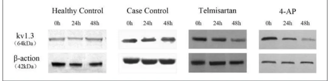
h in the telmisartan and 4-AP groups (Figures 3 and 4). Telmisartan inhibited the protein expression of the Kv1.3 potassium channel in the CD4+ T lymphocytes at 24 h and
48 h by 49.3% and 67.9%, respectively.
Discussion
EH is a chronic, low-grade inflammatory disease with abnormal activation of inflammatory cytokines and inflammatory signaling pathways.3,4,28 Expression of
inflammatory factors causes vascular inflammation and promotes development of hypertension, which further changes the structure and function of blood vessels and causes damage to the heart, kidney, and other target organs. Harrison and Gongora29 have proposed that the release of
various lymphocyte cytokines causes vasoconstriction and sodium and water retention, which further increases the blood pressure to promote the development of EH. Elevated IL-1β and CRP levels are also associated with hypertension. Thus, it has been confirmed recently that
tumor necrosis factor alpha increased the activity of reduced coenzyme II oxidase in polymorphonuclear leu-kocytes, which led to oxidative stress, an inflammatory response, and elevated blood pressure.30 Under various
conditions, activated T lymphocytes will produce multiple inflammatory cytokines, including IL-6 and IL-17. Consistent with most studies, here we found that the levels of IL-6 and IL-17 in peripheral blood were higher in Kazakh hypertensive patients than in related healthy per-sons, which indicated that there was an activation of inflammatory cytokines in the hypertensive patients.
The T lymphocyte function depends on the electrical activity of ion channels in their cell membrane.31 Studies
using patch clamps have suggested that Kv1.3 potassium channels are the key effectors of the sustained activation of CD4+T lymphocytes.32 The main effects of Kv1.3
potas-sium channels are to maintain the membrane potential and intracellular calcium signaling, which regulates the cell proliferation and cytokine production. Kv1.3 potassium channels are likely targets for the immune or bi-directional prevention of EH and are associated with many clinical diseases. Thus, Beeton and others33,34 studied peripheral
blood of multiple sclerosis patients and postmortem brain lesions and found that the antigen-specific CD4+ T cells
were mainly CCR7–CD45RA effector memory T cells, which exhibited increased expression of Kv1.3 channels. Guo et al.35 detected increased levels of Kv1.3 potassium
channel mRNA in rat lymphocytes, suggesting that the elevated expression of Kv1.3 on T lymphocytes plays an important role in atherosclerosis in rats. Xu et al.14 found
that CD4+/CD28− T lymphocytes were potential
immu-nomodulatory targets in patients with acute coronary syn-drome and suggested that selective blocking of CD4+/
CD28− T lymphocytes could be used to treat the disease.
Therefore, this study focused on Kv1.3 channels on CD4+
T lymphocytes derived from Kazakh hypertensive patients. As a Kvl.3 potassium channel inhibitor, 4-AP can inhibit the activation and proliferation of T lymphocytes and regulate the cytokine production and immune responses by inhibiting the Kvl.3 potassium channel. Some studies have shown that ARBs can effectively inhibit the Kvl.3 potassium channel, regulate the immunity through anti-inflammation and antioxidation, and play an important role as anti-atherosclerosis drugs. By observing Figure 3. Representative Western blots of protein samples extracted from activated peripheral blood CD4+ T lymphocytes from
Xinjiang Kazakh essential hypertension (EH) patients. Blots were probed with antibodies against the Kv1.3 potassium channel and β-actin: healthy control group; case control group; telmisartan group (100 μmol/l); 4-aminopytidine (4-AP) group (3 mmol/l).
Figure 4. Protein expression of the Kv1.3 potassium channel relative to β-actin in samples extracted from activated peripheral blood CD4+ T lymphocytes from Xinjiang Kazakh
blocking of Kvl.5/Kvl.3 potassium channels in Xenopus oocytes, Li et al.36 found that telmisartan blocked the
open-ing state of the Kv1.3 potassium channel, which is one of the mechanisms that can regulate the immune system and anti-atherosclerotic effects.
Data from our preliminary study37 have indicated that
the mRNA expression levels of the Kv1.3 potassium chan-nel are significantly increased in lymphocytes of SHRs, along with an elevated potassium current density, demon-strating an increase in functional potassium channels in SHR lymphocytes. Moreover, we have also previously reported that the mRNA and protein expression levels of the Kv1.3 potassium channel are significantly elevated in lymphocytes from hypertensive Kazakh patients from Xinjiang38 and that the Kv1.3 potassium current densities
in peripheral blood T lymphocytes of this population were also higher.39 These studies support the concept that
potassium channels of T lymphocytes are involved in the pathogenesis and progression of hypertension through their role in T lymphocyte activation. We have also per-formed studies demonstrating that telmisartan can block Kv1.3 potassium channels in lymphocytes of SHRs in a concentration-dependent manner.40 In addition,
telmisar-tan effectively inhibited potassium current densities in peripheral blood T lymphocytes of Kazakh patients with EH in a time-dependent manner.41 Hence, ARBs,
including telmisartan, have the potential to elicit potent anti-inflammatory effects by blocking Kv1.3 potassium channels on T lymphocytes, although the specific underly-ing mechanisms remain unclear.
In the present study, there were no significant differ-ences between the healthy control and case control groups, which indicated that the findings reflect a general phenom-enon characteristic of CD4+ T lymphocytes. Electron
microscopy revealed that after the incubation with 4-AP at an effective concentration for 0, 24, and 48 h, the numbers of CD4+ T lymphocytes were gradually reduced, their
cel-lular activity gradually lowered, the shape condensed and gradually shrank, and many apoptotic cells could be seen. qRT–PCR and western blotting revealed that after the incu-bation with 4-AP at an effective concentration for 0, 24, and 48 h, the mRNA and protein expression levels of the Kv1.3 potassium channel in CD4+ T lymphocytes were
sig-nificantly reduced. The results showed that 4-AP had an obvious inhibitory effect on T-lymphocytic proliferation, as well as on mRNA and protein expression of the Kv1.3 potassium channel in CD4+ T lymphocytes, which
indi-rectly indicated that the Kvl.3 potassium channel is the key factor of T lymphocyte activation. The 4-AP group was used as a positive control group, and the telmisartan group showed similar changes, although the changes in the T-lymphocytic proliferation and mRNA and protein expres-sion of the Kv1.3 potassium channel in CD4+ T
lympho-cytes were slower and weaker with telmisartan relative to those in the 4-AP group. This indicates that the anti-inflammatory function and inhibitory effects of telmisartan
on the T lymphocyte Kv1.3 potassium channel are unlikely to affect the normal immune function in hypertensive patients. The CCK-8 analysis, qRT–PCR, and Western blotting showed that the cellular activities and mRNA and protein expression of the Kv1.3 potassium channel in the telmisartan and 4-AP groups were significantly lower at 24 and 48 h than at 0 h, with a slow, time-dependent, descend-ing tendency. Compared with the data for the same time points in the healthy and case control groups, the cellular activities and mRNA and protein expression of the Kv1.3 potassium channel were significantly reduced in the tel-misartan group. This indicates that the drug can effectively inhibit the proliferation of CD4+ T lymphocytes, as well as
the mRNA and protein expression of the Kv1.3 potassium channel. Furthermore, the inhibitory effects of telmisartan on the T-lymphocytic proliferation and mRNA and protein expression of the Kv1.3 potassium channel were time-dependent over a 48-hour period.
Nataraj et al.42 demonstrated that the angiotensin II type
1 receptor (AT1R) is strongly expressed on T lymphocytes. AT1R can increase the intracellular Ca2+ concentration by
activating T lymphocyte potassium channels, which trig-gers the Ca2+-mediated activation of the calcineurin/
nuclear factor (CaN/NFAT) signalling pathway, leading to T lymphocyte activation and proliferation. We conclude that increased expression of potassium channels on CD4+
T lymphocytes enhances potassium ion efflux, and the increased gradient of the electric potential on both sides of the cytomembrane generates hyperpolarization to promote calcium ion influx. This causes a transient increase in the calcium concentration, with subsequent CaN-mediated dephosphorylation of NFAT and activation of the CaN/ NFAT signaling pathway. The activated CaN/NFAT signal-ling pathway mediates T lymphocyte proliferation and the release of inflammatory cytokines to promote develop-ment of hypertension. Telmisartan can inhibit T lympho-cyte potassium channels, which results in decreased influx of extracellular Ca2+ into the cytoplasm and inhibition of
the CaN/NFAT signalling pathway. Consequently, the acti-vation and proliferation of CD4+ T lymphocytes are
strongly suppressed, which promotes an anti-inflamma-tory environment.
Taken together, here we found that telmisartan could inhibit the culture and proliferation of CD4+ T
Declaration of conflicting interest
The author(s) declared no potential conflicts of interest with respect to the research, authorship, and/or publication of this article.
Funding
The author(s) disclosed receipt of the following financial support for the research, authorship, and/or publication of this article: This work was supported by the National Natural Science Foundation of China (grant number 81160039).
References
1. Mangin M. Hypertension and inflammation: The infection
connection. J Am Soc Hypertens 2014; 8: e7.
2. Harrison DG, Guzik TG, Lob HE, et al. Inflammation,
immunity, and hypertension. Hypertension 2011; 57:
132–140.
3. Paolo P and Marcello R. Inflammation and hypertension:
The search for a link. Nephrol Dial Transplant 2006; 21:
850–853.
4. Pietri P, Vyssoulis G, Vlachopoulos C, et al. Relationship between low-grade inflammation and arterial stiffness in
patients with essential hypertension. J Hypertens 2006; 24:
2231–2238.
5. Schmid-Schonbein GW, Seiffge D, Delano FA, et al. Leukocyte counts and activation in spontaneously
hyper-tensive and normohyper-tensive rats. Hypertension 1991; 17:
323–330.
6. Miguel CD, Rudemiller NP, Abais JM, et al. Inflammation and hypertension: New understandings and potential
thera-peutic targets. Curr Hypertens Rep 2015; 17: 1–10.
7. Dinh QN, Drummond GR, Sobey CG, et al. Roles of inflam-mation, oxidative stress, and vascular dysfunction in
hyper-tension. Biomed Res Int 2014; 7: 871–882.
8. Schiffrin EL. T lymphocytes: A role in hypertension. Curr
Opin Nephrol Hypertens 2010; 19: 181–186.
9. DeCoursey TE, Chandy KG, Gupta S, et al. Voltage-gated
K+ channels in human T lymphocytes: A role in
mitogen-esis? Nature 1984; 307: 465–468.
10. Rubiolo JA, Vale C, Martín V, et al. Potassium currents inhibition by gambierol analogs prevents human T
lympho-cyte activation. Arch Toxicol 2014; 89: 1119–1134.
11. Ohya S and Imaizumi Y. Intermediate-conductance Ca2+
-activated K+ channel KCa3.1 and its related molecules in
T-lymphocytes. Inflamm Cell Signal 2014; 1: 10–14.
12. Feng DY, Zhang CT, Ma YX, et al. Expression and sig-nificance of Kv1.3 potassium channel in peripheral blood CD4+ T cells and activated CD28null /CD28+ subtypes
from patients with acute coronary syndrome. Lin chuang
xin xue guan bing za zhi 2009; 37: 599–603.
13. Beeton C and Chandy KG. Potassium channels, memory
T cells, and multiple sclerosis. Neuroscientist 2005; 11:
550–562.
14. Xu R, Cao M, Wu X, et al. Kv1.3 channels as a potential target for immunomodulation of CD4+ CD28 null T cells in
patients with acute coronary syndrome. Clin Immunol 2012;
142: 209–217.
15. Lam J and Wulff H. The lymphocyte potassium channels
Kv1.3 and KCa3.1 as targets for immunosuppression. Drug
Dev Res 2011; 72: 573–584.
16. Di L, Srivastava S, Zhdanova O, et al. Inhibition of the K+
channel KCa3.1 ameliorates T cell-mediated colitis. Proc
Natl Acad Sci USA 2010; 107: 1541–1546.
17. Yu ZH, Xu JR, Wang YX, et al. Targeted inhibition of KCa3.1 channel attenuates airway inflammation and
remod-eling in allergic asthma. Am J Respir Cell Mol Biol 2013;
48: 685–693.
18. Hua X, Deuse T, Chen YJ, et al. The potassium chan-nel KCa3.1 as new therapeutic target for the prevention
of obliterative airway disease. Transplantation 2013; 95:
285–292.
19. Stühmer W and Pardo LA. K+ channels as therapeutic
tar-gets in oncology. Future Med Chem 2010, 2: 745–755.
20. Arcangeli A, Crociani O, Lastraioli E, et al. Targeting ion channels in cancer: A novel frontier in antineoplastic
ther-apy. Curr Med Chem 2009; 16: 66–93.
21. Manabe S, Okura T, Watanabe S, et al. Effects of angio-tensin II receptor blockade with valsartan on
pro-inflam-matory cytokines in patients with essential hypertension. J
Cardiovasc Pharmacol 2005; 46: 735–739.
22. Schieffer B, Bunte C, Witte J, et al. Comparative effects of AT1-antagonism and angiotensin-converting enzyme inhi-bition on markers of inflammation and platelet aggregation
in patients with coronary artery disease. J Am Coll Cardiol
2004; 44: 362–368.
23. Sanz-Rosa D, Oubiña MP, Cediel E, et al. Effect of AT1 receptor antagonism on vascular and circulating inflamma-tory mediators in SHR: Role of NF-kappaB/IkappaB system.
Am J Physiol Heart Circ Physiol 2005; 288: H111–H115. 24. Fliser D, Buchholz K, Haller H, et al. Anti-inflammatory
effects of angiotensin II subtype 1 receptor blockade in
hypertensive patients with microinflammation. Circulation
2004; 110: 1103–1107.
25. Liu F, Ma YT, Yang YN, et al. Current status of primary hypertension in Xinjiang: An epidemiological study of Han,
Uygur and Hazakh populations. Zhonghua Yi Xue Za Zhi
2010; 90: 3259–3263.
26. Liu LS. 2010 Chinese guidelines for the management
of hypertension. Zhonghua gao xue ya za zhi 2011; 19:
701–743.
27. Liu B, Li W, Hu B, et al. Prevalence and determinants of prehypertension in a Chinese population of 34–45 years old.
Chin J Hypertens 2010; 18: 187–192.
28. Sesso HD, Buring JE, RifaiN, et al. C-reactive protein and the
risk of developing hypertension. JAMA 2003; 290: 2945–2951.
29. Harrison DG and Gongora MC. Oxidative stress and
hyper-tension. Med Clin North Am 2009; 93: 621–635.
30. Mazor R, Itzhaki O, Sela S, et al. Tumor necrosis factor-alpha: A possible priming agent for the polymorphonuclear leuko-cyte reduced nicotinamide adenine dinucleotide phosphate
oxidase in hypertension. Hypertension 2010; 55: 353–362.
31. Panyi G, Varga Z, Gaspar R, et al. Ion channels and
lym-phocyte activation. Immunol Lett 2004; 92: 55–66.
32. Hu L, Pennington M, Jiang Q, et al. Characterization of the functional properties of the voltage-gated potassium
chan-nel Kv1.3 in human CD4+ T lymphocytes. J Immunol 2007;
179: 4563–4570.
33. Rus H, Pardo CA, Hu L, et al. The voltage-gated potassium channel Kv 1.3 is highly expressed on inflammatory
infil-trates in multiple sclerosis brain. Proc Natl Acad Sci USA
34. Beeton C, Wulff H, Standifer NE, et al. Kv1.3 channels are a therapeutic target for T cell-mediated autoimmune
dis-eases. Proc Natl Acad Sci USA 2006, 103: 17414–174149.
35. Guo LF, Zhang CT, Wu J, et al. T lymphocyte voltage dependent K+ channel is upregulated in patients with acute
coronary syndrome. Chin J Cardiol 2007, 35: 818–821.
36. Li MW, Wang XP, Gao CY, et al. Study of telmisartan blocking current on voltage-dependent channels Kvl.3 and
Kvl.5. J Cardiol 2009; 37: 165–168.
37. Luo J, Zhang YM, Ma KT, et al. Difference in the expres-sion of Kv channel in lymphocytes between spontaneously
hypertensive rats and Wistar rats. Sheng Li Xue Bao 2010;
62: 382–386.
38. Dai XJ, Zhang YM, Hou XL, et al. The expression of peripheral blood lymphocytes Kv1.3 channel in patients
with hypertension in Xinjiang Kazakh. Zhonghua gao xue
ya za zhi 2012; 20: 175–178.
39. Zhang QB, Zhang YM, Cheng LF, et al. Voltage-dependent potassium channel and calcium-activated potassium chan-nel current changes of peripheral blood T-lymphocytes from
hypertensive patients in Xinjiang Kazakh. Zhonghua Xin
Xue Guan Bing Za Zhi 2013; 41: 1020–1024.
40. Luo J, Zhang YM, Ma KT, et al. Effects of telmisartan on voltage dependent potassium channel expression in
lym-phocyte from spontaneously hypertensive rat. Zhonghua
gao xue ya za zhi 2010; 18: 639–642.
41. Zhang QB, Cheng LF, Zhang YM, et al. Effects of tel-misartan on the voltage-gated potassium channel following T cells activation and proliferation in peripheral blood of
patients with hypertension in Xinjiang Kazakh. Zhonghua
gao xue ya za zhi 2014; 22: 457–462.
42. Nataraj C, Oliverio MI, Mannon RB, et al. Angiotensin II regulates cellular immune responses through a