INTRODUCTION
Ureteroscopic lithotripsy (URSL) is a high-ly effective and minimal invasive procedure in the treatment of ureteric stones. Nowadays, most of the ureteric stones can be treated with URSL. Traditionally, staged URSL is performed for the management of bilateral ureteric stones. With the recent development of small-caliber uretero-scopes and with the advances in intracorporeal lithotripsy devices, it is now possible to perform bilateral single-session URSL in adults, and ure-teric stones may be fragmented successfully. The procedure may reduce costs and the need for a
second anesthetic procedure (1,2). There are few reports in the literature about single-session URSL for the management of bilateral ureteral stones. Deliveliotis et al. reported that bilateral ureteros-copy in single-session can be performed safely in selected patients (1). Günlüsoy et al. reported that bilateral single-session pneumatic lithotripsy can be performed safely and has high success rates with minimal morbidity and short hospital stay (2). In contrast, Hollenbeck et al. reported that bi-lateral ureteroscopy carries out an increased risk of postoperative morbidity (3). Thus, today, there is still no consensus on single-session URSL for the management of bilateral ureteric stones. Purpose: In nowadays there is no consensus on single-session ureteroscopic
litho-tripsy (URSL) for the management of bilateral ureteric stones. The aim of this study was to evaluate efficacy and safety of single-session URSL in patients with bilat-eral ureteric stones.
Materials and Methods: 41 patients who have undergone bilateral single-session URSL were evaluted in this study. A 8/9.8 Fr Wolf semi-rigid ureteroscope was used for the procedures, and the stones were fragmented with pneumatic lithotripter. Results: A high stone-free rate was achieved (90.2%) after single endoscopic proce-dure with a retreatment rate of 9.8%. The proceproce-dure was most successful for distal ureteric stones with a 96.2% stone-free rate followed by middle ureteric stones with a 81.8% stone-free rate while the least success was achieved for proximal ure-teric stones with a 77.7% stone-free rate (p < 0.05). A greater stone-free rate was obtained in those with stones less than 10 mm (93.7%) than in those with stones larger than 10 mm (77.7%) (p < 0.05). Ureteral perforation occurred in only one patient (2.4 %). No long-term complication was observed in any patient.
Conclusions: Bilateral single-session URSL can be performed effectively and safely with a low complication rate in patients with bilateral ureteric stones. It can reduce the need of anaesthetics and hospital stay.
Sıngle-sessıon ureteroscopıc pneumatıc lıthotrıpsy for
the management of bılateral ureterıc stones
_______________________________________________
Kenan Isen
Department of Urology, Ministry of Health, Diyarbakır Education and Research Hospital, Diyarbakır,Turkey
ABSTRACT
ARTICLE
INFO
_______________________________________________________________ _____________________
Key words:
Calculi; ureter; ureteroscopy; lithotripsy
Int Braz J Urol. 2012; 38: 63-68
________________
Submitted for publication: April 11, 2011
________________
Herein, experience of single-session URSL in the treatment of bilateral ureteric stones is presented and discussed with previous rele-vant publications.
MATERIALS AND METHODS
From February 2006 to May 2010, 41 pa-tients with bilateral ureteric stones were evaluted in this study. All patients were assessed by whole blood counts, BUN, serum creatinine, urinalysis, urine culture, plain abdominal X-ray (KUB), renal ultrasonography, non-contrast abdomino-pelvic CT or intravenous urography (IVU) if needed. The stone size was determined by the sum of the max-imum diameters of the calculi on KUB or non-contrast abdomino-pelvic CT. Informed consent was provided from all patients. The procedure was performed under spinal anesthesia or general anaesthesia. Cystoscopy was initially performed to evaluate the lower urinary tract and ureteral orifice. Ureteroscopic procedure was initially started at the side in which stone size was smaller than the other. Ureteroscopy was carried out with video guidance, (using a 8/9.8 Fr Wolf semi-rigid ureteroscope in all patients). Ureteral orifice di-lation was necessary in one patient. Pneumatic lithotripter (Karl Storz, Calcusplit 276300 20, Ger-many) and a 1.0 mm probe were used for stone fragmentation. After the identification of the stone, fragmentation was started with continuous mode and continued with single mode until the fragments became as small as three fold of the tip of probe. Stone forceps were used to remove stone fragments ≥ 4 mm. A stone cone™ Nitinol Retriev-al Device was used during pneumatic lithotripsy to prevent retrograde stone migration in all pa-tients who had proximal ureteral stones. Endo-scopic inspection was done at the end of the pro-cedure to rule out any residual calculi ≥ 4 mm or trauma. DJ stents (4.8 f) were placed through the ureteroscopic operative channel or over a guide-wire via the cystoscope. All patients received first generation cephalosporin preoperatively that was maintained until discharge. The operative time was calculated from the time the cystoscope was introduced to the final removal of all endoscopes. Stone fragments were sent for biochemical
analy-sis whenever possible. The stents were removed by using rigid or flexible cystoscope under local anesthesia. All patients were evaluated by KUB, ultrasonography, or non-conrast abdomino-pelvic CT if needed at postoperative one week. Follow-up non-contrast abdomino-pelvic CT, or IVU if needed was performed 3 months postoperatively. Fragmentation of stones < 4 mm was considered successful fragmentation, and complete remov-al of remov-all fragments was considered a stone-free outcome. Chi-square and Fisher exact tests were used for statistical analysis.
RESULTS
Patient’s characteristics, operative data and complications are shown in Table-1. Male/ female ratio was 0.7. The mean operative time
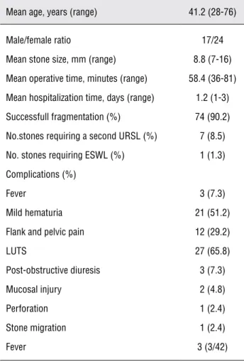
Table 1 - Patient’s characteristics, operative data and complications.
Mean age, years (range) 41.2 (28-76)
Male/female ratio 17/24 Mean stone size, mm (range) 8.8 (7-16) Mean operative time, minutes (range) 58.4 (36-81) Mean hospitalization time, days (range) 1.2 (1-3) Successfull fragmentation (%) 74 (90.2) No.stones requiring a second URSL (%) 7 (8.5) No. stones requiring ESWL (%) 1 (1.3) Complications (%)
was 58.4 minutes, and the mean hospital stay was 1.2 days. Successfull fragmentation (90.2 %) was achieved after single endoscopic procedure. A second URSL was performed in 7 (8.5%) of the stones. Stone forceps were performed to retrieve large stone fragments (≥ 4 mm) in 30 (36.5%) of the procedures. Minor complications such as LUTS, mild hematuria, flank and pelvic pain improved in one week after DJ stent removal. Perforation oc-cured in only one patient due to difficult uretero-scopic manipulation because of bleeding. Mucosal injury occurred in 2 patients, and the reasons for the mucosal injury were inadvertant positioning of an pneumatic probe and stone forceps. These pa-tients were treated with DJ stenting for 3 weeks. Although stone cone was used to prevent migra-tion of calculi, proximal migramigra-tion was observed in 1 patient. The patient was treated successfully with DJ stent insertion and subsequent ESWL after one
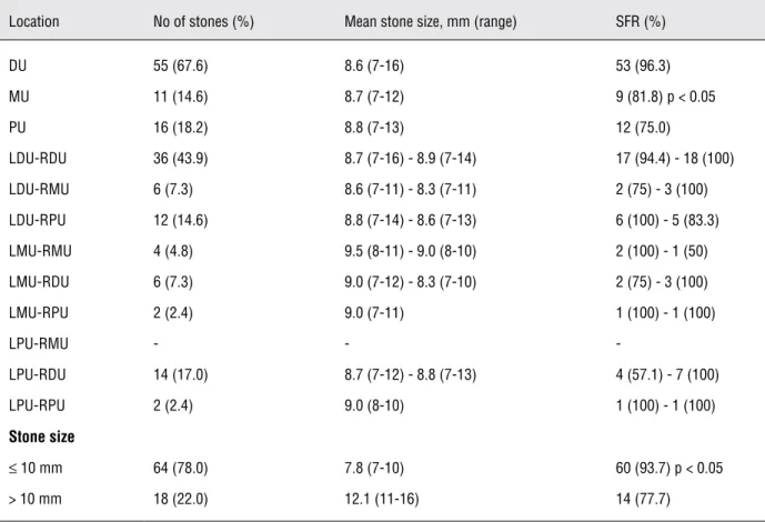
week. Fever (> 38º) was successfuly managed with antibiotic regimen in 3 patients. Post-obstructive diuresis was observed in 3 (7.3%) patients who had high serum creatinine level in a volume range of 6 to 10 liters in the first 24-48 hours, and serum cre-atinine level returned back to normal level within 2 to 3 days. The stone location and size and stone free rate are shown in Table-2. Aproximately, two third of stones were located in the distal ureter. The stone-free rate of distal ureter stone (96.2%) was significantly higher compared with those of middle (81.8%) and proximal (77.7%) ureter stones (p < 0.05). For patients with calculi less than 1 cm and greater than 1 cm, the initial stone-free rate after ureteroscopy was 93.7% and 77.7%, respectively (p < 0.05). Stone analysis results were available in 8 (19.5%) patients: calcium oxalate in 7, calcium phosphate in 2 and uric acid in 1. No long-term complication was observed in any patient.
Table 2 - Stone-free rate after bilateral single-session URSL according to stone location and stone size.
Location No of stones (%) Mean stone size, mm (range) SFR (%)
DU 55 (67.6) 8.6 (7-16) 53 (96.3) MU 11 (14.6) 8.7 (7-12) 9 (81.8) p < 0.05 PU 16 (18.2) 8.8 (7-13) 12 (75.0)
LDU-RDU 36 (43.9) 8.7 (7-16) - 8.9 (7-14) 17 (94.4) - 18 (100) LDU-RMU 6 (7.3) 8.6 (7-11) - 8.3 (7-11) 2 (75) - 3 (100) LDU-RPU 12 (14.6) 8.8 (7-14) - 8.6 (7-13) 6 (100) - 5 (83.3) LMU-RMU 4 (4.8) 9.5 (8-11) - 9.0 (8-10) 2 (100) - 1 (50) LMU-RDU 6 (7.3) 9.0 (7-12) - 8.3 (7-10) 2 (75) - 3 (100) LMU-RPU 2 (2.4) 9.0 (7-11) 1 (100) - 1 (100)
LPU-RMU - -
-LPU-RDU 14 (17.0) 8.7 (7-12) - 8.8 (7-13) 4 (57.1) - 7 (100) LPU-RPU 2 (2.4) 9.0 (8-10) 1 (100) - 1 (100)
Stone size
≤10 mm 64 (78.0) 7.8 (7-10) 60 (93.7) p < 0.05 > 10 mm 18 (22.0) 12.1 (11-16) 14 (77.7)
COMMENTS
Today, URSL is one of the daily urologists’ practices, and regardless of the location of the ureteric stone, access and definitive treatment is commonly achieved with a minimal risk of compli-cations. The main advantages of URSL are imme-diate relief of symptoms and stone fragmentation. Quick ureteral stone removal may be important in patients with bilateral ureteric stones because these patients are more likely to have acute ob-structive renal failure. The classic procedure for the management of bilateral ureteric stones is staged URSL. In recent years, some authors advo-cate single-session bilateral URSL for the manage-ment of bilateral ureteric stones due to successfull rates and minimal morbidity. The procedure may decrease the number of anaesthesia and surgical sessions, and hospital stay (1,2). In contrast, some authors reported that this procedure may also in-crease postoperative morbidity (3).
Single-session bilateral URSL for the management of bilateral ureteric stones has not been well documented. Only a few reports have been reported in the literature about single-session bilateral URSL for the management of bilateral ureteric stones. Deliveliotis et al. inves-tigated the possibility to perform bilateral ure-teroscopy in one session and to determine the procedure’s indications and complication rate. Twenty-two patients underwent bilateral ure-teroscopy in one session. No major complication was observed. They reported that bilateral ure-teroscopy in one session can be performed safely in selected patients (1). In contrast, Hollenbeck et al. reported that bilateral ureteroscopy carries out an increased risk of postoperative morbid-ity. The cumulative risk for staged and single-session bilateral URSL were 14% and 29%, re-spectively. However, there was no difference in cumulative morbidity and stone free rates at 1 month between the two approaches (3).
In a recent study, Günlüsoy et al. evalu-ated the feasibility and safety of bilateral sin-gle-session ureteroscopy in 38 patients for the management of bilateral ureteric stones with different localizations. The stones were locat-ed in the lower, middle and upper ureter in 44
(57.9%), 21 (27.6%) and 11 (14.5%) of the cases, respectively. Fifty-one stones (67.1%) were less than 1 cm. Of the 76 stones, 67 (88.1%) were fragmented in a single procedure. The stone free rate was 93.1% after the second session. Accord-ing to the localization of the stones, the stone clearance rate after single endoscopic session was 72.7% for upper ureteric stones, 80.9% for midureteric stones and 95.4% for lower ureteric stones. For patients with calculi less than 1 cm and greater than 1 cm, the initial stone-free rate after ureteroscopy was 94.1% and 76%, respec-tively. No major complication was observed. They reported that bilateral single-session pneu-matic lithotripsy can be performed safely and has high success rates with minimal morbidity and short hospital stay (2).
In the present study, 41 patients with bilateral ureteral stones were evaluted. A high stone-free rate was achieved (90.2%) after single endoscopic procedure with a retreatment rate of 9.8%. The stones were located in distal ureter (67.6%), in middle ureter (14.6%) and in proxi-mal ureter (18.2%). 78.0% of the stones were less than 1 cm. The procedure was most successful for distal ureteric stones with a 96.2% stone-free rate followed by middle ureteric stones with a 81.8% stone-free rate while the least success was achieved for proximal ureteric stones with a 77.7% stone-free rate. A greater stone-free rate was obtained in those with stones less than 10 mm (93.7%) than in those with stones larger than 10 mm (77.7%). Major complication was observed in only one patient (2.4%) during the procedures, and this patient was managed successfully with DJ stent. The results of this study indicate that the procedure can be performed in all ureteric stones; however, success rate can be affected by stone size and ureteric localization. Similar with the study of Günlüsoy et al. (2), single-session bilateral URSL can be performed effectively and safely with a low complication rate in patients with bilateral ureteric stones.
method for the treatment of ureteral stones espe-cially in proximal and impacted ureteral stones, but it is expensive and not available in most of the urologic centers (4-7). EAU-EBU update series reported that ballistic lithotripsy can be regarded as a standard for stones < 15 mm, because of its better efficacy and shorter operative time, while for stones > 15 mm a laser lithotripsy should be advised because of its minimal risk of ureteral in-jury (4). In the present study, a pneumatic lith-otripter was used for stone fragmentation in all patients, and high success rate and acceptable retreatment rates were achieved. However, pneu-matic lithotripsy has some disadvantages. It pro-duces larger fragments that potentially may cause more problems in terms of spontaneous passage or retropulsion during the procedure (8). There-fore, some authors recommended using forceps or stone cone to reduce re-treatment rate (9-11). Similarly, in this study, stone forceps were used to remove stone fragments ≥ 4 mm, and stone cone were used to reduce stone migration for proximal and middle ureteric stones.
Stents have been placed routinely after URSL to minimize the risk of flank pain and hy-dronephrosis due to ureteric edema, to facilitate the passage of residuel stone fragments and de-crease the risk of ureteric stricture. Recently, AUA and EAU guidelines on urolithiasis reported that stenting after uncomplicated URSL is optional (12). Generally, bilateral DJ stenting is performed on patients who had undergone single-session bi-lateral URSL. However, in this study, bibi-lateral DJ stenting was only performed in the patients who had high serum creatinine levels or bilateral ure-teral mucosal injury. The other indications for bi-lateral DJ stenting are bibi-lateral ureteral perforation and stone migration. Bilateral ureteric endoscopic procedures can cause bilateral ureteric edema, and to obtain normal serum creatinine levels may take longer periods of time. In my opinion, for patients with high serum creatinine level, bilateral DJ stenting are necessary to achieve normal se-rum creatinine level as soon as possible. In other patients, a DJ stent was placed on one-side due to minimize risk of acute obstructive renal failure due to bilateral ureteric edema and flank pain.
CONCLUSIONS
On the basis of my experience, single-ses-sion bilateral ureteroscopy with pneumatic litho-tripsy can be considered an acceptable treatment modality for bilateral ureteric stones. The proce-dure has high success rates with minimal morbid-ity and short hospital stay. It can reduce the need of anaesthetics and overall costs.
CONFLICT OF INTEREST
None declared.
REFERENCES
1. Deliveliotis C, Picramenos D, Alexopoulou K, Christois I, Kostakopoulos A, Dimopoulos C: One-session bilateral ure-teroscopy: is it safe in selected patients? Int Urol Nephrol. 1996; 28: 481-4.
2. Gunlusoy B, Degirmenci T, Arslan M, Kozacio lu Z, Nergiz N, Minareci S, et al.: Bilateral single-session ureteroscopy with pneumatic lithotripsy for bilateral ureter stones: feasible and safe. Urol Int. 2008; 81: 202-5.
3. Hollenbeck BK, Schuster TG, Faerber GJ, Wolf JS Jr.: Safety and eficacy of same-session bilateral ureteroscopy. J En-dourol. 2003; 17: 881-5.
4. Papadoukakis S, Stolzenburg JU, Truss MC: Treatment strategies of ureteral stones. Eur Urol EAU-EBU Update Ser. 2006; 4: 184-90.
5. Gettman MT, Segura JW: Management of ureteric stones: issues and controversies. BJU Int. 2005; 95(Suppl 2): 85-93.
6. Tan PK, Tan SM, Consigliere D: Ureteroscopic lithoclast lithotripsy: a cost-effective option. J Endourol. 1998; 12: 341-4.
7. Küpeli B, Biri H, Isen K, Onaran M, Alkibay T, Karao lan U, et al.: Treatment of ureteral stones: comparison of extracorpo-real shock wave lithotripsy and endourologic alternatives. Eur Urol. 1998; 34: 474-9.
8. El-Nahas AR, El-Tabey NA, Eraky I, Shoma AM, El-Hefnawy AS, El-Assmy AM, et al.: Semirigid ureteroscopy for ure-teral stones: a multivariate analysis of unfavorable results. J Urol. 2009; 181: 1158-62.
10. Isen K, Bogatekin S, Em S, Ergin H, Kilic V.: Is routine ure-teral stenting necessary after uncomplicated ureteroscopic lithotripsy for lower ureteral stones larger than 1 cm? Urol Res. 2008; 36: 115-9.
11. Gonen M, Cenker A, Istanbulluoglu O, Ozkardes H: Eficacy of dretler stone cone in the treatment of ureteral stones with pneumatic lithotripsy. Urol Int. 2006; 76: 159-62.
12. Preminger GM, Tiselius HG, Assimos DG, Alken P, Buck C, Gallucci M, et al.: 2007 guideline for the management of ureteral calculi. J Urol. 2007; 178: 2418-34.