Intrarenal Surgery vs Percutaneous Nephrolithotomy in the
Management of Lower Pole Stones Greater than 2 cm
_______________________________________________
Hakan Koyuncu
1, Faruk Yencilek
1, Mehmet Kalkan
2, Yavuz Bastug
3, Esin Yencilek
4, Ahmet Tunc
Ozdemir
11 Department of Urology, Yeditepe University Medical Faculty; 2 Department of Urology, Fatih University
Medical Faculty; 3 Department of Urology, Beykoz State Hospital, Istanbul, Turkey; 4 Department of Radiology, Haydarpasa Training and Research Hospital, Istanbul, Turkey
ABSTRACT
ARTICLE
INFO
______________________________________________________________ ______________________
Purpose: To compare the efficacy of RIRS and PNL in lower pole stones ≥2 cm. Materials and and Methods: A total of 109 patients who underwent PNL or RIRS for solitary lower pole stone between April 2009 and December 2012, were retrospectively analyzed. Lower pole stone was diagnosed with CT scan. Stone size was assessed as the longest axis of the stone. All patients were informed about the advantages, disad-vantages and probable complications of both PNL and RIRS before the selection of the procedure. Patients decided the surgery type by themselves without being under any influences and written informed consent was obtained from all patients prior to the surgery. Patients were divided into two groups according to the patients’ preference of surgery type. Group 1 consisted of 77 patients who underwent PNL and Group 2 consisted of 32 patients treated with RIRS. Stone free statuses, postoperative complica-tions, operative time and hospitalization time were compared in both groups.
Results: There was no statistical significance between the two groups in mean age, stone size, stone laterality, mean follow-up periods and mean operative times. In PNL group, stone-free rate was 96.1% at first session and 100% after the additional pro-cedure. In Group 2, stone-free rate was 90.6% at the first procedure and 100% after the additional procedure. The final stone-free rates and operative times were similar in both groups.
Conclusions: RIRS should be an effective treatment alternative to PNL in lower pole stones larger than 2 cm, especially in selected patients.
Key words:
Calculi; Nephrostomy, Percutaneous; Surgical Procedures, Operative; Retrograde Obturation
Int Braz J Urol. 2015; 41: 245-51
_____________________
Submitted for publication: April 30, 2014
_____________________
Accepted after revision: May 22, 2014
INTRODUCTION
Kidney stones greater than 2 cm have long been treated with percutaneous nephrolithotomy (PNL) (1, 2). PNL is also recommended as a primary treatment in the management of renal stones ≥2 cm by European Association of Urology (EAU) guidelines (3). Although PNL has stone-free rates higher than 90% regardless of stone size and location, PNL has
RIRS has become popular in the last decade with the technical advancements in endourologic equipments and increased surgeon experience. Today, in the management of renal stones, RIRS provides an alternative way to PNL by minimizing the risks related to PNL. Recent studies reported stone-free rates from 77% to >90% for RIRS of renal stones and 62% to 85% for the management of lower pole stones (2, 6-9). Furthermore, several studies have reported significant success rates with RIRS in the management of large renal stones (10). Recently, studies reporting the efficacy of RIRS in lower pole stones have increased (5). In addition, the complication rates of RIRS are lower and the only disadvantage of this technique is the possible need for repetition. To our knowledge, there is no study comparing the efficacy of RIRS and PNL in lower pole stones greater than 2 cm. In this study, our aim is to compare the efficacy of RIRS and PNL in lower pole stones ≥2 cm.
MATERIALS AND METHODS
A total of 109 patients who underwent PNL or RIRS for solitary lower pole stone between April 2009 and December 2012 were retrospecti-vely analyzed. Data were obtained from the pa-tients’ files which were recorded with electronic data management system. Patient assessment in-cluded detailed medical history, physical exami-nation and laboratory tests including urinalysis, urine culture, complete blood count, and serum biochemistry. Lower pole stone was diagnosed with computed tomography (CT) (including axial, sagittal and transverse sections). Stone size was assessed as the longest axis of the stone on CT scan. All patients were informed with the same diagrams and photos about the advantages, disad-vantages and probable complications of both PNL and RIRS before the selection of the procedure. Patients decided the surgery type by themselves without being under any influences and written informed consent was obtained from all patients prior to the surgery. Patients with the history of previous urinary stone surgery or urinary ano-maly were excluded. Patients were divided into two groups according to the patients’ preference of surgery type. Group 1 consisted of 77 patients
who underwent PNL and Group 2 consisted of 32 patients treated with RIRS. All patients were eva-luated with serum biochemistry and blood count at the day after surgery. In addition, all patients underwent CT for the stone clearance, at the first postoperative month. Treatment success was defi-ned as stone-free status or clinically insignificant residual fragments ≤2 mm. Patients were followed up every 3 months with urinalysis, urine culture and ultrasonography.
Stone-free status, postoperative compli-cations, operative time and hospitalization time were compared in both groups. Chi-square and t--test were used for statistical analysis and statis-tical significance was defined as p value <0.05 at 95% confidence interval.
PNL Technique
All procedures were performed under neral anesthesia. All patients received a third ge-neration cephalosporin at the induction of anes-thesia. A 6F ureteral catheter was placed within the cystoscope and the bladder was drained with a 16F urethral Foley catheter. After ureteral ca-theterization, patients were placed in the prone position, and percutaneous access was achieved under fluoroscopic guidance with the use of an 18-gauge needle and a guide wire. Tract dilation was accomplished by using Amplatz dilators up to 30F. Pneumatic lithotripter was used for fragmen-tation and stone removal was accomplished with retrieval graspers through a rigid 22F nephrosco-pe. The operations were completed when residual fragments were not detected on fluoroscopic ima-ging. After completion, a 16F re-entry catheter was inserted into the kidney and ureteral passage was controlled with antegrade pyelography. The re-entry catheter was removed on postoperative days 1 or 2 after removing the ureteral catheter and performing an antegrade pyelography confir-ming the ureteral passage. Then the patient was discharged on the next day.
RIRS Technique
anesthesia. Under general anesthesia, patients were placed in the lithotomy position on a fluoro--endoscopic table. Rigid ureteroscopy was routi-nely performed before flexible ureteroscopy in all patients for dilatation of the ureter and to place a hydrophilic guidewire into the renal pelvis. Af-ter passing a 0.038-inch safety guidewire into the renal pelvis, a ureteral access sheath (9.5/11.5 or 12/14Fr) was placed to allow for optimal visuali-zation, to maintain low intrarenal pressure, and to facilitate extraction of stone fragments. For the cases in which the 12/14Fr ureteral access sheath could not progress regularly under the fluorosco-pic control, 9.5/11.5Fr sheath was used. The stones were fragmented by a holmium: YAG laser (Lisa; Sphinx 30 W, Katlenburg University, Germany) (272µ caliber fiber) until they were deemed small enough to pass spontaneously. At the beginning of the laser lithotripsy, the laser functioning para-meters were 1.5 Joule/11 Hertz and when the sto-ne sizes decreased to 10 mm the parameters were changed to 10 J/12 H in order to avoid the pneu-matic effect of the laser, which could migrate the stone to other poles. Basket extraction of residual fragments was not routinely performed; however, some residual fragments were removed by tipless nitinol baskets for stone analysis. At the end of the procedure, a double-J stent was placed routi-nely in all patients. JJ stents of the patients were removed at the postoperative first month.
RESULTS
Stone caracteristics and demographic data of the patients in both groups are presented
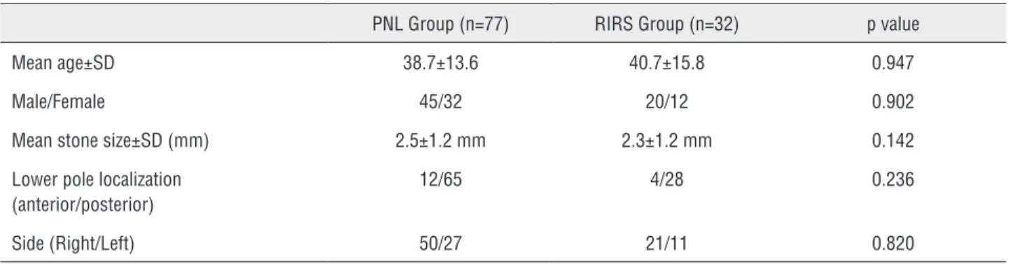
in Table-1. There was no statistical significance between the two groups in mean age of patients (p=0.947), stone size (p=0.142) and stone latera-lity (p=0.820). The mean follow-up period was 13.5±4.71 months (range 3 to 22 months) in Group 1 and 12.5±5.26 months (range 3 to 19 months) in Group 2, respectively. No statistical significance was observed in mean follow-up periods in both groups (p=0.270). Mean operative time in both groups were similar; 62.5±20.67 minutes (range 38 to 107 min) in Group 1 and 67.5±22.34 (range 42 to 110 min) min in group 2 (p=0.671).
In Group 1, all procedures were performed by a single access procedure. Stone-free rate was 96.1% (74/77) at first session. Since the three pa-tients had more than 3 residual fragments, they underwent an additional procedure (ESWL) and stone free rate increased to 100%. Thirty five (45.5%) patients were discharged at the postope-rative 2nd day and 45 (54.5%) in 3rd
day after con-firming the ureteral passage with antegrade pye-lography. Mean hospital stay was 2.4±0.49 days. One patient (0.9%) needed conservative manage-ment because of the persistent fever (Clavien grade I). Four patients (5.1%) needed blood transfusion because of hemorrhage (Clavien grade II) and one of them with significant bleeding (Clavien gra-de III) was treated with open surgical techniques (nephrolithotomy and primary renal parenchymal suturing). In Group 1, mean hemoglobin drop was 1.98±1.26 g/dL (range 0.3 to 8 g/dL). A JJ stent was placed into one patient (having persistent lumbar pain) (0.9%) because of the ureteral obs-truction and removed at the 7th
postoperative day. There was no urinary leakage, no adjacent organ
Table 1 - Stone Characteristics and Demographic Data of Patients.
PNL Group (n=77) RIRS Group (n=32) p value
Mean age±SD 38.7±13.6 40.7±15.8 0.947
Male/Female 45/32 20/12 0.902
Mean stone size±SD (mm) 2.5±1.2 mm 2.3±1.2 mm 0.142
Lower pole localization (anterior/posterior)
12/65 4/28 0.236
injury, no kidney loss or deaths. Chemical compo-sition of stones in Group 1 were calcium oxalate dehydrate (54/77, 70.1%), mixed (calcium oxalate dehydrate and monohydrate) (16/77, 20.7%), uric acid (5/77, 6.4%) and cystine stones (2/77, 2.5%).
In Group 2, stone-free rate was 90.6% (29/32) at the first procedure and 100% after the additional procedure (ureteroscopy). Three pa-tients (3.2%) needed an additional procedure be-cause of more than 3 residual fragments (three re-sidual fragments in two patients and four in one patient, sized approximately 2 mm, in the kidney), at the first month control. In the course of remo-ving the JJ stents of these three patients, flexible ureteroscopy was performed and all residual frag-ments were removed by tipless nitinol basket with no use of access sheath or holmium laser. Three patients (3.2%) with lumbar pain and persistent hematuria (Clavien grade I) were managed con-servatively and discharged at the postoperative 2nd
day. Recent patients (29/32, 90.6%) in RIRS group were discharged at the postoperative 1st
day. In Group 2, mean hemoglobin drop was 0.18±0.18 g/dL (range 0 to 0.8 g/dL) and mean hospital stay was 1.09±0.29 days. No intraoperative complica-tions such as ureteral perforation and no ureteral stricture at follow up period were observed. Stone analysis revealed calcium oxalate dehydrate in 23 patients (71.8%), mixed in 7 (21.8%) and uric acid in 2 (6.2%).
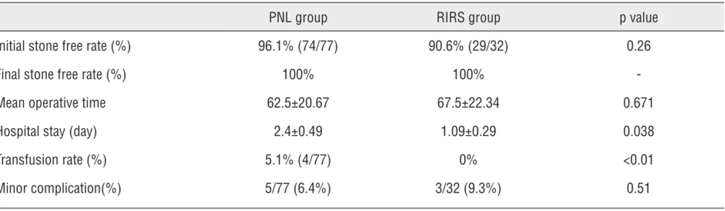
The treatment results of both groups are summarized in Table-2. The final stone-free ra-tes and operative times (p=0.671) were similar in both groups. Hospitalization time (p=0.038) and
hemorrhage (p<0.01) was higher in Group 1, ho-wever minor complications were similar in both groups (p=0.51).
DISCUSSION
Renal stones greater than 2 cm have tradi-tionally been treated with PNL (1, 2). PNL is also recommended as a first line treatment option in the management of renal stones ≥2 cm in EAU and American Urological Association guidelines (3, 11). Several studies concerning about the tre-atment of larger renal stones, have reported stone free rates of PNL up to 95% (4, 12). PNL has also proved to be highly effective in lower pole stones. In a study, the stone-free rate of PNL was reported as 92% and 86 % for lower pole stones 1 to 2 cm and more than 2 cm, respectively (4). In a compa-rative study, PNL was the most effective approach for the management of lower pole stones between 1 to 2 cm, compared with RIRS and shock wave lithotripsy (13). Similar success rate was confir-med in another comparative study with a stone--free rate of 83% in lower pole stones between 1.5 to 2 cm (14). Despite the reported stone-free rates, ranging from 85% to 95%, several complications of PNL constitute a concern. The incidence of pro-bable complications of PNL are reported in sig-nificant rates, including bleeding requiring blood transfusion 11.2% to 17.5%, fever 21% to 32.1%, sepsis 0.25% to 1.5%, pneumothorax 0% to 4% and colonic injury <1%. In consideration of other complications such as arteriovenous fistula, hy-pothermia, volume overload, colo-cutaneous
fis-Table 2 - Treatment Results in Both Groups.
PNL group RIRS group p value
Initial stone free rate (%) 96.1% (74/77) 90.6% (29/32) 0.26
Final stone free rate (%) 100% 100%
-Mean operative time 62.5±20.67 67.5±22.34 0.671
Hospital stay (day) 2.4±0.49 1.09±0.29 0.038
Transfusion rate (%) 5.1% (4/77) 0% <0.01
tula, electrolyte imbalance, pulmonary embolism and death, complication rate of PNL ranges from 0.03% to 10% in general (2, 10, 15). Additionally, in patients with significant comorbidities such as morbid obesity and bleeding diathesis, PNL is contraindicated due to the higher incidence of complications (11, 16). Finally, placement of the patient in a prone position increases the anesthe-tic risk because of the contractions of extremities and difficult airway.
Today, RIRS is an excellent minimally in-vasive treatment alternative for intrarenal stones smaller than 2 cm and reported stones-free rates are higher at this stone size (8, 17, 18). Increased experiences of the urologists and developments in the technology have created the substructure of this success. Development of new generation (bidirectional 270
º
flexion capacity, small calibershaft and improved optics) flexible ureteroscopes, improved flexibility of holmium laser fibers, di-fferent and small diameter stone retrieval devi-ces with the capability of facilitating intrarenal maneuvers have resulted in increased treatment success and decreased procedure related morbidi-ty, in the management of renal stones (19-21). In addition, ureteral access sheaths provided lower intrarenal pressure during prolonged procedu-res and facilitated the retrieval of multiple stone fragments (22, 23). All these innovations and es-pecially increased experience in RIRS aroused the urologists
’
interest to the success of this procedurein larger and lower calyceal renal stones.
Several studies reported their success rates of RIRS in the management of large renal stones. Grasso et al. reported an overall stone free rate of 91% for 66 renal stones >2 cm in 55 renal units. They reported that one third of patients have re-quired second procedure (8). Breda et al. achieved a 93.3% success rate after an average of 2.3 pro-cedures, in 15 patients with a single renal stone si-zed between 20 and 25 mm (24). In another study, authors showed an 87.5% stone free rate for renal stones between 2 and 3 cm with 43% of patients requiring second procedure (25). In a study inclu-ding 22 patients with renal stones larger than 2.5 cm, authors reported a 91.6% stone free rate with an average 1.9 procedures (18). Similarly, the suc-cess rate of RIRS was evaluated in a study
inclu-ding 90 patients with different sized (<10mm ≥20 mm) lower pole stones. They concluded an 82% final stone free rate for lower pole stones >2 cm, after a second procedure (9). Accordingly, recent studies report up to 85% stone free rates of RIRS for the management of lower pole stones (8, 17). With these similar results, all of these studies have showed that RIRS should be an efficient treatment modality for larger renal stones as PNL which is more invasive. Nevertheless, to our knowledge, there is no study comparing the success rates of RIRS and PNL in lower pole stones >2 cm. In the management of lower pole stones greater than 2 cm, we have demonstrated a final 100% stone-free rate of RIRS with similar stone free rates of PNL. We suggest that this higher success rate in RIRS group may be related with the increased experien-ce and the predominanexperien-ce of posterior localized lo-wer pole stones in the kidney.
Furthermore, the association of longer operative time and endoscopic management of large renal stones were emphasized in the lite-rature. However, recent reports demonstrated a rational operative time for ureteroscopy. Maria-ni et al. reported a mean operative time of 64 minutes (range 30 to 240 min) for the RIRS of renal stones between 2 and 4 cm (26). We also reported similar mean operative times in both groups, RIRS and PNL.
On the other hand, several limitations of our study must be addressed: 1. the number of pa-tients included is rather low (especially in Group 2) therefore, further multicentric series with lar-ger and equal number of study population have to be performed; 2. This study was a retrospective analysis. We suggest that a prospective study will exactly clarify the efficacy of RIRS in large lower pole stones.
CONCLUSIONS
RIRS can be an effective treatment alter-native to PNL in lower pole stones larger than 2 cm, especially in selected patients. Further, mul-ticentric comparative studies with larger study population are needed to confirm these results.
CONFLICT OF INTEREST
None declared.
REFERENCES
1. Segura J, Paterson D, LeRoy A, Williams HJ, Barret DM, Benson RC et al. Percutaneous removal of kidney stones: review of 1000 cases. J Urol 1985; 134: 1077-81.
2. Michel MS, Trojan L, Rasweiler JJ. Complications in percutaneous nephrolithotomy. Eur Urol 2007; 51: 899-906. 3. Türk C, Knoll T, Petrik A, Petrik A, Sarica K, Skolarikos A
et al. Members of the European Association of Urology (EAU) Guidelines Office. Guidelines on Urolithiasis. In: EAU Guidelines, edition presented at 28th Annual EAU Congress, Milano 2013, pp: 41-51.
4. Albala DM, Assimos DG, Clayman RV, Denstedt JD, Grasso M, Gutierrez-Aceves J et al. Lower pole I: A prospective randomized trial of extracorporeal shock wave lithotripsy and percutaneous nephrostolithotomy for lower pole nephrolithiasis-initial results. J Urol 2001; 166: 2072-80. 5. Lingeman JE, Siegel YI, Steele B, Nyhuis AW, Woods JR.
Management of lower pole nephrolithiasis: a critical analysis. J Urol 1994; 151: 663-7.
6. Unsal A, Resorlu B, Atmaca AF, Diri A, Goktug HN, Can CE et al. Prediction of morbidity and mortality after percutaneous nephrolithotomy by using the charlson comorbidity index. Urology 2012; 79: 55-60.
7. Deem S, Defade B, Modak A, Emmett M, Martinez F, Davalos J. Percutaneous nephrolithotomy versus extracorporeal shock wave lithotripsy for moderate sized kidney stones. Urology. 2011; 78: 439-43.
8. Grasso M, Conlin M, Bagley D. Retrograde ureteropyeloscopic treatment of 2 cm or greater upper urinary tract and minor staghorn calculi. J Urol 1998; 160: 346-51.
9. Grasso M, Ficazzola M. Retrograde ureteropyeloscopy for lower pole caliceal calculi. J Urol 1999; 162: 1904-8. 10. Gupta M, Oct Mc, Shah JB. Percutaneous management
of the upper urinary tract. Campbell-Walsh Urology, 9th ed. Philaselphia, PA: Saunders Elsevier, 2007; pp. 1544-8. 11. Preminger G, Assimos D, Lingeman J. AUA guideline on
management of staghorn calculi: diagnosis and treatment recommendations. J Urol 2005; 173: 1991-2000.
12. Segura JW, Preminger GM, Assimos DG, Dretler SP, Kahn RI, Lİngeman JE et al. Nephrolithiasis Clinical Guidelines Panel summary report on the management of staghorn calculi. The American Urological Association Nephrolithiasis Clinical Guidelines Panel. J Urol 1994; 151: 1648-51. 13. Ozturk U, Sener NC, Goktug G, Nalbant I, Gucuk
A, Imamoglu MA. Comparison of Percutaneous Nephrolithotomy, Shock Wave Lithotripsy, and Retrograde Intrarenal Surgery for Lower Pole Renal Calculi 10–20 mm. Urol Int 2014; 91: 345-9.
14. Haroon N, Nazım SM, Alter MH. Optimal Management of Lower Polar Calyceal Stone 15 to 20 mm. Korean J Urol 2013; 54: 258-62.
15. Unsal A, Resorlu B, Kara C, Bozkurt OF, Ozyuvali E. Safety and efficacy of percutaneous nephrolithotomy in infants, preschool age, and older children with different sizes of instruments. Urology 2010; 76: 247-52.
16. Pearle MS, Nakada SY, Womack JS, Kryger JV. Outcomes of contemporary percutaneous nephrostolithotomy in morbidly obese patients. J Urol 1998; 160: 669-73. 17. Mariani AJ. Combined electrohydraulic and holmium: YAG
laser ureteroscopic nephrolithotripsy of large (greater than 4 cm) renal calculi. J Urol 2007; 177: 168-73. 18. El-Anany FG, Hammouda HM, Maghraby HA. Retrograde
ureteropyeloscopic holmium: YAG laser lithotripsy for large renal calculi. BJU Int 2001; 88: 850-3.
19. Riley JM, Stearman L, Troxel S. Retrograde ureteroscopy for renal stones larger than 2.5 cm. J Endourol 2009; 23: 1395-8. 20. Bozkurt OF, Resorlu B, Yildiz Y, Can CE, Unsal A. Retrograde
intrarenal surgery versus percutaneous nephrolithotomy in the management of lower pole renal stones with a diameter 15 to 20 mm. J Endourol 2011; 25: 1131-5.
21. Johnson GB, Portela D, Grasso M. Advanced ureteroscopy: Wireless and sheathless. J Endourol 2006; 20: 552-5. 22. Kourambas J, Byrne RR, Preminger GM. Does a ureteral access
sheath facilitate ureteroscopy? J Urol 2001; 165: 789-93. 23. L’Esperance JO, Ekeruo WO, Scales CD, Marquet CG,
24. Breda A, Ogunyemi O, Leppert JT, Lam JS, Schulam PG. Flexible ureteroscopy and laser lithotripsy for intrarenal Stones 2 cm or greater. Is this the new frontier? J Urol 2008; 179: 981-4.
25. Ricchiuti DJ, Smaldone MC, Jacobs BL, Smaldone AM, Jackman SV, Averch TD. Staged retrograde endoscopic lithotripsy as alternative to PCNL in select patients with large renal calculi. J Endourol 2007; 21: 1421-4.
26. Mariani AJ: Combined electrohydraulic and holmium:YAG laser ureteroscopic nephrolithotripsy for 20 to 40 mm renal calculi. J Urol 2004; 172: 170-4.
27. Miller NL and Lingeman JE: Management of kidney stones. BMJ 2007; 334: 468-72.
28. Pevzner M, Stisser BC, Luskin J, Yeamans JC, Chend-Lucey M, Pahira JJ. Alternative management of complex renal stones. Int urol Nephrol 2011; 43: 631-8.
29. Harmon WJ, Sershon PD, Blute ML, Patterson DE, Segura JW. Ureteroscopy: current practice and long-term complications. J Urol 1997; 157: 28-32.
_______________________ Correspondence address: