ORIGINAL ARTICLE
842
New head-mounted display system applied to endoscopic
management of upper urinary tract carcinomas
_______________________________________________
Junichiro Ishioka
1, Kazunori Kihara
1, Saori Higuchi
1, Takayuki Nakayama
1, Hideki Takeshita
1, Soichiro
Yoshida
1, Yasukazu Nakanishi
1, Toshiki Kijima
1, Yoh Matsuoka
1, Noboru Numao
1, Kazutaka Saito
1,
Yasuhisa Fujii
11Department of Urology, Tokyo Medical and Dental University Graduate School, 1-5-45, Yushima,
Bunkyo-ku, Tokyo, Japan
ABSTRACT
ARTICLE
INFO
______________________________________________________________ ______________________
Purpose: We tested a new head-mounted display (HMD) system for surgery on the upper urinary tract.
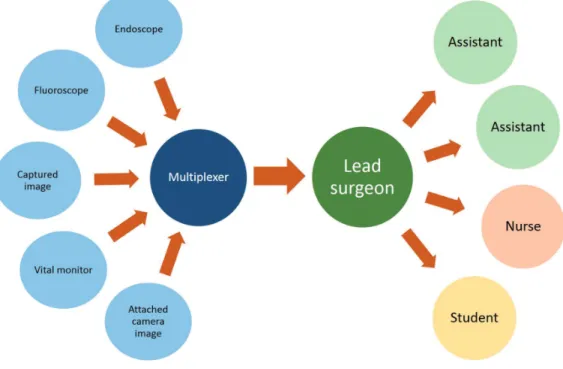
Surgical Technique: Four women and one man with abnormal findings in the renal pelvis on computed tomography and magnetic resonance imaging underwent surgery using this new system. A high definition HMD (Sony, Tokyo, Japan) is connected to a flexible ureteroscope (Olympus, Tokyo, Japan) and the images from the ureterosco-pe are delivered simultaneously to various participants wearing HMDs. Furthermore, various information in addition to that available through the endoscope, such as the narrow band image, the fluoroscope, input from a video camera mounted on the lead surgeon’s HMD and the vital monitors can be viewed on each HMD.
Results: Median operative duration and anesthesia time were 53 and 111 minutes, res-pectively. The ureteroscopic procedures were successfully performed in all cases. There were no notable negative outcomes or incidents (Clavien-Dindo grade ≥1).
Conclusion: The HMD system offers simultaneous, high-quality magnified imagery in front of the eyes, regardless of head position, to those participating in the endoscopic procedures. This affordable display system also provides various forms of information re-lated to examinations and operations while allowing direct vision and navigated vision.
Key words:
Urinary Tract; Ureteroscopy; Natural Orifice Endoscopic Surgery; Video-Assisted Surgery
Int Braz J Urol. 2014; 40: 842-5
_____________________
Submitted for publication: January 21, 2014
_____________________
Accepted after revision: April 06, 2014
INTRODUCTION
With the development of excellent smal-ler and flexible ureteroscopes, the ureteroscopic management of the upper urinary tract (UUT) and upper urinary tract urothelial carcinomas (UUTUC) has become more practical, and in-dications for such procedures have expanded from those patients with solitary kidney or re-nal insufficiency to patients with normal con-tralateral kidney (1). In order to make
ureteros-copy more feasible and effective, we applied a novel head-mounted display (HMD) system that displays simultaneous, high-quality magnified imagery in front of the eyes, regardless of head position, to those participating in endoscopic procedures. This affordable display system also provides various forms of information related to examinations and operations while allows direct vision and navigated vision. In this stu-dy, we describe an application of the system in UUT examinations.
IBJU| NEW HEAD-MOUNTED DISPLAY SYSTEM APPLIED TO URETEROSCOPY
843
SURGICAL TECHNIQUE
Four women and one man with abnormal findings in the renal pelvis on computed tomogra-phy (CT) and magnetic resonance imaging (MRI) underwent surgery using this new system. Each patient had irregular images in the renal pelvis on CT and low T2-weighted signals and diffuse high-intensity signals on diffusion-weighted MRI imaging, leading to suspicion of urothelial carci-noma of the renal pelvis. For further evaluation, ureteroscopy and, if possible, biopsy of the abnor-mal urothelium tissue with reference to the nar-row band image (NBI) was performed. The patients gave written informed consent to participate in a clinical trial to the institutional investigational re-view board of our institution.
The system presented here is used as follo-ws (Figure-1). A high definition HMD (Sony, Tokyo, Japan) is connected to a flexible urete-roscope (Olympus, Tokyo, Japan) and the images from the ureteroscope are delivered simultaneous-ly to various participants wearing HMDs. This mo-nitor is fitted with 0.7-inch (18.0mm diagonal)
Or-ganic Light-Emitting Diode panels with displayed pixel count of 1280x720. The device is already commercially available in Japan and Europe. Its purchase costs are €12300. Furthermore, the de-vices have four different input-output terminals, including Digital Video Interface and Serial Digi-tal Interface. Various informations in addition to that available through the endoscope, such as the NBI, the fluoroscope, input from a video camera mounted on the HMD and the vital monitors can be viewed on each head-mounted display (Figure--2A). Technical support was provided by the Sony Corporation. Two urologists (one lead surgeon and one assistant) performed the operation. Both the lead surgeon and the assistant each wore an HMD throughout the procedure. The imaging informa-tion obtained from the ureteroscope, captured narrow band image (NBI), images from the video camera attached to the HMD of the lead surgeon, and the patient’s vital signs monitor are split using an imaging splitter (400-VGA003, Sanwa Supply Incorporated, Okayama, Japan) and the composite image is outputted into two multiplexers (VPM--H1, MEDIAEDGE Corporation, Hyogo, Japan).
IBJU| NEW HEAD-MOUNTED DISPLAY SYSTEM APPLIED TO URETEROSCOPY
844
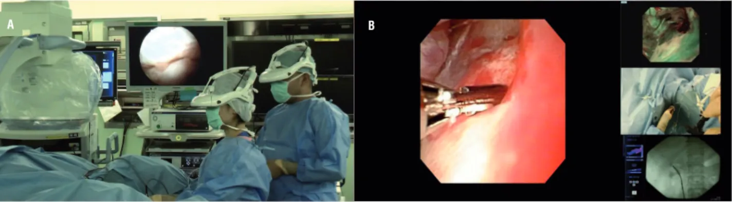
The images are integrated using a four-split screen technique on the multiplexer and are displayed on the HMDs (Figure-2B). The lead surgeon usually opts to arrange the ureteroscopic view as the main image displayed by the HMD during the observa-tion and biopsy, and the assistant may choose the fluoroscopic view as the main image.Median operative duration and anesthesia time were 53 and 111 minutes, respectively. The ureteroscopic procedure was successfully perfor-med in all cases. Tumor biopsy was perforperfor-med with a 3F cup biopsy forceps for flat or sessile lesions. There were no notable negative outcomes or incidents in the postoperative courses (Clavien--Dindo grade ≥1) of any patients (Table-1). During the procedures, neither the lead surgeon nor the
assistant surgeon experienced any HMD-related adverse effects no reported any discomfort. Of all the patients, 3 patients were diagnosed as having urothelial carcinoma and subsequently treated with radical nephroureterectomy.
COMMENTS
This is the first study to use the HMD sys-tem in ureteroscopy and we safely completed the procedures in a reasonable time by using the fea-tures of HMD system. There were no intra-opera-tive or postoperaintra-opera-tive complications.
Traditionally, nephroureterectomy has been the treatment of choice for UUTUC. As po-pulation of the elderly increases, the number of
Figure 2 - Photograph of ureteroscopy being performed using the head-mounted display (HMD) system. Both the lead surgeon and the assistant wear an HMD during all stages of the procedure (A). Captured images of the integrated image data include an ureteroscopic image, a fluoroscopic image, the patient’s vital signs, and the view from a camera attached to the HMD worn by the lead surgeon (B).
Table 1 – Patients’ clinical data and outcomes.
Age Gender Lesion ASA score (min.)
Operative duration (min.)
Duration of anesthesia
Clavien-Dindo grade ≥ 1
Pathological diagnosis
Subsequent treatment
62 Female Right
RP
2 73 139 none UC, G2 NU
45 Female Left RP 1 19 43 none no malignancy observation
69 Female Right
RP
1 62 136 none no malignancy observation
79 Female Right
RP
1 50 120 none UC, G2 NU
76 Male Right
RP
2 63 117 none UC, G1 NU
RP = Renal Pelvis; ASA = American Society of Anesthesiologists; UC = urothelial carcinoma; NU = nephroureterectomy
IBJU| NEW HEAD-MOUNTED DISPLAY SYSTEM APPLIED TO URETEROSCOPY
845
patients with a decreased estimated glomerular filtration rate (ill compromised contralateral kid-ney, solitary kidney) increase, and the importan-ce of endoscopic management is also increasing (1). Furthermore, the indications for endoscopic management have expanded to include those pa-tients without significant renal parenchymal dise-ase or comorbidities, and specifically those with a normal contralateral kidney as well as imperative cases (2).Although retrograde endoscopic procedu-res have become more practical and efficacious with the development of new endoscopic tools, ureteroscopy is still troublesome because the pro-cedure usually requires a variety of information from different screens. To offer better endoscopic management of UUT, we have tested a new HMD system, which we had already applied to mini-mally invasive endoscopic surgery (3). The HMD system has five visual functions: magnified vision, panoramic vision, multiple vision, shared vision, and navigated vision. The magnified view of the endoscopic image can be displayed in front of the user’s eyes. We have not experienced serious mal-function events during the procedure because the HMD also allows direct vision without the need to remove the headset. When the user looks do-wnward, direct unimpeded vision is possible. Mul-tiple and shared vision can be provided by using a signal changer. The HMD can display multiple informations from several imaging sources, and this can also be delivered to many participants simultaneously (4). Navigation with composite images, such as fluoroscopic images and NBI ima-ges, make it easier for surgeons to perform various procedures.
The present study has several limitations, including the small sample, the lack of control group and the lack of cost analysis. There is still a lot to be done in order to demonstrate the actual advantages in terms of oncological outcomes and cost-effectiveness. We think that much larger co-horts and longer follow-up would be needed.
In conclusion, the HMD system can be safely applied to ureteroscopy. The HMD system offers simultaneous, high-quality magnified ima-gery in front of the eyes, regardless of head posi-tion, to those participating in endoscopic procedu-res. This affordable display system also provides various forms of information related to examina-tions and operaexamina-tions while allowing direct vision and navigated vision.
CONFLICT OF INTEREST
Dr. Kihara has received research funding from Sony Corporation (Tokyo, Japan), but the sponsor had no control over the interpretation, writing, or publication of this work.
REFERENCES
1. Cutress ML, Stewart GD, Zakikhani P, Phipps S, Thomas BG, Tolley DA. Ureteroscopic and percutaneous management of upper tract urothelial carcinoma (UTUC): systematic review. BJU Int. 2012; 110: 614-28.
2. Coresh J, Selvin E, Stevens LA, Manzi J, Kusek JW, Eggers P, et al. Prevalence of chronic kidney disease in the United States. JAMA. 2007; 298: 2038-47.
3. Kihara K, Fujii Y, Masuda H, Saito K, Koga F, Matsuoka Y, et al. New three-dimensional head-mounted display system, TMDU-S-3D system, for minimally invasive surgery application: procedures for gasless single-port radical nephrectomy. Int J Urol. 2012; 19: 886-9; author reply 890. 4. Yoshida S, Kihara K, Takeshita H, Nakanishi Y, Kijima
T, Ishioka J, et al. Head-Mounted Display for Personal Integrated-Image Monitoring System: Ureteral Stent Placement. Urol Int. 2014. [Epub ahead of print]
_______________________ Correspondence address: