Endovascular treatment of aortic aneurysms in patients with
Behcet’s disease: report of two cases
Tratamento endovascular de aneurismas da aorta em pacientes com doença de Behçet: relato
de dois casos
Sergio Quilici Belczak1, Ricardo Aun2, Luisa Valentim3, Igor Rafael Sincos1, Luciano Dias Nascimento4, Pedro Puech-Leão5
Introduction
Behcet’s disease, a systemic vasculitis of unknown etio-logy, is characterized by recurrent oral and genital ulcers, ocular manifestations and skin lesions.1 Cardiovascular
manifestations may occur in around 7-38% of patients;1
it is, thus, the most common cause of death among these patients.2
Peripheral vascular lesion is rare, and arterial lesions are less common than venous disease; its prevalence is around 1.5-3% in the whole world.2 In venous involvement,
thrombosis is the most frequent event, occurring even in adequate anticoagulation.3 Aneurysm is more common
than occlusion in Behcet’s disease arterial involvement, and
the most common site for aneurysm formation is the abdo-minal aorta, followed by the femoral artery and the pulmo-nary arteries.4
Among all the vascular lesions that may occur in the-se patients, arterial aneurysms reprethe-sent the most diicult and challenging pathology for the vascular surgeon due to its technical diiculties and high recurrence.5 Due to its
ra-pid expansion, with a high rate of rupture, Behcet’s disea-se aneurysm should be readily treated. he most suitable treatment for aortic aneurysms in Behcet’s disease patients is still a controversial issue. Endovascular treatment is an important alternative for high-risk patients, once morbidity and mortality rates ater conventional surgery remain high.6
Abstract
Behcet’s disease, a systemic vasculitis of unknown etiology, may be the cause of aortic aneurysmal diseases in some patients. We report our experience with two Behcet’s disease patients who presented with aortic aneurysms and were submitted to endovascular therapy, and describe their respective follow-ups. Current pathophysiology, diagnosis, and treatment approaches were reviewed. Our experience suggests that the endovascular approach, combined with adequate immunosuppressive treatment, is an excellent therapeutic option for some patients with Behcet’s disease sufering from aneurysms.
Keywords: Behcet’s disease, aortic aneurysm, endovascular treatment.
Resumo
A doença de Behçet, uma vasculite sistêmica de causa desconhecida, pode ser causa de doença aneurismática da aorta em alguns portadores dessa patologia. Nós apresentamos nossa experiência com dois casos de aneurismas aórticos em pacientes com doença de Behçet submetidos à terapêutica endovascular, descrevendo seus respectivos seguimentos. A terapêutica atual, a patoisiologia e os critérios diagnósticos vigentes foram revisados. Concluímos que a técnica endovascular é uma excelente opção terapêutica para certos pacientes com doença de Behçet e que esta deve ser acompanhada de tratamento imunossupressivo adequado.
Palavras-chave: Doença de Behçet, aneurisma aórtico, terapia endovascular.
1 Residentes, Serviço de Cirurgia Vascular, Hospital das Clínicas (HC), Universidade de São Paulo (USP), São Paulo, SP, Brazil. 2 Livre-docente, Serviço de Cirurgia Vascular, HC, USP, São Paulo, SP, Brazil.
3 Residente, Serviço de Cirurgia Geral, HC, USP, São Paulo, SP, Brazil. 4 Médico preceptor, Serviço de Cirurgia Vascular, HC, USP, São Paulo, SP, Brazil. 5 Professor titular, Serviço de Cirurgia Vascular, HC, USP, São Paulo, SP, Brazil.
Two cases of aortic aneurysms in Behcet’s disease pa-tients, corrected with endovascular technique, are presen-ted, reporting their respective mid-term follow-ups, and current physiopathology, diagnostic criteria and treatment are reviewed.
Case 1
A 28-year-old Caucasian woman diagnosed with Behcet’s disease at 23 years of age, due to recurrent oral and genital ulcers, pseudofolliculitis and erythema nodosum, presented reporting pain in the lower posterior part of the abdomen for 6 months. She had no ophthalmologic abnor-malities or positive family history of the disease. At physical exam a pulsatile abdominal mass was found.
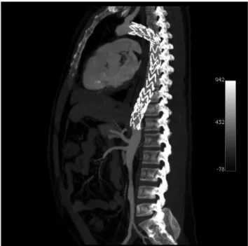
Initial ultrasonography revealed a saccular aneu-rysm of the abdominal aorta above the celiac trunk. Angiotomography conirmed a 5.7 x 4.7-cm saccular aortic aneurysm above the celiac trunk, in addition to occlusion of this ramus (Figures 1 and 2). Superior mesenteric artery (SMA) was patent with stenosis in its origin (Figure 3).
Distal abdominal aorta and iliac arteries had their low and diameter preserved.
Ater 1 month of optimized corticotherapic treatment (prednisone 30 mg/day) and normalized systemic inlam-matory signs, surgery was indicated. Endovascular tre-atment of aortic aneurysm was performed with a Zenith®
(Cook, USA), 22 x 115 mm endoprosthesis implant, as-sociated with SMA angioplasty with a Genius® (Eurocor,
Germany), 6 x 39 mm, stent grat. A proximal extension was necessary, due to the presence of type 1 endoleak (Zenith®,
24 x 115 mm). Intraoperative arteriography at the second procedure evidenced no leaks and unobstructed artery.
he patient had a favorable evolution during postope-rative. A control angiotomography was performed, which did not evidence any abnormalities. She was discharged on the 8th postoperative day, maintaining immunosuppressive treatment.
On the sixth postoperative month, an angiotomography was performed to conirm aneurysm exclusion (Figure 4) and SMA stent patency (Figure 5). Ater a 1-year follow up, the patient remains asymptomatic and has no complaints.
Case 2
A 33-year-old man, with a history of visual loss re-sulting from successive posterior and anterior uveitides,
Figure 1 - Angiotomography of the aorta: 5.7-cm diameter saccular
an-eurysm of abdominal aorta above the celiac trunk
Figure 2 - Angiotomographic reconstruction: stenosis in the origin of
superior mesenteric artery
Figure 3 - Tridimensional reconstruction: thoracabdominal aorta,
presented with progressive deterioration of the renal func-tion. A systemic vasculitis was suspected and the patient underwent arteriograpy, which demonstrated a descending thoracic aortic aneurysm with 5-cm-diameter (Figures 6, 7 and 8). he Department of Rheumatology of the FMUSP deined the diagnosis as Behcet’s disease.
While hospitalized, the patient complained about pain in the chest and non-speciic thoracic pain. Physical exam
Figure 4 - Tridimensional angiotomography: well-placed
endoprosthe-ses, absence of leaks
Figure 5 - Axial cut: patent stent graft of the superior mesenteric artery
Figure 6 - Aortic arch: aneurysm below subclavian artery
Figure 7 - Aortography: 5-cm aneurysm in descending thoracic aorta
Figure 9 - Angiotomography after 18 months: absence of leaks
Figure 10 - Angiotomographic reconstruction: patent subclavian artery
and celiac trunk
Figure 11 - Tridimensional reconstruction: overlapping well-placed
en-doprostheses indicated bilaterally decreased femoral and distal pulses and
normal pulses in the upper limbs. here was no signiicant diference in blood pressure between the arms. Emergency intervention was chosen due to acute symptoms.
An endovascular treatment of the thoracic aortic aneu-rysm was performed with the implant of a Talent® (Medtronic,
USA), 22 x 130 mm endoprosthesis, and a Talent®, 24 x 130
mm distal extension, maintaining the let subclavian artery’s patency. At control arteriography, a type 1 distal leak was found, which was corrected with the implant of a third Talent®, 24 x 130 mm prosthesis, 3 cm above the celiac trunk.
Postoperative had no intercurrences. Follow-up angio-tomography was performed on the seventh postoperative day, revealing neither leaks nor patency of the prosthesis (Figures 8, 9, 10 e 11).
he patient was discharged on the eighth postope-rative day with prednisone 30 mg/day and monthly pul-se therapy with cyclophosphamide for dipul-seapul-se remission. Angiotomography performed at 18 month’s follow-up sho-wed absence of leaks, endoprosthesis patency and absence of recurrent aneurysm signs.
Discussion
Behcet’s disease was irst described in 1937 by Turkish dermatologist Hulusi Behcet as a systemic inlammatory disease which classically causes oral and genital ulcers, in addition to ocular inlammation.7 It is more common in
men. In Japan it is the second most common cause of ac-quired blindness.8 And it may also afect the vascular
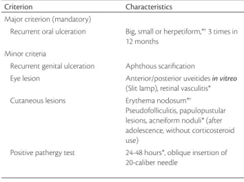
Criterion Characteristics
Major criterion (mandatory)
Recurrent oral ulceration Big, small or herpetiform,*† 3 times in
12 months Minor criteria
Recurrent genital ulceration Aphthous scariication
Eye lesion Anterior/posterior uveitides in vitreo
(Slit lamp), retinal vasculitis* Cutaneous lesions Erythema nodosum*†
Pseudofolliculitis, papulopustular lesions, acneiform noduli* (after adolescence, without corticosteroid use)
Positive pathergy test 24-48 hours*, oblique insertion of 20-caliber needle
* Observed by the physician.
† Reliable report by the patient.
Table 1 - International criteria for Behcet’s disease pathognomonic laboratorial tests or speciic histological
inding for Behcet’s disease. herefore, the diagnosis is ba-sed on clinical indings, according to criteria established by the International Study Group for Behcet’s Disease (Table 1).
he spectrum of the vascular disease is broad and sin-gular in Behcet’s disease. he frequency of vascular involve-ment is estimated in 7-38% of cases.1 Vascular lesions may
be arterial or venous. Usually the most afected artery is the aorta, followed by femoral and pulmonary arteries; 65% of these involvements are aneurysms and 35%, occlusions.9,10
Aneurysms in Behcet’s disease are in many aspects di-ferent from degenerative aneurysms. Incidence is higher among younger persons; the suprarenal type is the most common one and the aneurysm’s format is, as described in case 1, usually saccular. hey are oten multiple, presen-ting urgent symptoms.11-13 he commonest complication of
aneurysm is rupture, which is the most frequent cause of death related to vascular complication.
Pathogenesis of arterial lesions is considered to be rela-ted to arterial vessels and wall vasculitis. hen, the forma-tion of a pseudoaneurysm caused by the obliteraforma-tion of the vessels of the vasa vasorum due to the inlammatory pro-cess follows, which results in the rupture of the nutrition low and in necrosis of the aortic wall.14
Immunohistochemical studies have conirmed an ac-cumulation of complement and immunoglobulin in the medial and adventitial layer. here is a thickening of the intima, where the internal and the external elastic lamina, medial and adventitial are ruptured. he destruction of the medial layer seems to be responsible for the development of aneurysm dilation. Lesions of the aortic wall are frequently located and observed in the saccular type.13 Reports
indi-cate that these aneurysms are usually ruptured, with no cor-relation with the size of the dilation.15
External manifestations, such as active uveitis, aphthas and genital ulcers, and also internal manifestations, such as cardiovascular complications, are strongly related to vascu-lar disease exacerbation. If really necessary, surgeries should be performed when the disease is totally controlled with corticoids and immunosuppressants, because any interven-tion may lead to posterior complicainterven-tions, such as new oc-clusions, aneurysms or pseudoaneurysms.16 In emergency
cases, as a rupture, the remission state of the patient should be reevaluated. It has been shown that, before intervention, the attack dose of corticoids/immunosupressants in bolus (e.g., prednisolone) may decrease perioperative and posto-perative vascular complications.17 However, the role of
im-munosuppressant and antiinlammatory medications in the
prevention of recurrent arterial lesion ater intervention in Behcet’s disease also needs posterior evaluations. In both cases, pharmacological control of the disease’s activities in-volves the use of corticosteroids and immunosupressants.
Open surgical repair was previously the deinitive tre-atment for vascular lesions in Behcet’s disease patients, but surgery’s poor result in vascular/Behcet’s disease is usually attributed to postoperative vascular complications, such as pseudoaneurysm and grat occlusion.18,19 Aiming at
pre-venting surgical complications, endovascular methods, e.g. endoprostheses, have recently been recommended due to being less invasive.20
Recent studies reveal that patients receiving endovas-cular treatment present lower operative time, hospital stay and blood loss than those who undergo open surgical re-pair, with a lower mortality rate (0.6-3.5%). Success rate in prostheses release is high (90% in low-risk patients and 80% in moderate-to-high-risk patients), although some limitations still exist, e.g. the size of the delivery systems, leaks and the position of main trunks.21 here is a clear
possibility of relapse in Behcet’s disease ater endovascular treatment, in which interrupted immunosuppressive the-rapy is a risk factor.22 However, Park et al.9 indicate that
relapsing may be controlled through the repeated use of endoprostheses.
References
1. Sakane T, Takeno M, Suzuki N, Inaba G. Behcet’s disease. N Engl J Med. 1999;341:1284-91.
2. Park JH, Chung JW, Joh JH, et al. Aortic and arterial aneurysms in Behcet disease: management with stent-grafts – initial experience. Radiology. 2001;220:745-50.
3. Silva Júnior OF, Araújo RHS, Freire EAM, et al. Doença de Behçet cursando com trombose de veia cava superior. J Vasc Bras. 2006;5:74-7.
4. Gouëic Y, Pistorius M, Heymann MF, Chaillou P, Patra P. Association of aneurysmal and occlusive lesions in Behçet’s dise-ase. Ann Vasc Surg. 2005;19:276-9.
5. Barlas S. Behçet’s disease. An insight from a vascular surgeon’s point of view. Acta Chir Belg. 1999;99:274-81.
6. Nitecki SS, Ofer A, Karram T, Schwartz H, Engel A, Hofman A. Abdominal aortic aneurysm in Behçet’s disease: new treatment options for an old and challenging problem. Isr Med Assoc J. 2004;6:152-5.
7. Behcet H. Üeber rezidivierende Aphtose durch ein vírus verur-sachte Geschwüre am Mund, am Auge und an den Genitalien. Dermatol Wochenschr. 1937;105:1152-7.
8. Behçet‘s disease research committee of Japan. Behçet’s disease: gui-de to diagnosis of Behçet’s disease. Jpn J Ophthalmol. 1974;18:291-4.
9. Park JH, Han MC, Bettmann MA. Arterial Manifestations of Behcet’s disease. AJR Am J Roentgenol. 1984;143:821-5.
10. Koç Y, Güllü I, Akpek G, et al. Vascular involvement in Behçet’s disease. J Rheumatol. 1992;19:402-10.
11. Kwon TW, Park SJ, Kim HK, Yoon HK, Kim GE, Yu B. Surgical treat-ment result of abdominal aortic aneurysm in Behçet’s disease. Eur J Vasc Endovasc Surg. 2008;35:173-80.
12. Gürler A, Boyvat A, Türsen U. Clinical manifestations of Behçet’s disease: an analysis of 2147 patients. Yonsei Med J. 1997;38:423-7.
13. Okita Y, Ando M, Minatoya K, Kitamura S, Matsuo H. Multiple pseudoaneurysms of the aortic arch, right subclavian artery, and abdominal aorta in a patient with Behçet’s disease. J Vasc Surg. 1998;28:723-6.
14. Matsumoto T, Uekusa T, Fukuda Y. Vasculo-Behcet’s disease: a pa-thologic study of eight cases. Hum Pathol. 1991;22:45-51.
15. Vasseur MA, Haulon S, Beregi JP, Le Tourneau T, Prat A, Warembourgh H. Endovascular treatment of abdominal aneurys-mal aortitis in Behçet’s disease. J Vasc Surg. 1998;27:974-6
16. D’Alessandro GA, Machietto RF, Silva SM, et al. Aneurisma de arté-ria poplítea como manifestação da doença de Behçet descompen-sada. J Vasc Bras. 2006;5:215-9.
17. Ugurlucan M, Sayin OA, Surmen B, et al. Complication of Behcet’s disease: spontaneous aortic pseudoaneurysm. J Card Surg. 2006;21:589-91.
18. Kingston M, Ratclife JR, Altree M, Merendino KA. Aneurysm after arterial puncture in Behçet’s disease. Br Med J. 1979;30:1766-7.
19. Ascione R, Modi P, Triggiani D, Iannelli G, Spampinato N. Detection of postoperative pseudoaneurysms following abdominal aortic aneurysm repair in Behçet’s disease by MRA. J Cardiovasc Surg (Torino). 2002;43:251-4.
20. Kasirajan K, Marek JM, Langsfeld M. Behçet’s disease: Endovascular management of a ruptured peripheral arterial aneurysm. J Vasc Surg. 2001;34:1127-9.
21. Kutlu R, Gulcan O, Akbulut A, Turkoz R, Baysal T. Endovascular treat-ment of huge saccular abdominal aortic aneurysm in a young Behcet patient: mid-term result. BMC Med Imaging. 2002;2:1.
22. Kwon KB, Shim WH, Yoon YS, et al. Endovascular therapy com-bined with immunosuppressive treatment for pseudoaneurys-ms in patients with Behcet’s disease. J Endovasc her. 2003;10: 75-80.
Correspondence:
Sergio Quilici Belczak Rua Sabará, 47/4, Higienópolis CEP 01239-011 – São Paulo, SP, Brazil Tel.: +55 (11) 83837803 E-mail: [email protected]
Author contributions