Repair of ruptured abdominal aortic aneurysms with
bifurcated endografts: a single-center study
Andre´ Brito Queiroz, Karina Paula Domingos Rosa Schneidwind, Grace Carvajal Mulatti,
Fa´bio Rodrigues Ferreira Espirito Santo, Paulo Sassaki Neto, Inez Ohashi Torres, Nelson De Luccia
Faculdade de Medicina da Universidade de Sa˜o Paulo, Divisa˜o de Cirurgia Vascular e Endovascular, Sa˜o Paulo/SP, Brazil.
OBJECTIVE:The aim of this study was to describe our early experience in the treatment of ruptured abdominal aortic aneurysms with bifurcated endografts. We report on our initial twelve-month experience using this approach.
METHODS: Clinical data on patients with ruptured abdominal aortic aneurysms treated at a single tertiary center in Brazil were prospectively recorded. The eligibility for endovascular treatment was evaluated by computed tomography scanning and anatomical features were determined based on the method of treatment. RESULTS:From February 2012 to January 2013 (12 months), 28 consecutive patients (mean age 67.2 years, range 45-85 years) underwent treatment for ruptured abdominal aortic aneurysms at our hospital. Eighteen patients (64.3%) were suitable for and underwent endovascular treatment with bifurcated endografts (16 patients) or aortouniiliac endografts (two patients). Ten patients who were considered unsuitable for endograft repair underwent open repair. Seven patients were classified as hemodynamically unstable (Endovascular, 5; Open, 2), and 21 were classified as stable (Endovascular, 13; Open, 8). The overall 30-day mortality rate associated with endovascular treatment was 27.8% (stable, 18.7%; unstable, 40%) and the rate associated with open repair was 50% (stable, 37.5%; unstable, 100%).
CONCLUSIONS:In this study, the suitability of patients for endovascular repair of ruptured abdominal aortic aneurysms was high and the overall results of endovascular treatment remain encouraging. Indeed, bifurcated endografts are a feasible option for treating anatomically eligible ruptured abdominal aortic aneurysms.
KEYWORDS: Aortic Aneurysms; Aneurysm Rupture; Endovascular Repair.
Queiroz AB, Schneidwind KP, Mulatti GC, Santo FR, Neto PS, Torres IO, et al. Repair of ruptured abdominal aortic aneurysms with bifurcated endografts: a single-center study. Clinics. 2014;69(6):420-425.
Received for publication onAugust 4, 2013;First review completedSeptember 18, 2013;Accepted for publication onDecember 13, 2013 E-mail: [email protected]
Tel.: 55 11 97260-0774
& INTRODUCTION
The worldwide mortality rate for patients undergoing conventional repair of ruptured abdominal aortic aneur-ysms (RAAAs) is up to 50% (1). Despite advances in critical care and surgical techniques, the results of conventional repair have been relatively poor in the last few decades (2). Since 1994, endovascular aneurysm repair (EVAR) has been used to treat RAAAs. Initially, the endovascular technique used for EVAR was aortouniiliac (AUI) endografting with femorofemoral crossover (3,4). Since its initial use, EVAR has been increasingly used to treat RAAAs (REVAR), yielding encouraging results and a possible improvement in the number of operative deaths,
although no results from completed randomized trials are available to date (5).
Conventional repair may be challenging, it is associated with the risk of injury to the vascular system and other organs and requires general anesthesia in potentially unstable patients. EVAR offers several potential advantages, namely, it is less invasive, eliminates damage to periaortic and abdominal structures, decreases bleeding from surgical dissection and reduces the requirement for deep anesthesia (6). However, EVAR has some limitations and is not feasible in all cases. Eligibility for EVAR is mainly related to anatomical factors such as infrarenal neck morphology and iliac artery patency. Furthermore, optimal planning for EVAR requires preoperative imaging tests, which may delay the treatment. This approach requires both a permanent vascular team that is well trained in endovas-cular techniques and the availability of a wide variety of endografts in the hospital.
Bifurcated endografting is the standard EVAR approach in elective cases worldwide and has been increasingly used in REVAR in many centers (6,7). Our group has extensive experience with EVAR in elective cases and has been using
Copyrightß2014CLINICS– This is an Open Access article distributed under the terms of the Creative Commons Attribution Non-Commercial License (http:// creativecommons.org/licenses/by-nc/3.0/) which permits unrestricted non-commercial use, distribution, and reproduction in any medium, provided the original work is properly cited.
No potential conflict of interest was reported.
EVAR for RAAAs for approximately five years. Almost 50 cases had been completed at the time this study was initiated, most of which had been treated with AUI endografts. The experience and skills acquired with EVAR in elective cases have been useful in emergency cases. Therefore, in February 2012, we decided to repair all the anatomically eligible RAAAs with bifurcated endografts. We report our initial 12-month experience using this approach.
& METHODS
This was a nonrandomized, single-center study. Clinical data were prospectively recorded and analyzed. A surgical team consisting of two vascular surgery residents and a staff vascular surgeon is permanently present in the emergency department of our hospital and the chief resident and a senior surgeon are permanently on call for aortic emergen-cies. In addition, we have a wide variety of endovascular materials for EVAR available in the surgical supply storage room. These characteristics make it possible to offer full-time endovascular service in our hospital.
All procedures were performed in the operating room, and even the EVAR patients were prepared for open repair if necessary. Fluoroscopic monitoring was performed with a mobile C-arm (OEC 9900, GE Medical system, USA; or BV Pulsera; Philips, USA).
The computed tomography (CT) room of the emergency department is connected to the emergency room; therefore, CT scans can be performed within approximately 15 minutes after admission in emergency situations, especially when the patient is expected from another hospital after prior phone contact. CT scans were performed in all patients admitted without this exam, even in patients with a systolic blood pressure less than 80 mmHg, i.e., unstable patients. During CT scanning, the operating room was prepared to further reduce delay. Patients with previously known or suspected renal function impairment underwent a CT scan without contrast enhancement. Each patient received intravenous N-acetylcysteine and sodium bicarbonate to reduce contrast-induced nephropathy (8).
Immediately after CT scanning, patients were individu-ally evaluated to identify their suitability for endovascular repair. Anatomical suitability was assessed in all patients with aortic aneurysm rupture on CT. Unruptured sympto-matic aneurysms were excluded from this study.
Patients were monitored by a member of the vascular team from the time of arrival at the emergency department until they entered the operating room. Fluid restriction was applied in all patients, even in the hypotensive cases, leading to permissive hypotension to keep systolic blood pressure at approximately 80 mmHg and avoid bleeding onset or increase (9).
Patients who were eligible for EVAR were positioned supine on the angiographic table, and the right upper limb was angled 90
˚
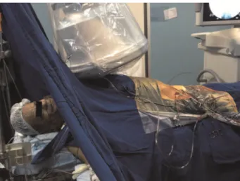
to the body, providing venous access and an arterial line for the anesthesiologists. The left arm was positioned along the body to permit free movements of the C-arm. A central venous line and a diuresis catheter were also inserted. Short-term antibiotic prophylaxis was per-formed with cefazolin.Direct surgical exposure of the femoral arteries was achieved by using short oblique groin incisions. This step was performed under local anesthesia (lidocaine 0.5%) in
most patients (Fig. 1). A Superstiff Lunderquist guidewire (Cook Inc., Bloomington, IN, USA) was introduced through one side until the thoracic aorta was reached. A Pig Tail catheter (Cook Inc., IN, USA) was introduced through the other side until reaching the level of the renal arteries, as previously identified on the CT scan. A compliant balloon (Reliant; Medtronic, World Medical Manufacturing Corp., FL, USA) and an introducer sheath (12F, 45 cm; Flexor, Cook, IN, USA) were placed over the Lunderquist guide-wire in the suprarenal aorta to promote aortic occlusion in case of severe circulatory collapse, as previously described (10). When the chosen device, according to preoperative CT results, was ready for insertion and deployment, general anesthesia was initiated to eliminate patient movement, which improves fluoroscopic imaging and enables precise graft deployment. All patients underwent an intraoperative completion angiography to confirm aneurysm exclusion.
Most of these patients did not receive systemic heparin due to the increased risk of bleeding. Heparin was preferably used locally in femoral arteries only at the end of the procedure.
Fluid resuscitation was instituted after the surgeon informed the anesthesiologists that the aneurysm had been excluded from circulation, and blood pressure was cor-rected to more physiological values. After the procedure, the patients were transferred to the intensive care unit for standard postoperative care.
& RESULTS
A total of 28 patients with RAAAs were consecutively treated at our hospital from February 2012 to January 2013 (12 months). A total of 18 patients (64.3%) were considered suitable for (based on their anatomy) and underwent EVAR. In 2 of these 18 patients, EVAR was performed with AUI grafts due to unilateral iliac occlusions.
The other 16 EVAR cases were treated with bifurcated endografts. Contralateral gate cannulation and a complete endovascular procedure were possible in all patients. Furthermore, aneurysm exclusion, which was evaluated on the angiogram and performed at the end of the procedure, was achieved in 100% of cases in the endovas-cular group. No patient required emergent conversion of bifurcated stent grafts to AUI devices or open surgery.
The remaining 10 patients (35.7%) were considered unsuitable for endovascular repair and were treated with the open repair approach (Table 1). Anatomical contra-indications for EVAR in these cases were an unsuitable or absent infrarenal neck in seven cases, bilateral iliac obstruc-tion in two cases and a complex infrarenal aortic dissecobstruc-tion in one case.
CT scans were performed in 16 patients. The other 12 patients arrived at our emergency department with a previous CT exam from other hospitals. No patient died or exhibited a worse hemodynamic status during transpor-tation or performance of the CT scan.
The patients’ mean age was 67.2 years (45-85 years), and the mean aneurysm diameter was 77.7 mm (50-120 mm). Seven patients (25.9%) were hemodynamically unstable, 5 (29.4%) in the endovascular group and 2 (20%) in the open repair group. The mean length of the proximal neck was 18.8 mm (6-48 mm) in the endovascular group and 6.7 mm (0-33 mm) in the open repair group. Four patients in the REVAR group had a proximal neck that was shorter than 10 mm (6, 7, 7 and 8 mm).
Of the 28 patients, 10 (35.7%) had been previously diagnosed with AAA prior to rupture, 4 had not previously undergone surgery due to poor clinical conditions and high risks related to the procedure, 3 had refused surgery and 3 were waiting for elective repair. No patient refused surgery after the diagnosis of RAAA.
A supraceliac aortic occlusion balloon was successfully used in two patients because of severe circulatory collapse during REVAR with bifurcated endografts.
The devices used were Endurant (Medtronic, CA, USA) in 14 patients (77.8%), Zenith (Cook, IN, USA) in 1 patient (5.5%), AFX (Endologix, CA, USA) in 2 patients (11.1%) and Anaconda (Vascutek, Inchinnan, UK) in 1 patient (5.5%).
All REVAR procedures in unstable patients were initiated under local anesthesia, which was combined with intrave-nous sedation for pain relief and patient immobility when necessary (Table 2). All procedures were completed under general anesthesia. Intravenous heparin was not used in unstable patients and it was used in 54% (7/13) of the stable patients based on the surgeon’s discretion.
The 30-day mortality rate was 27.8% (5/18) in the endovascular group, and 2 of these deaths occurred in unstable patients. In the open repair group, the periopera-tive mortality rate was 50% (5/10), and 2 of the deaths occurred in unstable cases (Fig. 2). The overall mortality rate was 35.7% (10/28), and the difference in this rate between the OPEN and EVAR groups was not statistically significant
(p= 0.221). The median total hospital stay of the survivors in
the REVAR group was 9 days (3-26 days), whereas that in the open repair group was 15.6 days (7-40 days;p= 0.208).
The following major complications occurred in 7 patients in the REVAR group (38.8%): acute renal failure requiring dialysis (n = 3); pneumonia (n = 2); myocardial infarction (n = 3); intra-abdominal compartment syndrome (n = 2) in two unstable patients in the endovascular group, one of whom was treated with surgical evacuation immediately after surgery and the other of whom underwent delayed laparotomy and died due to multiple organ failure before drainage; colon ischemia (n = 1); lower limb ischemia (n = 1); and prolonged ileus (n = 1). Six patients (60%) in the open repair group had the following major complications: acalculous cholecystitis (n = 1), evisceration (n = 1), lower limb ischemia (n = 1), myocardial infarction (n = 2) and acute renal failure requiring dialysis (n = 1).
& DISCUSSION
In this study, we included only patients with evidence of ruptured aneurysms and showed that approximately 64.2% of patients admitted with RAAA were deemed suitable for REVAR. Most patients who were deemed unfit had an unsuitable infrarenal aortic neck (7/10). We did not intend to compare open and endovascular repair because of the absence of randomization and the heterogeneity of the groups, especially considering the more complex anatomy in the open repair group.
AUI endografts were adopted in the early phase of REVAR worldwide due to its rapid deployment, which is possible because there is no need to cannulate the contralateral gate (3,4,11). This procedure has a shorter learning curve, which makes it attractive to some groups that are beginning to use REVAR. We believe that the greatest advantage of AUI endografts may be achieved in unstable patients because of the faster hemorrhage control afforded by these grafts. However, this procedure requires an occluder device in the contralateral common iliac artery to achieve complete control of any intra-abdominal bleed-ing, which could be time-consuming in an emergency situation. This procedure also requires a femorofemoral prosthetic bypass, carrying risks such as late occlusion or infection.
Recent publications from experienced centers have demonstrated a preference for bifurcated endografts with encouraging results (7,12). Verhoeven et al. reported a short mean total operating time with this approach in
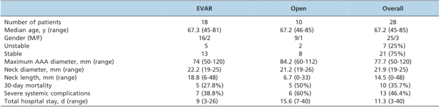
Table 1 -Summary of all patients’ clinical and anatomical features.
EVAR Open Overall
Number of patients 18 10 28
Median age, y (range) 67.3 (45-81) 67.2 (46-85) 67.2 (45-85)
Gender (M/F) 16/2 9/1 25/3
Unstable 5 2 7 (25%)
Stable 13 8 21 (75%)
Maximum AAA diameter, mm (range) 74 (50-120) 84.2 (60-112) 77.7 (50-120)
Neck diameter, mm (range) 22.2 (19-25) 21.2 (19-26) 21.9 (19-25)
Neck length, mm (range) 18.8 (6-48) 6.7 (0-33) 14.5 (0-48)
30-day mortality 5 (27.8%) 5 (50%) 10 (35.7%)
Severe systemic complications 7 (38.8%) 6 (60%) 13 (46.4%)
Total hospital stay, d (range) 9 (3-26) 15.6 (7-40) 11.3 (3-40)
acute situations (13), similar to that reported in some AUI studies (14). Acquired experience and some techniques, such as aortic balloon occlusion, have enabled the safe use of bifurcated grafts in REVAR. Indeed, in the present study, AUI endografts were used only if the anatomy represented a contraindication for the use of the bifurcated system.
Patients with ruptured aneurysms appear to have larger aneurysm diameters and shorter proximal necks (15). With increasing experience, there seems to be a common tendency in endovascular centers to extend EVAR indica-tions to more challenging cases. Consistent with other authors, we performed REVAR in patients with proximal necks that were shorter than 10 mm (7,16). When the intention was to use bifurcated grafts, we successfully cannulated the contralateral gate in all cases, with no instances of conversion to AUI devices.
Performing a preoperative CT scan is one of the most interesting issues regarding REVAR. It is an extremely important imaging exam for surgical planning but may cause a delay in unstable cases or in patients who are at risk of instability. Hinchcliffe et al. reported that the concern that CT scanning prior to surgery could be detrimental to patients seems to be unfounded (5). Patients who were considered unfit for CT scanning due to hemodynamic instability had bad outcomes with open surgery. It seems that early surgery does not confer a survival benefit, and, conversely, a delay does not translate to increased mortality. Lloyd et al. reported a survival time longer than 2 hours in 87.5% of untreated patients with ruptured aneurysms and a median time between admission and death of more than 10 hours (17). In most cases, there is sufficient time available to safely image the patients prior to endovascular repair (18). As in other referral centers, in our emergency department,
Table 2 -Summary of endovascular cases.
Stable Unstable Overall
Number of patients 13 5 18
Product
Endurant 9 (7 BIF, 2 AUI) 5 14 (77.8%)
Zenith 1 - 1 (5.5%)
Endologix 2 - 2 (11.1%)
Anaconda 1 - 1 (5.5%)
Anesthesia
Local to general 6 (46%) 5 (100%) 11 (61%)
General 7 (54%) - 7 (39%)
Intravenous heparin 7 (54%) - 7 (39%)
30-day mortality 3 (18.7%) 2 (40%) 5 (27.8%)
Severe systemic complications 4 (30.7%) 3 (60%) 7 (38.8%)
Acute renal failure (dialysis) 1 2{{ 3
Pneumonia 1 1{ 2
Myocardial infarction 1* 2{1 3
Intra-abdominal compartment syndrome - 2{1 2
Colon ischemia - 1{ 1
Lower limb ischemia 1 - 1
Prolonged ileus 1* - 1
BIF: bifurcated endograft, AUI: aortouniiliac endograft. *,{,{,1represent the same patient.
CT scanning can be promptly performed, with minimum patient transportation (19). We decided to perform CT scans in all patients admitted without this exam, even in those with a systolic blood pressure less than 80 mmHg, i.e., unstable patients, because of the importance and speed of this exam. Moreover, with regards to REVAR, the decision about the feasibility of this approach can be made in real-time during image processing. We believe that maintaining permissive hypotension is a major factor associated with the safe performance of this exam.
Hemodynamic instability during induction of general anesthesia remains the most challenging issue associated with RAAA. The use of local anesthesia and avoidance of a loss of abdominal tone are important advantages of REVAR, but it also carries some potential disadvantages, including patient discomfort and movement artifacts that may cause inaccurate deployment of the grafts. We addressed this issue by using a hybrid approach. In the majority of our patients, the femoral arteries were accessed, and the guidewires and catheters were placed under local anesthe-sia. Based on a previous CT scan, a selected endograft was prepared, and a compliant balloon and a 12F introducer sheath were always ready for use in case of hemodynamic collapse. General anesthesia was then initiated. Although some authors have reported using the occluding balloon in up to 25% of cases, we only used it in two patients (11.1%). We agree with other authors who believe that the main goal is to achieve rapid deployment of the endograft and reserve the occluding balloons for severely unstable patients (40-50 mmHg systolic blood pressure) who are unable to maintain their blood pressure without the balloon (20).
Hemodynamic instability has been the most widely used criterion to define the severity of RAAA, although it is difficult to designate a single parameter to define the hemodynamic status because it depends on the amount and rate of bleeding and the previous conditions of the patient. Similar to other studies, we selected a blood pressure cutoff of 80 mmHg (6,17,18) to define the hemodynamic status.
We obtained a survival rate of 66.6% (4/6) among the unstable patients in the endovascular group and we reinforce the importance of performing CT scans and assessing REVAR suitability in these patients. Consistent with other authors, we believe that this group of patients could be the most likely to benefit from a less invasive procedure (18,20).
Three patients in the REVAR group developed renal failure requiring dialysis; one stable patient had previously known severe renal function impairment and required postoperative dialysis despite the completion of preopera-tive CT scanning without contrast enhancement and with restriction of the use of contrast during the procedure, and two unstable patients developed renal failure due to contributing factors such as intra-abdominal compartment syndrome and postoperative myocardial infarction requir-ing cardiac catheterization with the use of more iodinated contrast (Table 2). Although the stable patient who required dialysis had an aneurysm neck shorter than 10 mm, no patient in this series experienced coverage of the renal arteries with the endografts.
Although there are several studies on REVAR, most of them used very different methods of patient selection and included less than 30 patients, making it difficult to compare them. The mortality rate among such studies ranges from 9.5-46% (5,7). We obtained a mortality rate of 27.7% (5/18)
in the REVAR group, which is comparable to that in other specialized centers. Our overall mortality rate was 35.7% (10/28), which is lower than the historical mortality associated with the open approach (1,2,7). Indeed, we believe that bifurcated endografts are a feasible option for treating anatomically eligible ruptured aneurysms and may represent a valuable approach in experienced centers.
& AUTHOR CONTRIBUTIONS
De Luccia N, Queiroz AB, Mulatti GC and Schneidwind KP were responsible for data collection. De Luccia N, Queiroz AB, Mulatti GC, Schneidwind KP and Neto PS conceived and designed the study. Queiroz AB, Santo FR and Neto PS were responsible for statistical analysis and interpretation. Queiroz AB, De Luccia N, Santo FR, Torres IO and Schneidwind KP were responsible for the final approval.
& REFERENCES
1. Akkersdijk GJ, van der Graaf Y, van Bockel JH, de Vries AC, Eikelboom BC. Mortality rates associated with operative treatment of infrarenal abdominal aortic aneurysm in The Netherlands. Br J Surg. 1994;81(5): 706-9.
2. Bown MJ, Sutton AJ, Bell PR, Sayers RD. A meta-analysis of 50 years of ruptured abdominal aortic aneurysm repair. Br J Surg. 2002;89(6):714-30.
3. Yusuf SW, Whitaker SC, Chuter TAM, Wenham PW, Hopkinson BR. Emergency endovascular repair of leaking aortic aneurysm. Lancet. 1994;344:1645, http://dx.doi.org/10.1016/S0140-6736(94)90443-X. 4. Ohki T, Veith FJ, Sanchez LA, Cynamon J, Lipsitz EC, Wain RA,
et al. Endovascular graft repair of ruptured aortoiliac aneurysms. J Am Coll Surg. 1999;189(1):102-12, http://dx.doi.org/10.1016/S1072-7515(99) 00051-4.
5. Hinchliffe RJ, Bruijstens L, MacSweeney ST, Braithwaite BD. A randomised trial of endovascular and open surgery for ruptured abdominal aortic aneurysm - results of a pilot study and lessons learned for future studies. Eur J Vasc Endovasc Surg. 2006;32(5):506-13, http:// dx.doi.org/10.1016/j.ejvs.2006.05.016.
6. Mehta M. Endovascular aneurysm repair for ruptured abdominal aortic aneurysm: the Albany Vascular Group approach. J Vasc Surg. 2010;52(6):1706-12, http://dx.doi.org/10.1016/j.jvs.2010.06.103. 7. Lachat ML, Pfammatter T, Witzke HJ, Bettex D, Kunzli A, Wolfensberger
U, et al. Endovascular repair with bifurcated stent-grafts under local anaesthesia to improve outcome of ruptured aortoiliac aneurysms. Eur J Vasc Endovasc Surg. 2002;23(6):528-36, http://dx.doi.org/10.1053/ ejvs.2002.1622.
8. Brown JR, Block CA, Malenka DJ, Oconnor GT, , Schoolwerth AC, Thompson CA. Sodium bicarbonate plus N-acetylcysteine prophylaxis: a meta-analysis. JACC Cardiovasc Interv. 2009;2(11):1116-24, http://dx. doi.org/10.1016/j.jcin.2009.07.015.
9. van der Vliet JA, van Aalst DL, Schultze Kool LJ, Wever JJ, Blankensteijn JD. Hypotensive hemostatis (permissive hypotension) for ruptured abdominal aortic aneurysm: are we really in control? Vascular. 2007;15(4):197-200, http://dx.doi.org/10.2310/6670.2007.00028. 10. Berland TL, Veith FJ, Cayne NS, Mehta M, Mayer D, Lachat M.
Technique of supraceliac balloon control of the aorta during endovas-cular repair of ruptured abdominal aortic aneurysms. J Vasc Surg. 2013;57(1):272-5, http://dx.doi.org/10.1016/j.jvs.2012.09.001.
11. Ohki T, Veith FJ. Endovascular grafts and other image-guided catheter-based adjuncts to improve the treatment of ruptured aortoiliac aneurysms. Ann Surg. 2000;232(4):466-79, http://dx.doi.org/10.1097/ 00000658-200010000-00002.
12. Mehta M, Kreienberg PB, Roddy SP, Paty PS, Taggert JB, Sternbach Y, et al. Ruptured abdominal aortic aneurysm: endovascular program development and results. Semin Vasc Surg. 2010;23(4):206-14, http://dx. doi.org/10.1053/j.semvascsurg.2010.10.003.
13. Verhoeven EL, Prins TR, van den Dungen JJ, Tielliu IF, Hulsebos RG, van Schilfgaarde F. Endovascular repair of acute AAAs under local anesthesia with bifurcated endografts: a feasibility study. J Endovasc Ther. 2002;9(6):729-35, http://dx.doi.org/10.1583/1545-1550(2002)009,0729: EROAAU.2.0.CO;2.
15. Hinchliffe RJ, Alric P, Rose D, Owen V, Davidson IR, Armon MP, et al. Comparison of morphologic features of intact and ruptured aneurysms of infrarenal abdominal aorta. J Vasc Surg. 2003;38(1):88-92, http://dx. doi.org/10.1016/S0741-5214(03)00079-X.
16. Mehta M, Taggert J, Darling RC 3rd, Chang BB, Kreienberg PB, PAty PS, et al. Establishing a protocol for endovascular treatment of ruptured abdominal aortic aneurysms: outcomes of a prospective analysis. J Vasc Surg. 2006;44(1):1-8, http://dx.doi.org/10.1016/j.jvs.2006.02.057. 17. Lloyd GM, Bown MJ, Norwood MG, Deb R, Fishwick G, Bell PR, et al.
Feasibility of preoperative computer tomography in patients with ruptured abdominal aortic aneurysm: a time-to-death study in patients without operation. J Vasc Surg. 2004;39(4):788-91, http://dx.doi.org/10. 1016/j.jvs.2003.11.041.
18. Boyle JR, Gibbs PJ, Kruger A, Shearman CP, Raptis S, Phillips MJ. Existing delays following the presentation of ruptured abdominal aortic aneurysm allow sufficient time to assess patients for endovascular repair. Eur J Vasc Endovasc Surg. 2005;29(5):505-9, http://dx.doi.org/10. 1016/j.ejvs.2005.01.027.
19. Willmann JK, Lachat ML, von Smekal A, Turina MI, Pfammatter T. Spiral-CT angiography to assess feasibility of endovascular aneurysm repair in patients with ruptured aortoiliac aneurysm. Vasa. 2001;30(4):271-6, http://dx.doi.org/10.1024/0301-1526.30.4.271. 20. Veith FJ, Cayne NS, Berland TL, Mayer D, Lachat M. Current role for