ORIGINAL
RES
EAR
Correspondence to: Mariana Tirolli Rett – Rua Claudio Batista, s/n – Santo Antonio – CEP: 49060-100 – Aracaju (SE), Brazil – E-mail: [email protected]
Presentation: aug. 2012 – Accepted for publication: dec. 2012 – Financing source: Programa Especial de Inclusão em Iniciação Científica (PIIC) and Programa Institucional de Bolsas de Iniciação à Extensão (PIBIX)-UFS – Conflict of interest: nothing to declare – Approval report from the Ethics Committee n° CAAE – 0090.0.107.000-11.
ABSTRACT | This study compared the pulmonary function and fatigue in patients before and after adju-vant radiotherapy (RT) and correlated the pulmonary function with the radiotherapy dose and fatigue. A longitudinal and observational study was conducted involving 20 women. Pulmonary function was evalu-ated by digital lung spirometry (ClementClarke®) and manometry (GlobalMed®, model MVD 300) and fatigue was analyses by the Functional Assessment of Cancer Therapy Fatigue (FACT-F). All evaluations were con-ducted before the first RT session and up to one week after this treatment. Statistical analyses were conduct-ed by the Wilcoxon Signconduct-ed Rank Test and Spearman, considering p<0.05. There was significant reduction at spirometry parameters: forced vital capacity (23.52%), forced expiratory volume in the first second (26.23%), peak expiratory flow (10.12%) (p=0.001). Maximal expira-tory pressure (25.45%) and maximal inspiraexpira-tory pres-sure (32.92%) also showed significant reduction at ma-nometry. There was a significant reduction on physical well-being and functional well-being and a significant increase in fatigue evaluated by the FACT-F (p=0.001). There was no correlation between pulmonary function, radiation dose and fatigue. Short-term effects of radio-therapy showed reduction of pulmonary function, but the values were considered similar to normal. There was
and fatigue of women undergoing treatment for
breast cancer
Efeito da radioterapia na função pulmonar e na fadiga de mulheres em tratamento
para o câncer de mama
Efecto de la radioterapia en la función pulmonar y en la fatiga de mujeres en
tratamiento para el cáncer de mama
Dayane Evellyn dos Santos1, Mariana Tirolli Rett2, Andreza Carvalho Rabelo Mendonça3,
Thaysa Samanta Bezerra1, Josimari Melo DeSantana2, Walderi Monteiro da Silva Júnior2
Study carried out at Fundação de Beneficência Hospital Cirurgia (FHBC) and in Hospital de Urgências de Sergipe(HUSE) – Aracaju (SP), Brazil.
1Physical Therapist by Universidade Federal de Sergipe (UFS) – Aracaju (SE), Brazil. 2Physical Therapist; Adjunct Professor at UFS – Aracaju (SE), Brazil.
3Physical Therapist; Master’s degree student at the Postgraduate Program in Health Sciences at UFS – Aracaju (SE), Brazil.
a significant increase in fatigue, and significant decrease of physical well-being and functional well-being. Keywords | breast neoplasms; radiotherapy; respiratory function tests; radiation effects; fatigue; physical therapy.
INTRODUCTION
Breast cancer is the main cause of death due to malig-nant diseases among Brazilian women, and 52,680 new cases were expected for 20121. Despite the evolution in
diagnosis and treatment, the surgical procedures and complementary therapies, such as radiotherapy, chemo-therapy and hormone chemo-therapy are still prevalent2-4.
Radiotherapy (RT) is capable of destroying tumor cells, reducing the risk of local recurrence and increa-sing survival rates5,6. he most common application
technique is the external RT (teletherapy), in which ionizing radiation goes through diferent tissues be-fore reaching the tumor area; therebe-fore, organs and normal tissues are subjected to the toxic efects of the emitted rays7. he absorption of radiation can cause
biochemical changes and cell damage both immedia-tely and later on8-10.
After RT, some side efects are likely to occur, such as: pain, skin changes, mobility restriction, local sen-sitive change and fatigue8,9. Pulmonary changes with
radiological abnormalities, such as the increased den-sity, symptomatic radioactive pneumonitis, pulmonary ibrosis, ventilation deicit and quantitative reduction in pulmonary function tests can also be expected7,10-14..
Added to the mentioned changes, fatigue after RF can compromise the execution of daily activities, social and family contact, besides causing work loss15. his
symptom has a multifactorial causes and can be related to the RT itself, as well as physical and psychological factors16,17.
In national literature, the evaluation of fatigue con-comitant with the assessment of pulmonary function during treatment with RT for breast cancer was not found. hus, knowing the potential efects of RT can enable the prevention and treatment of such disorders. herefore, the objective of this study was to compare the pulmonary function and fatigue of women submitted to surgery for breast cancer before and after adjuvant RT. It also aimed at correlating pulmonary function with the radiation dose and fatigue.
METHODOLOGY
An observational and longitudinal study was perfor-med from August to October 2011. Women submit-ted to mastectomy and quadrantectomy, with axil-lary lymphadenectomy and prescription for adjuvant RT were included. he excluded patients were those submitted to bilateral surgery, breast reconstruction or placement of breast prosthesis, with history of pneumopathy (lung cancer, pulmonary emphysema, chronic obstructive pulmonary disease, bronchiecta-sis), concomitant chemotherapy and neoadjuvant RT. Twenty-ive women were eligible for the study, ho-wever 5 were excluded for interrupting radiotherapy, so 20 women remained.
RT was performed from ive to six weeks (depen-ding on the dose) with daily applications. he irradiated
aumento significativo da fadiga no FACT-F (p=0,001). Não foram observadas correlações entre as variáveis da função pulmonar com a dose de radiação e fadiga. Em curto prazo, a RT promo-veu redução na função pulmonar, mas a mesma permaneceu próxima à normalidade para a amostra estudada. Observou-se aumento significativo da fadiga e diminuição dos escores dos do-mínios bem-estar físico e funcional.
Descritores | neoplasias da mama; radioterapia; testes de função respiratória; efeitos de radiação; fadiga; fisioterapia.
RESUMEN | El presente estudio comparó la función pulmonar y la fatiga de mujeres antes y después de la radioterapia (RT) como ayudante para el tratamiento del cáncer de mama, y se corre-lacionó la función pulmonar con la dosis de radiación y fatiga. Fue realizado un estudio observacional longitudinal involucran-do 20 mujeres. La función pulmonar fue evaluada por espirome-tría (ClementClarke®) y manovacuomeespirome-tría (GlobalMed®, modelo MVD 300) y, la fatiga fue evaluada por la Functional Assessment of Cancer Therapy Fatigue (FACT-F). Todas las evaluaciones fue-ron realizadas antes de la primera sesión y una semana después del término de la RT. Para el análisis estadístico fueron utilizados los tests Wilcoxon Signed Rank Test y correlación de Spearman, adoptando un nivel de significancia p<0,05. En la espirometría, se encontró reducción significativa de la capacidad vital forzada (23,52%), del volumen espiratorio forzado en el primer segundo (26,23%) y del peak de flujo espiratorio (10,12%) (p=0,001). Las pre-siones espiratorias e inspiratorias máximas también disminuye-ron significativamente (25,45% y 32,92%, respectivamente). Se ob-servó disminución significativa del bienestar físico y del bienestar funcional, y un aumento significativo de la fatiga en el FACT-F (p=0,001). No fueron observadas correlaciones entre las variables de la función pulmonar con la dosis de radiación y fatiga. En cor-to plazo, la RT promueve la reducción de la función pulmonar, pero los valores son considerados similares a los normales. Se observó aumento significativo de la fatiga y disminución de los puntajes en los dominios de bienestar físico y funcional.
regions were: breast region (or plastron), and the axil-lary region and clavicular fossa with lower doses.
Pulmonary function was assessed by the spiro-metry (ClementClark®) and manovacuometry tests
(GlobalMed®, MVD 300 model), which measure
ca-pacities and volumes, and respiratory muscle strength, respectively.
Spirometry was conducted according to the criteria by the American horacic Society18. he forced vital
capacity (FVC) was assessed in liters (L), the forced expiratory volume (FEV1), in L, the FEV1/FVC ratio, in %, and the peak expiratory low (PEF), in liters per minute (L/min). For this test, patients were told to perform maximal inspiration, until the full lung ca-pacity (FLC), followed by a continuous and forced expiration, until the residual volume (RV), in the mouthpiece of the device. he obtainment of three acceptable (with variation of PEF values lower than 10%) and reproducible curves (with the two highest values of FEV1 and FVC, with variation lower than 0.15 L) were considered, adopting the highest values measured from each variable.
Manovacuometry followed the procedure proposed by Neder et al.19. A deep and prolonged inspiration was
required, until FLC, followed by forced expiration on the mouthpiece of the device to obtain the maximal ex-piratory pressure (EPmax). For the maximal insex-piratory pressure (IPmax), a forced expiration was requested,
until RV, followed by deep inspiration. hree EPmax and IPmax maneuvers were performed with a one mi-nute interval. he highest obtained value was registered, considering the performance of three acceptable and reproducible maneuvers, with the maximum of 10% of diference between them.
To assess fatigue, the Functional Assessment of Cancer herapy Fatigue (FACT-F) was used, which consists of 40 items, being 27 related to the Functional Assessment of Cancer herapy-General (FACT-G) and 13 speciic items about fatigue20. he questions in
FACT-G are distributed into domains about physi-cal well-being, social/family well-being and functional well-being (each with 7 items and a 0 to 28 score) and emotional well-being (7 items and a 0 to 24 score). In these domains, the higher the score, the better the assessed well-being. he fatigue subscale has a 0 to 52 score, and the higher the score, the lower the fatigue. here is a total score corresponding to the sum of do-mains and the fatigue subscale, ranging from 0 to 160, and the higher the score, the lower the fatigue.
All evaluations were performed by the same evalua-tor in two moments: before the irst RT session and a week after its conclusion. he study was approved by the Research Ethics Committee of Universidade Federal de Sergipe (UFS) – CAAE 0090.0.107.000-11. he sta-tistical analysis was conducted by BioEstat 5.0 and the Wilcoxon Signed Rank test and Spearman’s correlation test were used, with a p<0.05 signiicance level.
RESULTS
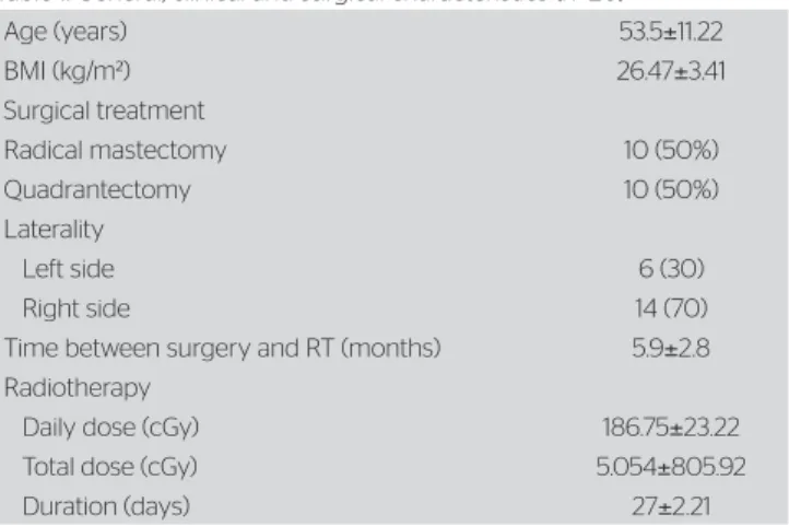
he general, clinical and surgical characteristics of the 20 women are described in Table 1. Only one had wor-ked in an environment with dust, two were smokers and three had smoked for about four years.
Spirometry showed a signiicant decrease in FVC (23.52%), FEV1 (26.23%) and PEF (10.12%), (p=0.001). he FEV1/FLC ratio did not present signi-icant changes (p=0.430) (Table 2).
Age (years) 53.5±11.22
BMI (kg/m²) 26.47±3.41
Surgical treatment
Radical mastectomy 10 (50%)
Quadrantectomy 10 (50%)
Laterality
Left side 6 (30)
Right side 14 (70)
Time between surgery and RT (months) 5.9±2.8
Radiotherapy
Daily dose (cGy) 186.75±23.22
Total dose (cGy) 5.054±805.92
Duration (days) 27±2.21
Table 1. General, clinical and surgical characteristics (n=20)
BMI: body mass index; kg/m²: kilogram/meter2; RT: radiotherapy; cGy: centigrays.
Spirometry Before After % reduction p value
FVC (L) 2.56±0.38 1.95±0.37 23.52±5.98 0.001*
FEV1 (L) 2.04±0.37 1.50±0.31 26.23±10.15 0.001*
PEF (L/min) 315.05±26.45 282.25±20.34 10.12±6.32 0.001*
FEV1/FVC (%) 76.05±3.73 72.20±3.73 0.7±7.53 0.43
Table 2. Comparison of spirometry before and after radiotherapy (n=20)
Signiicant reduction in respiratory muscle strength was found: EPmax was initially 73±12.47, and after RT, 55±7.90 cmH2O, which is similar to a 25.45% reduction. IPmax went from 69.50±10.41 to 46.25±5.32 cmH2O, corresponding to a 32.92% reduction (p=0.001) (Figure 1).
he signiicant aggravation of the physical wel-l-being was observed, decreasing from 22.45±5 to 16.75±5.5, as well as of the functional well-being, from 17.35±5.5 to 14.95±5.2 and the fatigue subsca-le, from 46.1±4.9 to 26.1±6.8 (p=0.001) (Figure 2).
he total FACT-F score also presented signiicant re-duction, from 114.70±12.60 to 85.10±14.00 (p=0.001). No signiicant correlation was found between the pul-monary function after RT and the total radiation dose, with the fatigue subscale and with the total FACT-F score (Table 3).
DISCUSSION
A reduction in the main pulmonary function measu-res was observed (FVC, FEV1, EPmax and IPmax), as well as the aggravation of the physical and functional well-being and fatigue. Despite the decreased pulmo-nary function and the reduction of more than 20% of FVC and FEV1, the values remained within norma-lity, according to weight, age and height of the studied sample18,19. Changes in lung capacity and volume are
expected after RT11,21-25, since there are potential risks
of damaging the pulmonary parenchyma, losing type ii pneumocytes, losing surfactant and edema in the base-ment membrane26. But there is also the possibility that
the patient can remain asymptomatic or never present any changes, be it in the parenchyma or in the pulmo-nary function, due to the “compensation in relation to the health lung”, which did not receive radiation24.
In the short term, the efects of RT did not cause an impact on the pulmonary function, even with the decrease of some spirometry and manovacuometry pa-rameters, maybe because of the radiation dose, or the short follow-up time, or even because of the “compen-sation of the health lung”. In the study by Schettino, Jotta e Cassali25, the authors used the same instruments
to assess pulmonary function, but they did not ind any changes immediately after RT. However, this study in-volved only ten women.
More damage in the pulmonary function and in the capacity of alveolar difusion is demonstrated by studies
Pulmonary function Total RT dose Fatigue subscale score Total FACT-F score
r p value r p value r p value
FVC -0.36 0.11 -0.07 0.75 0.32 0.12
FEV1 0.20 0.37 -0.14 0.54 -0.03 0.86
PEF -0.28 0.21 -0.09 0.70 0.02 0.29
FEV1/FVC -0.29 0.90 -0.16 0.49 0.18 0.42
EPmax -0.41 0.17 -0.32 0.15 0.41 0.15
IPmax -0.32 0.16 -0.22 0.34 0.38 0.11
Table 3. Correlation of pulmonary function values (spirometry and manovacuometry) after radiotherapy with the total radiotherapy dose, the fatigue subscale score and the total score of the Functional Assessment of Cancer Therapy Fatigue (n=20)
RT: radiotherapy; FACT-F: Functional Assessment of Cancer Therapy Fatigue; FVC: forced vital capacity; FEV1: forced expiratory volume; PEF: peak expiratory flow; EPmax: maximal expiratory pressure; IPmax: maximal inspiratory pressure; r: Spearman’s correlation
Figure 2. Scores of the Functional Assessment of Cancer Therapy Fatigue domains and of the fatigue subscale before and after radiotherapy
60
50
40
30
20
10
0
Physical Well-being
Family Well-being
Emotional Well-being
Functional Well-being
Fatigue
Before After
Figure 1. Comparison of manuvacometry (EPmax and IPmax) before and after radiotherapy.
PRE
SSURE (cmH
2
O)
EPmax IPmax
90
80
70
60
50
40
30
20
10
0
Before After
EPmax: maximal expiratory pressure; IPmax: maximal inspiratory pressure *p<0.05, comparison before and after radiotherapy
with longer follow-up and higher number of reassess-ments after RT4,5,23-25. Ooi et al.24, in a prospective study
performed 1, 3, 6 and 12 months after the conclusion of RT, showed that RT can be associated with an irreversi-ble reduction of FCV, FEV1 and the carbon monoxide difusing capacity.
he deicit in ventilation and in the capacity of al-veolar difusion that occurs in the irradiated lung can be related to the used technique, to the total dose, to the fractionated dose and the volume of the lung which received radiation8,12,13. Lung disorders can be
temporarily classiied as actinic pneumonitis, which appears from three to six months, and pulmonary i-brosis, which occurs about one year after RT26. Due
to the period in which they were reassessed, until one week after the conclusion of RT, the patients did not present with any characteristic symptom of the afo-rementioned chronic lesions. Besides, no signiicant correlation between pulmonary function and total ra-diation dose was found, which is in accordance with the proile of the studied patient23.
Imaging tests can also be useful5,6,12, because at an
acute phase, radiation can lead to the appearance of at-tenuation or consolidation areas in lung x-rays, while at a late stage, bronchiectasis areas and loss of volume can be observed10,12. However, it was not possible to
de-monstrate radiological changes in this study, because only tests of pulmonary function and fatigue assessment were conducted.
It is worth to mention that even without clinical repercussion, the decrease of EPmax and IPmax was observed. EPmax is connected with the promotion of an efective coughing and elimination of secretions25,
so, its aggravation can compromise the clinical picture of these patients if, in the long term, they present with some pulmonary disorder. Such reduction in the respi-ratory muscle strength can be associated with changes that take place in the pulmonary parenchyma, besides tiredness and physical exhaustion reported by the pa-tients after RT.
Besides pulmonary repercussions, RT can cause fatigue. his has been shown by the decrease of the fatigue subscale, the FACT-F general score and the aggravation of the physical and functional well-being. hese domains had complaints of indisposition, lack of energy, somnolence, loss in the work activities, pain, among others. According to literature and the clinical practice, the presence of fatigue, tiredness and physical exhaustion as a secondary efect of RT8 has a
negati-ve repercussion on the performance of daily life tasks,
socialization, recreational activities and in the global physical conditioning15,26.
Even though there have been correlations between pulmonary function and fatigue, the reduction of values such as volume, capacities and respiratory muscle stren-gth can also inluence the appearance of fatigue26. he
symptoms of lack of energy, tiredness, and intolerance to efort can be a relex of the RT itself, of psychological factors, of the need to sleep/rest during the day8,15,21,26,
and they can even be increased by the negative reper-cussions of RT on the respiratory function26. Clinically,
it is expected that when the pulmonary function is compromised, even if temporarily or without great re-percussions, the patients can experiment situations of indisposition or exhaustion.
he scores of the family and emotional well-being domains are in accordance with those of literature, when considering women who are being treated for breast cancer20 and did not present diferences after RT.
It is believed that these women have proper family su-pport and receive susu-pport from their friends, at home and in their social environment, which justiies the lack of compromise in this aspect. Besides, they may work with strategies to face the disease, which does not com-promise the emotional well-being.
Even though the values of capacities, volumes and respiratory muscle strength are within normality for the studied sample, and considering the spirometry as an important indicator of risk for the development of pulmonary diseases18, the reduction of the assessed
pa-rameters conirms the potentially negative efect caused by RT, in the short term, on the pulmonary functional reserve6. It is clear that these results should be
conside-red important, since the life expectancy of women with breast cancer has increased, they have been adopting a more active life style, and due to the fact that this sam-ple represents diferent women who, in the same age group, one day can be submitted to RT.
CONCLUSION
It was observed that, in the short term, RT promoted a negative impact on pulmonary function, a signiicant increase in fatigue and the compromise of physical and functional well-being. However, no signiicant correla-tions were observed between pulmonary function, total radiation dose and fatigue.
ACKNOWLEDGEMENTS
To the patients, to Fundação de Beneicência Hospital Cirurgia, to the Ministry of Education, to Programa Especial de Inclusão em Iniciação Cientíica (PIIC/ POSGRAP/PROEST/UFS) and Programa Institucional de Bolsas de Iniciação à Extensão (PIBIX).
REFERENCES
1. Instituto Nacional de Câncer José Alencar Gomes da Silva. Rio de Janeiro: INCA [cited 2012 Feb 12] Estimativas 2012: incidência de câncer no Brasil. Available from: http://www.inca.gov.br/estimativa/2012/ estimativa20122111.pdf
2. Bregagnol KR, Dias SA. Alterações funcionais em mulheres submetidas à cirurgia de mama com linfadenectomia axilar total. Rev Bras Cancerol. 2010;56(1):25-33.
3. Campbell A, Mutrie N, White F, MCguire F, Kearney N. A pilot study of a supervised group exercise programme as a rehabilitation treatment for women with breast cancer receiving adjuvant treatment. Eur J Oncol Nurs. 2005;9(1):56-63.
4. Camargo MC, Marx AG (ed). Reabilitação física no câncer de mama. São Paulo: Roca; 2000. p. 9-15.
5. Järvenpää R, Holli K, Pitkänen M, Hyödynmaa S, Rajala J, Lahtela SL, et al. Radiological pulmonary findings after breast cancer irradiation: A prospective study. Acta Oncol. 2006;45(1):16-22.
6. Kahán Z, Csenki M, Varga Z, Szil E, Cserháti A, Balogh A, et al. The risk of early and late lung sequelae after conformal radiotherapy in breast cancer patients. Int J Radiat Oncol Biol Phys. 2007;68(3):673-81.
7. Gomide LB, Filho JT, Matheus JP, Milani JG, Carrara HH, Reis FJ. The long-term impact of breast radiotherapy on dyspnea and pulmonary function. Breast J. 2009;15(5):560-1.
8. Schnur JB, Graf Zivin J, Mattson Jr DM, Green S, Jandorf LH, Wernicke AG, et al. Acute skin toxicity-related, out-of-pocket expenses in patients with breast cancer treated with external beam radiotherapy: a descriptive, exploratory study. Support Care Cancer. 2012;20(12):3105-13.
9. Dagnelie PC, Pijls-Johannesma MC, Lambin P, Beijer S, De Ruysscher D, Kempen GI. Impact of fatigue on overall quality of life in lung and breast cancer patients selected for high-dose radiotherapy. Ann Oncol. 2007;18(5):940-4.
10. Coggle JE, Lambert BE, Moores SR. Radiation efects in the lung. Environ Health Perspect. 1986;70(1):261-91.
11. Botterman J, Tasson J, Schelstraete K, Pauwels R, Van der Straeten M, De Schryver A. Scintigraphic, spirometric, and roentgenologic efects of radiotherapy on normal lung tissue. Short-term observations in 14 consecutive patients with breast cancer. Chest. 1990;97(1):97-102.
12. Choi YW, Munden RF, Erasmus JJ, Park KJ, Chung WK, Jeon SC, et al. Efects of radiation therapy on the lung: radiologic appearances and diferential diagnosis. Radiographics. 2004;24(4):985-97.
13. Cudkowicz L, Cunningham M, Haldane EV. Efects of mediastinal irradiation upon respiratory function following mastectomy for carcinoma of breast. A five-year follow-up study. Thorax. 1969;24(3):359-67.
14. Lee TS, Kilbreath SL, Refshauge KM, Pendlebury SC, Beith JM, Lee MJ. Quality of life of women treated with radiotherapy for breast cancer. Support Care Cancer. 2008;16(4):399-405.
15. Schmidt ME, Chang-Claude J, Vrieling A, Heinz J, Flesch-Janys D, Steindorf K. Fatigue and quality of life in breast cancer survivors: temporal courses and long-term pattern. J Cancer Surviv. 2012;6(1):11-9.
16. Bower JE, Ganz PA, Desmond KA, Rowland JA, Meyerowitz BE, Belin TR. Fatigue in breast cancer survivors: occurrence, correlates, and impact on quality of life. J Clin Oncol. 2000;18(4):743-53.
17. Tchen N, Jufs HG, Downie FP, Yi QL, Hu H, Chemerynsky I, et al. Cognitive function, fatigue, and menopausal symptoms in women receiving adjuvant chemotherapy for breast cancer. J Clin Oncol. 2003;21(22):4175-83.
18. Miller MR, Hankinson J, Brusasco V, Burgos F, Casaburi R, Coates A, et al. Standardisation of spirometry. Eur Respir J. 2005;26(2): 319-38.
19. Neder JA, Andreoni S, Lerario MC, Nery LE. Reference values for lung function tests. II. Maximal respiratory pressures and voluntary ventilation. Braz J Med Biol Res. 1999;32(6):719-27.
20. Ishikawa NM, Thuler LC, Giglio AG, Baldotto CS, de Andrade CJ, Derchain SF. Validation of the Portuguese version of functional assessment of cancer therapy-fatigue (FACT-F) in Brazilian cancer patients. Support Care Cancer. 2010;18(4):481-90.
21. Rutqvist LE, Rose C, Cavallin-Stahl E. A systematic overview of radiation therapy efects in breast cancer. Acta Oncol. 2003;42(5-6): 532-45.
22. Marta GN, Hanna SA, Martella E, Silva JL, Carvalho HA. Early stage breast cancer and radiotherapy: update. Rev Assoc Med Bras. 2011;57(4):459-64.
23. Tokatli F, Kaya M, Kocak Z, Ture M, Mert S, Unlu E, et al. Sequential pulmonary efects of radiotherapy detected by functional and radiological end points in women with breast cancer. Clin Oncol (R Coll Radiol). 2005;17(1):39-46.
24. Ooi GC, Kwong, DL, Ho JC, Lock DT, Chan FL, Lam WK, et al. Pulmonary sequelae of treatment for breast cancer: a prospective study. Int J Radiat Oncol Biol Phys. 2001;50(2): 411-9.
25. Schettino RC, Jotta LM, Cassali GD. Função pulmonar em mulheres com câncer de mama submetidas à radioterapia: um estudo piloto. Fisioter Pesqui. 2010;17(3):248-52.