ISSN 1806-3713 © 2016 Sociedade Brasileira de Pneumologia e Tisiologia
http://dx.doi.org/10.1590/S1806-37562016000000151
Prominent bronchial vasculature, hemoptysis,
and bilateral ground-glass opacities in a
young woman with mitral stenosis
Fabian Aigner1, Rudolf Speich1, Macé Matthew Schuurmans11. Division of Pulmonology, University Hospital Zurich, Zurich, Switzerland. E-mail: [email protected] A 22-year-old woman presented with hemoptysis and
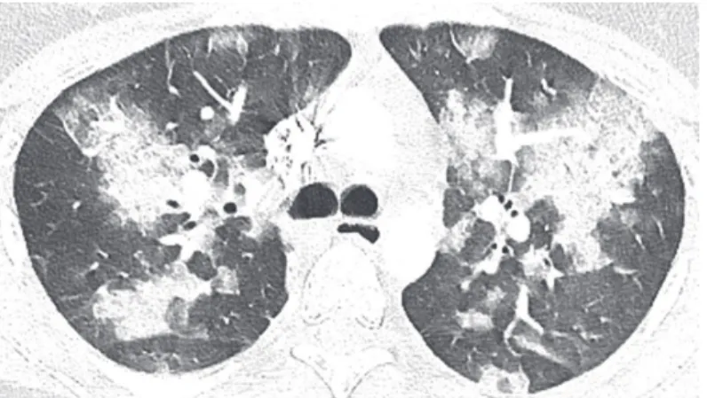
dyspnea (respiratory rate, 38 breaths/min; peripheral oxygen saturation, 81%; pro-brain natriuretic peptide, 2,073 ng/L), together with bilateral inspiratory crackles and a diastolic murmur over the apex. Imaging showed multifocal consolidations surrounded by ground-glass opacities, predominantly in the upper lobes (Figure 1), and no pulmonary embolism. Bronchoscopy showed no endobronchial blood but prominent bronchial vasculature
on the left (Figure 2). The BAL luid was consistently
bloody (90% hemosiderin-laden alveolar macrophages). Echocardiography detected severe mitral stenosis with
classic “hockey stick” morphology of the anterior lealet
and mild aortic regurgitation. The mean pulmonary
artery pressure was 48 mmHg, and the pulmonary artery wedge pressure was 32 mmHg. Mitral valve reconstruction was performed.
Mitral stenosis can provoke hemoptysis.(1) Dilatation of the bronchial vasculature may be the irst hint of
elevated left atrial pressure. The bronchial venous plexus arising from the bronchial arterial circulation is connected to the pulmonary venous circulation. Two thirds of the blood from the venous plexus returns to the pulmonary veins and then to the left atrium.(2,3) An increase in pulmonary venous pressure leads to a
reverse low of blood from the pulmonary veins to the
bronchial venous plexus, visible as engorged bronchial vasculature.
A B C
Figure 1. CT of the chest at the level of the main carina showing bilateral consolidations surrounded by ground glass opacities.
Figure 2. In A, iberoptic bronchoscopy image of the main left bronchus. In B, a closer view at the left secondary upper lobe carina. In C, a close-up of the left superior bronchus showing the lingula and left upper lobe segments. The bronchial mucosa is edematous and contains a network of prominent blood vessels. The vessels are dilated and engorged to a variable extent. At some sites, the vessels are focally distributed, although most parts of the bronchial mucosa exhibit a dense and partially conluent vascular network.
RECOMMENDED READING
1. Wood P. An appreciation of mitral stenosis. I. Clinical features. Br Med J. 1954;1(4870):1051-63; contd. http://dx.doi.org/10.1136/ bmj.1.4870.1051
2. Ohmichi M, Tagaki S, Nomura N, Tsunematsu K, Suzuki A.
Endobronchial changes in chronic pulmonary venous hypertension. Chest. 1988;94(6):1127-32. http://dx.doi.org/10.1378/chest.94.6.1127 3. Baile EM. The anatomy and physiology of the bronchial circulation. J
Aerosol Med. 1996;9(1):1-6. http://dx.doi.org/10.1089/jam.1996.9.1 J Bras Pneumol. 2016;42(5):386-386
386