Radiol Bras. 2015 Nov/Dez;48(6):391–395
391
Aspiration pneumonia in children: an iconographic essay
*
Pneumonia por aspiração na infância: ensaio iconográfico
Oliveira GA, Pessanha LB, Guerra LFA, Martins DLN, Rondina RG, Silva JRP. Aspiration pneumonia in children: an iconographic essay. Radiol Bras. 2015 Nov/Dez;48(6):391–395.
Abstract
R e s u m o
In most cases of aspiration pneumonia in children, the disease is specific to this age group. Clinical and radiological correlation is essential for the diagnosis. The present pictorial essay is aimed at showing typical images of the most common etiologies.
Keywords: Pneumonia; Aspiration; Childhood.
A maioria das pneumonias por aspiração na infância é específica deste grupo etário. Para diagnosticá-las é essencial a correlação clinicorradiológica. O objetivo deste ensaio é mostrar imagens típicas das causas mais frequentes.
Unitermos: Pneumonia; Aspiração; Infância.
* Study developed in the Unit of Radiology and Imaging Diagnosis at Universidade Federal do Espírito Santo (UFES), Vitória, ES, Brazil.
1. MD, Radiologist at Hospital Infantil Nossa Senhora da Glória, Vitória, ES, Bra-zil.
2. MDs, Residents of Radiology at Universidade Federal do Espírito Santo (UFES), Vitória, ES, Brazil.
supplemented by computed tomography and
esophageal-gastroduodenal seriography (EGDS) are almost always
enough to make the diagnosis
(13,18).
DISCUSSION
The function of conducting food from the mouth to the
stomach involves a joint action of the muscles innervated by
the IX, X, XI and XII cranial pairs
(12,19). Due to immaturity,
central nerve system injuries or drugs effects, this mechanism
may be disturbed, and part of the food is diverted into the
airways (Figures 1, 2 and 3). In such situations, radiological
findings are similar to those observed in adult individuals
(13).
Gabriel Antonio de Oliveira
1, Laís Bastos Pessanha
2, Luiz Felipe Alves Guerra
2, Diego Lima Nava Martins
2,
Ronaldo Garcia Rondina
2, Jamine Ronacher Passos Silva
3INTRODUCTION
Recently, the Brazilian radiological literature has been
worried a lot about the relevance of imaging methods in the
improvement of the diagnosis in pediatrics
(1–11). Aspiration
pneumonias result from passage of the oropharyngeal,
esoph-ageal or stomach contents into the lower respiratory tract
(12).
The resulting compromise of the lungs depends on the
na-ture and amount of aspirated material
(12). In the pediatric
group, aspiration occurs most frequently because of
degluti-tion abnormality, congenital malformadegluti-tions and
gastroesoph-ageal reflux. Lipoid pneumonia is more rarely observed and
is always iatrogenic
(13–17). Chest radiography, sometimes
3. MD, Resident of Neurology at Universidade Federal do Espírito Santo (UFES), Vitória, ES, Brazil.
Mailing Address: Dra. Laís Bastos Pessanha. Departamento de Clínica Médica/ CCS/UFES. Avenida Marechal Campos, 1468, Nazareth. Vitória, ES, Brazil, 29043-900. E-mail: [email protected].
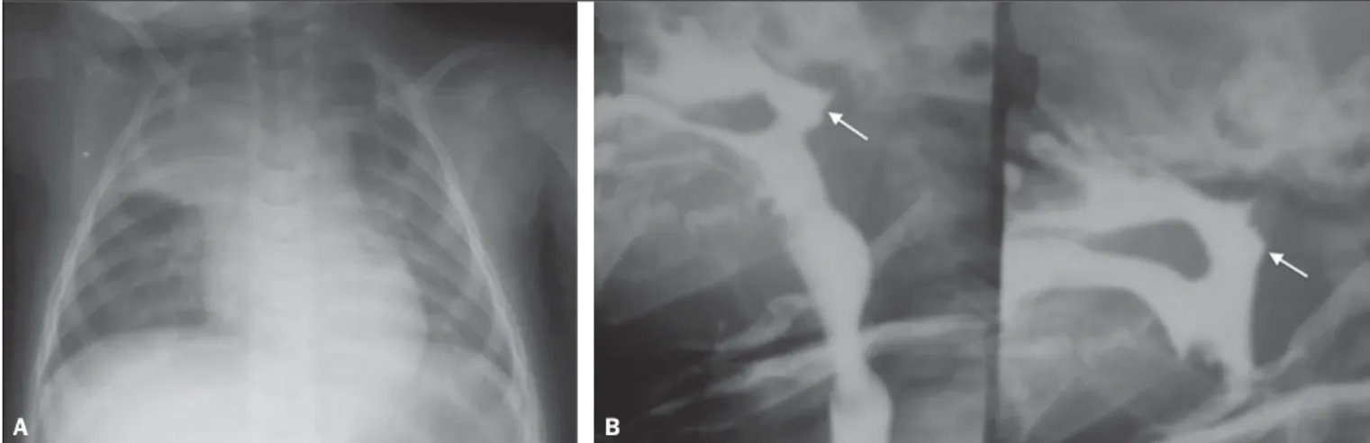
Received February 5, 2014. Accepted after revision September 3, 2014. Figure 1. A neonate with encephalopathy caused by perinatal anoxia, presenting with respiratory symptoms. A: Anteroposterior chest radiography showing opacity in the upper third of the right lung, limited by the horizontal scissure, characterizing involvement of the right upper lobe. B: Deglutition study demonstrating the contrast agent transit into the nasopharynx (arrows), characterizing lack of motor coordination.
Any stasis resulting from narrowing of the esophageal
lumen may lead to aspiration
(18–20). Usually, this does not
occur in cases of acquired achalasia and stenosis, because
children frequently adapt themselves to such conditions.
Esophageal atresia usually is detected and surgically corrected
before causing significant aspiration
(18,19). Amongst those
cases of compression by anomalous vessels, compression by
double aortic arch is the one that most frequently causes
symptoms
(13,21)(Figure 4). The diagnosis of H-type
trache-oesophageal fistula may be late, as contrast-enhanced images
not always can easily demonstrate it
(13,18)(Figure 5).
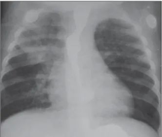
Figure 2. A three-month-old male child with neurological sequelae of congenital toxoplasmosis. Inadvertent contrast aspiration into the bronchial three during EGDS, resulting from non-coordinated deglutition. Chest radiography demonstrating paracardiac opacities corresponding to aspiration bronchopneumonia.
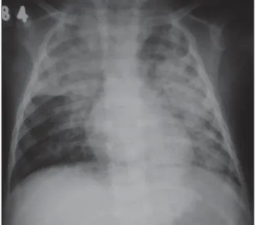
Figure 3. A previously healthy 18-month-old, afebrile male child, presented vom-iting during recovering from anesthesia for palpebral injury suture, progressing to respiratory failure requiring ventilatory assistance. Anteroposterior view of the chest demonstrating opacities in the right upper lobe and in the upper segment of the left lower lobe, which represent usual sites in cases of aspiration occurring with the child in dorsal decubitus.
Figure 4. A seven-month-old male child with repetition pneumonia. Anteroposte-rior view of the chest with esophageal contrast-enhancement. Opacity is observed in the right upper lobe, compatible with pneumonia. Concentric narrowing of the lumen of the proximal esophageal third, with upstream dilatation. Such findings are strongly suggestive of extrinsic compression by double aortic arch. After sur-gical correction, the respiratory symptoms and the esophageal compression dis-appeared.
Figure 5. A six-year-old boy with repetition pneumonia. A: Chest radiography, anteroposterior view showing subtle diffuse opacities in both lungs, confluent in the middle lobe. The caudal sift of the horizontal scissure (arrows) characterizes the presence of atelectatic component. B: Esophagography demonstrating H-type fistula to the trachea (arrow). C: High resolution computed tomography, axial section at the level of the fistula characterized by the dark dot (arrow) between the esophagus (with air) and the trachea. Centrilobular opacities, some of them branch-ing, demonstrating involvement of small airways.
A
C
Figure 6. A twenty-month-old female child presenting with fever and cough. A: Anteroposterior chest radiography revealing the presence of bilateral, diffuse, ill defined, coalescent opacities in the middle lobe, conditioning the partial fading of the cardiac silhouette. B: Computed tomography, axial section identifying bilateral, predomi-nantly central consolidations with air bronchograms. C: EGDS demonstrating reflux. As no clinical and radiological improvement was observed after antibiotic therapy, lung biopsy was indicated and showed foreign body granulomas and vegetal fibers presumably coming from gastroesophageal reflux. After appropriate treatment, clinical and radiological healing was observed.
A
B
C
Figure 7. A neonate with Down syndrome and respiratory symptoms. A: Anteroposterior chest radiography showing left lung with decreased volume and transparency. Fading of the left cardiac silhouette indicates upper lobe atelectasis. B: EGDS. Small bowel transit shows partial obstruction at the level of the second duodenal portion, with appearance suggestive of duodenal diaphragm (windsock sign). C: Secondary gastroesophageal reflux.
A
B
C
Respiratory manifestations stand out in the wide
spec-trum of gastroesophageal reflux disease
(18–21). More than
highlighting the presence of reflux – whose diagnosis is
es-sentially clinical –, EGDS plays a relevant role in the
dem-onstration of either normal or pathological anatomy
(19–21).
In the absence of anatomical alterations, reflux is considered
to be primary, resulting from generally transient
immatu-rity of the distal esophageal high pressure zone
(19,20)(Fig-ure 6). Surgical intervention is indicated in cases of reflux
secondary to partial or total obstruction – usually
hyper-trophic pyloric stenosis or malformations of the second
por-tion of the duodenal arch (Figure 7)
(13,19,20).
Lipoid pneumonia is not related to anatomical or
func-tional anomalies
(13,15). Aspiration occurs because of the use
of mineral oil in the treatment of intestinal constipation
(Figure 8) or as an adjuvant in cases of intestinal subocclusion
caused by
Ascaris lumbricoides
(4). The oil inhibits the cough
reflex and ciliary motion, and silently reaches the alveoli.
Because of the difficulty in removing the oil from the lungs,
such pneumonias present a slow evolution pattern
(14,15).
IMAGING FINDINGS
Aspiration pneumonias involve the alveoli
(12,20,21). The
literature reports a most frequent involvement of the
poste-rior segments of the upper lobes and the upper segments of
the lower lobes
(12,13,18). This happens as aspiration occurs with
the child in dorsal decubitus, like in most gastroesophageal
reflux and vomiting episodes
(12,13). In other situations, such
as tracheoesophageal fistula and lack of motor coordination,
other pulmonary segments may be affected
(19–21)(Figures 2
resolution computed tomography is useful
(13). Aspiration may
result in atelectasis or pneumonia, the latter with or without
atelectatic component
(13). The absence of fever suggests pure
atelectasis
(22)(Figures 3 and 9).
REFERENCES
1. Araújo BCL, Motta MEA, Castro AG, et al. Clinical and video-fluoroscopic diagnosis of dysphagia in chronic encephalopathy of childhood. Radiol Bras. 2014;47:84–8.
2. Sakuno T, Tomita LM, Tomita CM, et al. Sonographic evaluation
of visceral and subcutaneous fat in obese children. Radiol Bras. 2014;47:149–53.
3. Lederman HM. Visceral and subcutaneous fat. Radiol Bras. 2014; 47(3):ix.
4. Fonseca JM, Borém LMA. Cloverleaf skull syndrome: case report. Radiol Bras. 2014;47:189–90.
5. Berdichevski EH, Mattos SG, Bezerra S, et al. Prevalence of acute pyelonephritis and incidence of renal scarring in children under the age of two with urinary tract infection evaluated by 99m
Tc-DMSA renal scintigraphy: the experience of a university hospital. Radiol Bras. 2013;46:30–4.
6. Daud DF, Campos MMF, Fleury Neto LAP. Cardiac tamponade in an infant during contrast infusion through central venous cath-eter for chest computed tomography. Radiol Bras. 2013;46:385– 6.
7. Gun S, Ciantelli GL, Takahashi MAU, et al. Complete renal fusion in a child with recurrent urinary tract infection. Radiol Bras. 2012; 45:233–4.
8. Monteiro AMV, Lima CMAO, Medina P. Is there any influence of breastfeeding on the cerebral blood flow? A review of 256 healthy newborns. Radiol Bras. 2012;45:263–6.
9. Bitencourt AGV, Pinto, PNV, Almeida MFA, et al. Incidence and imaging findings of lymphoma after liver transplantation in chil-dren. Radiol Bras. 2012;45:7–11.
10. Valente M, Oliveira LAN, Carneiro-Sampaio M. Pediatric radiol-ogy: the necessity of a child-friendly diagnosis. Radiol Bras. 2012; 45(5):v.
11. Araújo Filho JAB, Martines JAS, Martines BMR, et al. Inflamma-tory myofibroblastic tumor of the bladder in a child: a case report. Radiol Bras. 2012;45:230–2.
12. Marik PE. Aspiration pneumonitis and aspiration pneumonia. N Engl J Med. 2001;344:665–71.
13. Franquet T, Giménez A, Rosón N, et al. Aspiration diseases: find-ings, pitfalls, and differential diagnosis. Radiographics. 2000;20:673– 85.
14. Oliveira GA, Lamego CM, Vargas PR. Pneumonias lipóides exóge-nas na infância. Rev Assoc Med Bras. 1984;30:34–6.
15. Oliveira GA, Del Caro SR, Bender Lamego CM, et al. Radiographic
Figure 8. Anteroposterior view of chest in a three-year-old boy undergoing treat-ment for constipation with mineral oil e diagnosis of bronchopneumonia refractory to antibiotics. Coalescent opacities in both lungs, with “butterfly wing” distribu-tion. In the clinical context, such a finding allows for the diagnosis of lipoid pneu-monia, with no need for biopsy.
Figure 9. A six-year-old male child said to be asthmatic, presented with dyspnea and sudden chest pain. A: Anteroposterior view showing left hemithorax with decreased volume and transparency, right lung herniation and mediastinal displacement to the left, characterizing left lung atelectasis. B: EGDS demonstrating reflux. After four-day anti-reflux treatment, the symptoms disappeared and chest radiography was normal. C: Normal anteroposterior chest radiography after treatment for gastroesophageal reflux disease.
plain film and CT findings in lipoid pneumonia in infants follow-ing aspiration of mineral oil used in treatment of bowel obstruction by Ascaris lumbricoides. Pediatr Radiol. 1985;15:157–60. 16. Sias SMA, Quirico-Santos TQ. Pneumonia lipóide na infância –
revisão de literatura. Pulmão RJ. 2009;Supl 1:S9–S16.
17. Wolfson BJ, Allen JL, Panitch HB, et al. Lipid aspiration pneumo-nia due to gastroesophageal reflux. A complication of nasogastric lipid feedings. Pediatr Radiol. 1989;19:545–7.
18. Vaughan D, Katkin JP. Chronic and recurrent pneumonias in chil-dren. Semin Respir Infect. 2002;17:72–84.
19. Weir K, McMahon S, Barry L, et al. Oropharyngeal aspiration and pneumonia in children. Pediatr Pulmonol. 2007;42:1024–31. 20. Loughlin GM. Respiratory consequences of dysfunctional
swallow-ing and aspiration. Dysphagia. 1989;3:126–30.
21. Toufen Junior C, Camargo FP, Carvalho CRR. Pneumonia aspira-tiva associada a alterações da deglutição. Relato de caso. Rev Bras Ter Intensiva. 2007;19:118–22.