Racial Differences in Hypogonadal Improvement and
Prostate-Specific Antigen Levels in Hypogonadal Men Treated With
Testosterone Replacement Therapy
Robert M. Coward, Jay Simhan, Culley C. Carson III
Division of Urologic Surgery, The University of North Carolina, Chapel Hill, North Carolina, USA
ABSTRACT
Purpose: To observe hypogonadal men undergoing testosterone replacement therapy (TRT) and assess racial differences
in hypogonadal improvement and prostate-speciic antigen (PSA) levels.
Materials and Methods: In a retrospective analysis, 75 hypogonadal men were followed for an average 34 months after
initiating TRT. Total testosterone and PSA levels were assessed every 6 months, and patients diagnosed with prostatitis or prostate cancer during treatment were excluded.
Results: For 16 African American men, the average age at diagnosis of hypogonadism was 53.5 years, compared with
57.8 years in 59 Caucasian men (p = NS). Pre- and post-treatment testosterone was 219 ng/dL and 310 ng/dL in African American men, and 247 ng/dL and 497 ng/dL in Caucasian men (p = NS). Symptomatic response was 81% in African American men and 93% in Caucasian men (p = NS). Baseline PSA level was 1.32 ng/mL in African American men and 1.27 ng/mL in Caucasian men, and there was no signiicant difference in PSA between racial groups at 6-month intervals, although there was a small decreasing trend in the PSA of African Americans compared with Caucasians.
Conclusions: Hypogonadal African American men have a similar normalization of testosterone and symptomatic response
as hypogonadal Caucasian men, and PSA levels remain stable over time in both groups. In this hypogonadal cohort, in contrast to studies of eugonadal men, higher PSA levels in African Americans were not observed.
Key words: prostate-speciic antigen; hypogonadism; testosterone; continental population groups Int Braz J Urol. 2010; 36: 700-9
INTRODUCTION
Testosterone deiciency syndrome (TDS) is a condition characterized by serum androgen deiciency
that adversely affects the function of multiple organ
systems and negatively impacts quality of life. The
clinical symptoms include sexual dysfunction such as erectile dysfunction and decreased libido, as well as fatigue, depressed mood, impaired cognition, and
decreased muscle mass (1). Although the majority
doi: 10.1590/S1677-55382010000600008
of cases of TDS occur in aging men, for which the
syndrome is commonly referred to as late-onset
hy-pogonadism or andropause, TDS can also occur in younger men. Treatment of TDS with testosterone
replacement therapy (TRT) is appropriate when the testosterone level is below the lower limit of normal,
generally accepted to be 300 ng/dL (2).
An extrapolation from the Massachusetts Male Aging Study found a prevalence of TDS of
their ifth, sixth and seventh decades of life, and as a
result of the expanding population of the elderly, the
incidence of TDS is on the rise (3). Racial differences of the prevalence and incidence of TDS are unknown because epidemiologic series on the subject have been primarily comprised of Caucasian men.
The beneicial effects of TRT in the treatment of TDS are well characterized. As levels of serum tes
-tosterone are normalized, the desired improvements
in sexual function, mood, cognition, and muscle mass
can be achieved. The question of how TRT affects
racial groups differently has not been addressed in
previous studies. It is unknown if different racial groups normalize testosterone levels with the same
vigor, or if the same symptomatic response can be
achieved.
Racial differences of androgen levels of eugonadal men have been previously examined,
although the results of many studies are discordant. While several previous studies have shown African American men to have higher baseline testosterone levels than Caucasian men (4-7), others have shown no differences (8-12). One of the largest prospective
studies of racial differences of serum androgen levels
in eugonadal men found no signiicant differences
in total testosterone, bioavailable testosterone, or
sex-hormone binding globulin levels in 538 African American, 651 Hispanic, and 710 Caucasian men (13).
The average prostate-speciic antigen (PSA) level of African American men has been shown in multiple studies to be higher than the average PSA level of Caucasian men, both in eugonadal men with
a new diagnosis of prostate cancer (14), as well as in
eugonadal men without prostate cancer (15-17). Moul and colleagues examined the PSA level of 541 men
with newly diagnosed prostate cancer and reported
that after adjusting for stage, grade, and age, the mean PSA value for 133 African American men was 14.00 ng/mL compared with 8.29 ng/mL for 408 Caucasian men (p < 0.001) (14). To study racial differences in PSA level and PSA density in men without prostate cancer, a retrospective study of 826 consecutive men who underwent prostate biopsy was performed. Of the 498 men with negative biopsies, serum PSA lev
-els (7.97 ng/mL versus 4.30 ng/mL, p < 0.0001) and calculated PSA density (0.19 versus 0.11, p < 0.0001)
were signiicantly higher in African American men than in Caucasian men (15).
This difference in PSA has been shown to not be the result of racial differences in prostate size. In a study of 11,011 men who underwent prostatectomy, race and serum PSA values were correlated to prostate weight, and although serum PSA was 15% higher in African American men there were no signiicant associations between race and prostate size (17). As described above, racial differences of androgen levels have not been deinitely proven to be higher in African Americans even though this potential dif
-ference may partially explain the dif-ference in PSA
levels given the strong positive association between
prostate size and serum PSA value (18). Racial dif -ferences in hormones such as estrogen, insulin, and
insulin-like-growth factor that also regulate prostate size may augment or even negate the potential an
-drogenic difference. Other theories that have been investigated to explain the racial difference in PSA include genetic polymorphisms. African Americans are more likely to possess polymorphisms favoring increased androgen activity, including shorter CAG
trinucleotide repeats in the androgen receptor gene
and a less active CYP3A4 gene which is involved with the deactivation of testosterone (19,20).
Both short-term and long-term changes in the PSA levels of hypogonadal men undergoing TRT have been reported. A review of 18 short-term TRT trials found an average initial increase in PSA level of 0.3 ng/mL, and the same authors reported that in six
studies of short-term TRT in slightly older men with
TDS, there was a higher average increase in PSA level of 0.43 ng/mL (21). Rhoden and Morgentaler reported that 579 hypogonadal men on TRT from nine differ -ent studies, with a duration of treatm-ent of about one
year, had a mean PSA level increase of 0.46 ng/mL (22). Wang et al. reported that in 163 hypogonadal
men on TRT for 3 years, after a small initial increase
in PSA level of 0.26 ng/mL at 6 months, there were no further PSA level increases with continued TRT over the following 3 years (23). In another study of 187 hypogonadal men on TRT, there were no signii
-cant differences in PSA after one year (24). With the
longest follow-up data available, a recent study of
least 5 years (25). The racial differences in PSA levels of hypogonadal men undergoing TRT are unknown.
The aim of the present study was to
retro-spectively review patients undergoing TRT for TDS
to evaluate the racial differences in hypogonadal
improvement and PSA levels. We hypothesized that the normalization of testosterone levels, as well as
the symptomatic responses, will be similar among
hypogonadal Caucasian and African American men undergoing TRT. Based on previous studies of race and PSA, we also hypothesized that the PSA levels of African American men with TDS will be higher than the PSA levels of Caucasian men.
MATERIALS AND METHODS
The medical records of all men evaluated in
the Urology Clinic at our institution from January 2000 through June 2006 were queried for a diag
-nosis of hypogonadism by International Center for Diseases-9 (ICD-9) code 257.2, yielding 267 men initially. In order to be diagnosed with hypogonadism,
the patient must have reported symptoms in addition
to having had a serum testosterone level below 300 ng/dL. Men then must have been seen in our clinic at least two or more times with the diagnosis.
For inclusion in the study, full demographic information must have been available, and each
pa-tient must have had an initial testosterone and PSA level. The patients must have been started on TRT
and then had at least one subsequent testosterone
level and PSA level measured within one year of initiating TRT. All men included in the study had a normal baseline PSA level prior to initiating TRT and
underwent routine digital rectal examinations during
treatment. Patients with a biopsy-proven diagnosis or
recent history of prostatitis prior to treatment were
ex-cluded from the study. Four men were diagnosed with prostate cancer while on TRT and were excluded. The
decision to perform prostate biopsy during TRT was
made at the discretion of the clinician (CCC) without
strict criteria, but all four men with diagnosed cancer
had an abnormally increased PSA velocity with two consecutive PSA elevations. Further details of the men
diagnosed with prostate cancer have been previously
published (25). One Asian male, and one male with
race listed as “other,” were excluded secondary to
insuficient numbers. After applying these criteria, 75 men with TDS were selected for a complete chart review.
Each patient’s age at the time of diagnosis,
and race, were identiied as demographic data. The
patient’s symptomatic response to treatment was
determined subjectively by the patient in
follow-up clinic visits as either a positive response or no
response. Comorbid conditions including benign prostatic hyperplasia (BPH), coronary artery disease, diabetes mellitus type 2, hypertension, hyperlipid
-emia, and obstructive sleep apnea were also assessed. BPH was speciically assessed by the clinician prior
to initiating TRT by querying for lower urinary tract
symptoms and by digital rectal examination. The other comorbid conditions were identiied through chart review rather than ICD-9 coding, and were not deined by strict criteria.
Testosterone and PSA levels were drawn during a morning clinic, generally between 9 a.m. and 12 p.m., although the exact time varied and was not recorded. The PSA level was performed by UNC McLendon Laboratories (Chapel Hill, NC), and the
total and free testosterone levels were performed by
Mayo Medical Laboratories (Rochester, MN). The
free testosterone was calculated with a direct
radioim-munoassay. The form of TRT was selected by patient
preference, and both the form and dose were not
con-sistently available. The target testosterone level was a eugonadal level (> 300 ng/dL) with symptomatic
improvement, and the dose was titrated accordingly
by the clinician.
The patients were evaluated in regular six-month intervals, and rarely if the treatment dates
were not exactly at 6 months they were rounded to the closest six-month interval. Basic statistical data was obtained at each 6-month interval for all variables. The data were compared using a Student’s-t-test, with p < 0.05 considered statistically signiicant.
RESULTS
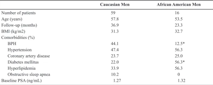
-pared with 57.8 years (range 25-82) in 59 Caucasian men (p = NS). The mean follow-up for Caucasian men was 36.9 months compared with 23.3 months for African American men (p = NS). Among comorbid conditions, Caucasian men had more BPH (44.1% versus 12.5%, p < 0.05), and African American men had a higher prevalence of diabetes mellitus type 2 (56.3% versus 22%, p < 0.05).
Pre- and post-treatment (after 1 year) total testosterone levels were 247 ng/dL and 497 ng/dL in Caucasian men (p < 0.05), and 219 ng/dL and 310 ng/dL in African American men (p < 0.05). When the
pre-treatment total testosterone level of 247 ng/dL for the Caucasian cohort was compared with the pre-treat
-ment level of 219 ng/dL in African Americans, there was no signiicant difference (p = NS). Similarly,
the post-treatment total testosterone levels were not statistically different when the two racial cohorts
were compared, 497 ng/dL in Caucasian men versus 310 ng/dL in African American men (p = NS). Total
testosterone levels remained eugonadal at 3 years
after initiating TRT, with 355.3 ng/dL in Caucasian men and 326.3 ng/dL in African American men. Free testosterone levels were 7.68 ng/dL and 9.14
Table 1 – Patient characteristics.
Caucasian Men African American Men
Number of patients 59 16
Age (years) 57.8 53.5
Follow-up (months) 36.9 23.3
BMI (kg/m2) 31.3 32.7
Comorbidities (%)
BPH 44.1 12.5*
Hypertension 47.4 56.3
Coronary artery disease 23.7 25.0
Diabetes mellitus 22.0 56.3*
Hyperlipidemia 33.9 56.3
Obstructive sleep apnea 10.2 0
Baseline PSA (ng/mL) 1.27 1.32
*p < 0.05 BMI = body mass index; BPH = benign prostatic hyperplasia; PSA =prostate-speciic antigen.
Table 2 – Racial differences in biochemical and symptomatic responses to TRT.
Caucasian Men African American Men Total testosterone (ng/dL)
Baseline 246.6 218.9
1 year 496.6 310.1
3 years 355.3 326.3
Free testosterone (ng/dL)
Baseline 7.68 9.14
1 year 15.63 9.97
3 years 13.23 8.95
Symptomatic Response (%) 93.2 81.3
ng/dL in Caucasian men and African American men, respectively. At one year, free testosterone after TRT increased to 15.63 ng/dL in Caucasians and 9.97 ng/dL in African Americans, and these values were consis
-tent at 3 years, with 13.23 ng/dL for Caucasians and 8.95 ng/dL in African Americans. The symptomatic response was 81% in African American men and 93% in Caucasian men, although this was not statistically different. The racial differences in the biochemical and symptomatic response are listed in Table-2.
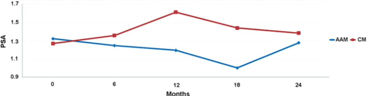
The baseline PSA level was 1.32 ng/mL in African American men and 1.27 ng/mL in Caucasian men. The changes in PSA levels in 6-month intervals are listed in Table-3 and are displayed in Figure-1. At 6-month intervals, there was a small decreasing trend in the PSA level of African American men over 2 years of treatment compared with Caucasian men. However, none of the differences in PSA levels be -tween the two racial cohorts at each time point were
statistically signiicant, and the PSA levels remained stable over the 2 year period in all men.
COMMENTS
In hypogonadal men treated with TRT, ra-cial differences in hypogonadal improvement have
not been previously reported. In the present study, symptomatic hypogonadal African American men had comparable baseline testosterone levels as Caucasian men. Additionally, the symptomatic and biochemical
responses of total testosterone achieved at 1 year and
at 3 years were similar between the two cohorts. The free testosterone levels of African American men did
not respond to TRT with the same vigor as the total
testosterone. Sex hormone binding globulin levels
were unavailable in this study, and therefore the dis-crepancy between total and free testosterone cannot
be further explained. These data suggest that there
are no racial differences in hypogonadal
improve-ment among hypogonadal Caucasian and African American men undergoing TRT. This is consistent
with eugonadal men, as studies vary as to whether a
racial difference in androgen levels exists (4-12).
Table 3 –Racial differences in PSA level.
*p = NS; PSA = prostate-speciic antigen; SD = standard deviation. Caucasian Men
PSA Level (ng/mL) ± SD PSA Level (ng/mL) ± SDAfrican American Men
Baseline 1.27 ± 1.04 1.32 ± 0.78
6 months 1.39 ± 0.98 1.24 ± 0.64
12 months 1.61 ± 1.47 1.19 ± 0.64
18 months 1.44 ± 1.43 1.00 ± 0.53
24 months 1.38 ± 1.04 1.28 ± 0.49
Figure 1 – Comparing prostate-speciic antigen (PSA) change of African American men (AAM) versus Caucasian men (CM) treated
Population-based studies have shown that African American men have higher PSA levels than Caucasian men (14-17). The racial differences in PSA levels of hypogonadal men, and how the PSA
changes during TRT among different races, have not
been published before this study. Several previous studies have reported that PSA either remains stable, or has a small initial increase that stabilizes over time (21-25). In the small cohort of hypogonadal men in the present study, the baseline PSA was not different between the racial groups, with 1.32 ng/mL in African American men and 1.27 ng/mL in Cauca
-sian men (p = NS). After initiation of TRT, the PSA was followed every 6 months for a two year period,
and there was a small decreasing trend without
statistical signiicance in the PSA levels of African American men over 2 years of treatment compared with Caucasian men, although the PSA remained stable over time in all men. The higher mean PSA level of eugonadal African American men that has
been previously reported was not observed in our
cohort of hypogonadal men. Furthermore, after two
years of TRT resulting in a corrected, eugonadal
total testosterone level, the PSA did not increase in 6-month intervals. These data suggest that androgens are less likely to play a role in the higher baseline PSA levels of eugonadal African American men;
however, further research is necessary to explain
the observed racial differences in the PSA levels of eugonadal men.
This study has several important
limita-tions. All of the standard limitations inherent to a retrospective chart review apply to this study. The sample size was small with 75 men, and the
two racial groups were not well matched, with
only 16 African American men compared with 59 Caucasian men. It can be argued that with only 16 African American men that the study is under -powered to identify a true racial difference if there
indeed is one. This discrepancy can lead to selec
-tion bias that can adversely affect the results. The follow-up period for the Caucasian men was longer with a mean 36.9 months versus 23.3 months for the African Americans. The reason for the differ
-ent follow-up period is simply that the African American men were lost to follow-up earlier, and this is a limitation of a retrospective study. It is
unclear whether the discrepancy in follow-up time affects the results, although ideally the follow-up
period would be equal between the two cohorts. The
doses of testosterone and delivery system were not consistently available and therefore were not report-able, and these data would be useful correlative data
to the biochemical and symptomatic response data.
The assessment of a symptomatic response was only
quantiied in this study by either a positive response
or no response, and ideally a validated questionnaire to assess the response would be preferred, although to
our knowledge one does not exist. The prevalence of BPH was higher in the Caucasian group, 44.1% versus 12.5% (p < 0.05), and because of the positive association between prostate size and serum PSA value, these differences in BPH could affect the base
-line PSA level (18). Additionally, diabetes mellitus occurs in a higher incidence in the African American group at 56.3% versus 22.0% in the Caucasian group (p < 0.05). Because hypogonadism is a component
of metabolic syndrome and can be associated with insulin resistance and diabetes mellitus, the racial difference in the prevalence of diabetes mellitus may
confound the analysis (26). There are too few subjects to adjust for the difference in diabetes mellitus, but it
can explain the difference of the baseline testosterone
level and ultimately the PSA.
CONCLUSIONS
With these limitations in mind, and under-standing that this is a small, retrospective study, we
can make several useful conclusions based on the fact that this is the irst study to report the racial differ
-ences in hypogonadal improvement and PSA changes in men undergoing TRT. Hypogonadal African American men had a similar normalization of total testosterone and symptomatic response as Caucasian men undergoing TRT. PSA levels over time trended slightly lower in hypogonadal African American men on TRT compared with Caucasian men, although the PSA remained stable in all men. In this hypogonadal
cohort, in contrast to other studies of eugonadal men,
CONFLICT OF INTEREST
None declared.
REFERENCES
1. American Association of Clinical Endocrinologists: American Association of Clinical Endocrinologists
medical guidelines for clinical practice for the evalua-tion and treatment of hypogonadism in adult male
pa-tients – 2002 update. Endocr Pract 2002; 8: 439-56. 2. Bhasin S, Cunningham GR, Hayes FJ, Matsumoto AM,
Snyder PJ, Swerdloff RS, et al.: Testosterone therapy in adult men with androgen deiciency syndromes: an endocrine society clinical practice guideline. J Clin Endocrinol Metab. 2006; 91: 1995-2010. Erratum in: J Clin Endocrinol Metab. 2006; 91: 2688.
3. Araujo AB, O’Donnell AB, Brambilla DJ, Simpson WB, Longcope C, Matsumoto AM, et al.: Prevalence and incidence of androgen deiciency in middle-aged and older men: estimates from the Massachusetts Male Aging Study. J Clin Endocrinol Metab. 2004; 89: 5920-6.
4. Ellis L, Nyborg H: Racial/ethnic variations in male testosterone levels: a probable contributor to group differences in health. Steroids. 1992; 57: 72-5. 5. Gapstur SM, Gann PH, Kopp P, Colangelo L, Long
-cope C, Liu K: Serum androgen concentrations in young men: a longitudinal analysis of associations with age, obesity, and race. The CARDIA male hormone study. Cancer Epidemiol Biomarkers Prev. 2002; 11: 1041-7.
6. Winters SJ, Brufsky A, Weissfeld J, Trump DL, Dyky MA, Hadeed V: Testosterone, sex hormone-binding globulin, and body composition in young adult African American and Caucasian men. Metabolism. 2001; 50: 1242-7.
7. Wu AH, Whittemore AS, Kolonel LN, John EM, Gal
-lagher RP, West DW, et al.: Serum androgens and sex
hormone-binding globulins in relation to lifestyle
fac-tors in older African-American, white, and Asian men in the United States and Canada. Cancer Epidemiol Biomarkers Prev. 1995; 4: 735-41.
8. Abdelrahaman E, Raghavan S, Baker L, Weinrich M, Winters SJ: Racial difference in circulating sex hormone-binding globulin levels in prepubertal boys. Metabolism. 2005; 54: 91-6.
9. Asbell SO, Raimane KC, Montesano AT, Zeitzer KL, Asbell MD, Vijayakumar S: Prostate-speciic antigen
and androgens in African-American and white normal subjects and prostate cancer patients. J Natl Med Assoc. 2000; 92: 445-9.
10. Platz EA, Rimm EB, Willett WC, Kantoff PW, Giovan
-nucci E: Racial variation in prostate cancer incidence and in hormonal system markers among male health professionals. J Natl Cancer Inst. 2000; 92: 2009-17. 11. Ross R, Bernstein L, Judd H, Hanisch R, Pike M,
Henderson B: Serum testosterone levels in healthy young black and white men. J Natl Cancer Inst. 1986; 76: 45-8.
12. Rohrmann S, Nelson WG, Rifai N, Brown TR, Dobs A, Kanarek N, et al.: Serum estrogen, but not testos
-terone, levels differ between black and white men in a nationally representative sample of Americans. J Clin Endocrinol Metab. 2007; 92: 2519-25.
13. Litman HJ, Bhasin S, Link CL, Araujo AB, McKinlay JB: Serum androgen levels in black, Hispanic, and white men. J Clin Endocrinol Metab. 2006; 91: 4326-34. 14. Moul JW, Sesterhenn IA, Connelly RR, Douglas T,
Srivastava S, Mostoi FK, et al.: Prostate-speciic an -tigen values at the time of prostate cancer diagnosis in
African-American men. JAMA. 1995; 274: 1277-81. 15. Henderson RJ, Eastham JA, Culkin DJ, Kattan MW,
Whatley T, Mata J, et al.: Prostate-speciic antigen (PSA) and PSA density: racial differences in men without prostate cancer. J Natl Cancer Inst. 1997; 89: 134-8.
16. Morgan TO, Jacobsen SJ, McCarthy WF, Jacobson DJ, McLeod DG, Moul JW: Age-speciic reference ranges for prostate-speciic antigen in black men. N Engl J Med. 1996; 335: 304-10.
17. Mavropoulos JC, Partin AW, Amling CL, Terris MK, Kane CJ, Aronson WJ, et al.: Do racial differences in prostate size explain higher serum prostate-speciic antigen concentrations among black men? Urology. 2007; 69: 1138-42.
18. Roehrborn CG, Boyle P, Gould AL, Waldstreicher J: Serum prostate-speciic antigen as a predictor of pros
-tate volume in men with benign prostatic hyperplasia. Urology. 1999; 53: 581-9.
19. Irvine RA, Yu MC, Ross RK, Coetzee GA: The CAG and GGC microsatellites of the androgen receptor gene are in linkage disequilibrium in men with prostate cancer. Cancer Res. 1995; 55: 1937-40.
EDITORIAL COMMENT
In the current article the authors evaluated the effects of testosterone replacement therapy (TRT) assessing racial differences regarding symptomatic
response and prostate speciic antigen (PSA) varia
-tion.
Regarding symptomatic response it is inter-esting to observe that satisfactory clinical response
was reported by 81 and 93% (p > 0.05) of African American and Caucasian men, respectively. Even with
a potential selected bias related to a higher number
of Caucasian men (59 vs. 16) when compared with African American individuals as well as a numeri
-cally, but not signiicantly, period of follow-up in the Caucasian Group (36.9 vs. 23.3 months, respectively).
21. Bhasin S, Singh AB, Mac RP, Carter B, Lee MI, Cun
-ningham GR: Managing the risks of prostate disease during testosterone replacement therapy in older men: recommendations for a standardized monitoring plan. J Androl. 2003; 24: 299-311.
22. Rhoden EL, Morgentaler A: Inluence of demographic
factors and biochemical characteristics on the
pros-tate-speciic antigen (PSA) response to testosterone replacement therapy. Int J Impot Res. 2006; 18: 201-5.
23. Wang C, Cunningham G, Dobs A, Iranmanesh A, Matsumoto AM, Snyder PJ, et al.: Long-term testos
-terone gel (AndroGel) treatment maintains beneicial
effects on sexual function and mood, lean and fat mass,
and bone mineral density in hypogonadal men. J Clin Endocrinol Metab. 2004; 89: 2085-98.
24. El-Sakka AI, Hassoba HM, Elbakry AM, Hassan HA: Prostatic speciic antigen in patients with hypogonad
-ism: effect of testosterone replacement. J Sex Med. 2005; 2: 235-40.
25. Coward RM, Simhan J, Carson CC 3rd: Prostate-speciic antigen changes and prostate cancer in hy -pogonadal men treated with testosterone replacement
therapy. BJU Int. 2009; 103: 1179-83.
26. Makhsida N, Shah J, Yan G, Fisch H, Shabsigh R: Hypogonadism and metabolic syndrome: implications for testosterone therapy. J Urol. 2005; 174: 827-34.
Accepted after revision: May 24, 2010
Correspondence address: Dr. Robert Matthew Coward
2113 Physicians Ofice Bldg CB#7235 170 Manning Dr
Chapel Hill, NC, 27599-7235, USA Fax: + 1 919 966-0098
E-mail: [email protected]
This is a very high rate of clinical response and may
relect strict inclusion criteria, specially related to the levels of baseline testosterone (218 and 246 ng/dL). An important limitation of the current study is the fact that the authors considered only subjective evaluation to characterize clinical response. This aspect limits major considerations regarding clinical response evaluation. Certainly, the major improvements were related to patient sexual concerns. However, appropri
-ate objective evaluation is recommended and there are several tools available in the literature for this purpose. A high rate of clinical response (overall 70%) to TRT
could also be observed in a recent study, Rhoden and
study the irst 3 months response was a pivotal issue in maintaining men on TRT.
Another aspect analyzed in the current study was the inluence of TRT on PSA levels regarding race. It is well recognized that African America men are of greater risk for developing prostate cancer and that PSA is the most important marker for early diag
-nosis. There was no signiicant increase in PSA levels
before and after initiating TRT independently of the
race group. In a review of the literature of 579 hypo
-gonadal men under TRT the mean increase in PSA levels was 0.46 ng/mL (2). Some authors observed
in their series that after a small initial increase (mean
0.26 ng/mL), no further increases in general was observed (3). Also other authors (2) demonstrated,
that in a selected population free of prostate cancer,
based in a prior biopsy before starting TRT, that PSA decreased in 21%, unchanged in 22%, and increased in 57% cases. All of this information supports the fact of the limited inluence of testosterone levels and PSA levels (4). One of the possible explanations regarding
this issue is the fact that exogenous testosterone does not raise intraprostatic concentrations of testosterone
or dihydrotestosterone, suggesting a saturation model. Based on this concept androgens exert their prostatic
effects primarily via binding to the androgen receptor
(AR), and maximal androgen-AR binding is achieved
at serum testosterone concentrations well below
the physiologic range. Finally, prostate function is
exquisitely sensitive to variations in androgen con-centrations at very low concon-centrations, but becomes insensitive to changes in androgen concentrations at
higher levels (5). Based on this concept the current
study demonstrates that testosterone levels have a
lim-ited effect on changes in PSA levels independently of
race when hypogonadal (non-castrated) are submitted to TRT restoring testosterone levels to an eugonadal
status.
REFERENCES
1. Rhoden EL, Morgentaler A: Symptomatic response rates to testosterone therapy and the likelihood of completing 12 months of therapy in clinical practice. J Sex Med. 2010; 7: 277-83.
2. Rhoden EL, Morgentaler A: Inluence of demographic
factors and biochemical characteristics on the
pros-tate-speciic antigen (PSA) response to testosterone replacement therapy. Int J Impot Res. 2006; 18: 201-5.
3. Wang C, Cunningham G, Dobs A, Iranmanesh A, Mat
-sumoto AM, Snyder PJ, et al.: Long-term testosterone gel (AndroGel) treatment maintains beneicial effects
on sexual function and mood, lean and fat mass, and
bone mineral density in hypogonadal men. J Clin Endocrinol Metab. 2004; 89: 2085-98.
4. Morgentaler A, Schulman C: Testosterone and prostate safety. Front Horm Res. 2009; 37: 197-203.
5. Morgentaler A, Traish AM: Shifting the paradigm of testosterone and prostate cancer: the saturation model and the limits of androgen-dependent growth. Eur Urol. 2009; 55: 310-20.
Dr. Ernani L. Rhoden
Section of Urology Porto Alegre Federal University of Health Sciences Rio Grande do Sul, BrazilE-mail: [email protected]
EDITORIAL COMMENT
The diminution in serum androgen levels in
aged men has been extensively studied. The mecha -nisms of this phenomenon have not been fully illus-trated, and are probably multifactorial, involving the
hypothalamic-pituitary-testicular axis. The greatest
concerns regarding testosterone replacement therapy
is the fear of causing or promoting prostate cancer. A
decline in testicular function with a consequent
function and growth of the prostate and may contrib-ute to the development of prostate cancer and benign
prostatic hypertrophy (2).
The effect of parenteral testosterone
replace-ment therapy on prostatic speciic antigen (PSA) level
or the development or growth of prostate cancer is
unclear. This has prompted many investigators to investigate the effect of this treatment on serum PSA levels in hypogonadal men with erectile dysfunction. Previous studies have shown no signiicant differ -ences between the baseline levels of mean, median,
and range of PSA and categories of PSA level (nor -mal, borderline, high) at one year post-testosterone
therapy (3). In the present study, the authors elegantly demonstrated that Hypogonadal African American men have a similar normalization of testosterone and symptomatic response as hypogonadal Cauca
-sian men, and PSA levels remained stable over time in both groups. However, more long-term studies
are warranted to further investigate the relationship
between testosterone replacement and PSA level and
to better clarify the effects of parenteral testosterone replacement therapy on the development or growth
of prostate cancer.
REFERENCES
1. Gray A, Berlin JA, McKinlay JB, Longcope C: An
examination of research design effects on the
asso-ciation of testosterone and male aging: results of a meta-analysis. J Clin Epidemiol. 1991; 44: 671-84. 2. Bhasin S, Singh AB, Mac RP, Carter B, Lee MI, Cun
-ningham GR: Managing the risks of prostate disease during testosterone replacement therapy in older men: recommendations for a standardized monitoring plan. J Androl. 2003; 24: 299-311.
3. El-Sakka AI, Hassoba HM, Elbakry AM, Hassan HA: Prostatic speciic antigen in patients with hypogonad
-ism: effect of testosterone replacement. J Sex Med. 2005; 2: 235-40.