Flagella and Head Plasma Membrane Proteins from Sea
Bream Spermatozoa: Effect of Antifreeze Proteins
Loredana Zilli1*, Jose´ Beira˜o2, Roberta Schiavone1, Maria Paz Herraez2, Antonio Gnoni3, Sebastiano Vilella1
1Department of Biological and Environmental Sciences and Technologies, University of Salento, Lecce, Italy,2Department of Molecular Biology, University of Leo´n, Leo´n, Spain,3Department of Basic Medical Sciences, Neurosciences and Sense Organs, University of Bari, Bari, Italy
Abstract
Cryopreservation induces injuries to fish spermatozoa that in turn affect sperm quality in terms of fertilization ability, motility, DNA and protein integrity and larval survival. To reduce the loss of sperm quality due to freezing-thawing, it is necessary to improve these procedures. In the present study we investigated the ability of two antifreeze proteins (AFPI and AFPIII) to reduce the loss of quality of sea bream spermatozoa due to cryopreservation. To do so, we compared viability, motility, straight-line velocity and curvilinear velocity of fresh and (AFPs)-cryopreserved spermatozoa. AFPIII addition to cryopreservation medium improved viability, motility and straight-line velocity with respect to DMSO or DMSO plus AFPI. To clarify the molecular mechanism(s) underlying these findings, the protein profile of two different cryopreserved sperm domains, flagella and head plasma membranes, was analysed. The protein profiles differed between fresh and frozen-thawed semen and results of the image analysis demonstrated that, after cryopreservation, out of 270 proteins 12 were decreased and 7 were increased in isolated flagella, and out of 150 proteins 6 showed a significant decrease and 4 showed a significant increase in head membranes. Mass spectrometry analysis identified 6 proteins (4 from isolated flagella and 2 present both in flagella and head plasma membranes) within the protein spots affected by the freezing-thawing procedure. 3 out of 4 proteins from isolated flagella were involved in the sperm bioenergetic system. Our results indicate that the ability of AFPIII to protect sea bream sperm quality can be, at least in part, ascribed to reducing changes in the sperm protein profile occurring during the freezing-thawing procedure. Our results clearly demonstrated that AFPIII addition to cryopreservation medium improved the protection against freezing respect to DMSO or DMSO plus AFPI. In addition we propose specific proteins of spermatozoa as markers related to the procedures of fish sperm cryopreservation.
Citation:Zilli L, Beira˜o J, Schiavone R, Herraez MP, Gnoni A, et al. (2014) Comparative Proteome Analysis of Cryopreserved Flagella and Head Plasma Membrane Proteins from Sea Bream Spermatozoa: Effect of Antifreeze Proteins. PLoS ONE 9(6): e99992. doi:10.1371/journal.pone.0099992
Editor:Chris D. Wood, Universidad Nacional Auto´noma de Me´xico, Mexico ReceivedOctober 30, 2013;AcceptedMay 21, 2014;PublishedJune 18, 2014
Copyright:ß2014 Zilli et al. This is an open-access article distributed under the terms of the Creative Commons Attribution License, which permits unrestricted
use, distribution, and reproduction in any medium, provided the original author and source are credited.
Funding:This work was supported by Apulia Region (Italy) under the program between the Apulia Region and the University of Salento. The funders had no role in study design, data collection and analysis, decision to publish, or preparation of the manuscript.
Competing Interests:The authors have declared that no competing interests exist. * Email: [email protected]
Introduction
Fish sperm cryobanks are considered a useful tool for genetic management and artificial reproduction [1,2]. For this purpose it is necessary to preserve only high quality gametes and to develop freezing methods, which allows the maintenance of the charac-teristics of spermatozoa. Although cryopreservation protocols of sea bream spermatozoa have been well established [3,4], some degradation of sperm quality compared to fresh sperm are still reported [4,5]. The freezing–thawing procedure affects DNA integrity [4,6–8], membrane lipids [5,9,10], sperm motility [11– 14], fertilization ability [1,15,16] and larval survival [17].
Antifreeze proteins (AFPs) are present in the body fluid of cold water marine fish and some terrestrial invertebrates [18–20]. AFPs are a class of peptides that reversibly inhibit ice crystal growth through a non-colligative mechanism [21–23]. Three types are commercially available: namely, AFP types I and III, and antifreeze glycoproteins (AFGP). AFPs have a dual effect in low-temperature storage: inducing ice nucleation and inhibiting
re-crystallization. In the absence of the aggregation effect, AFPs act as re-crystallization inhibitors and may mitigate cryoinjury; on the other hand, when aggregation occurs, the AFPs serve as ice nucleators and lead to cell membrane damage [24]. AFPs have been applied during low-temperature preservation in different cell types [25,26]. AFPs can also interact with plasma membrane at low-temperatures, as demonstrated for AFPI in liposomes [27].
phosphoryla-tion state) due to the freezing-thawing procedure may contribute to the observed changes in spermatozoa quality [12,32–34].
The aim of our work was to evaluate the ‘‘protective’’ effect of the AFPs on sperm proteins during freezing-thawing procedure and, also, to identify specific proteins of flagella and head plasma membranes spermatozoa as potential markers related to the procedures of fish sperm cryopreservation. In particular, we analyzed by 2-DE associated with Nano-LC mass spectrometry the effect of AFPs on the pattern of sperm proteins extracted from isolated flagella and head plasma membranes. Our results show a higher ability of AFPIII, as compared to AFPI, in the protection of sperm proteins during the freezing-thawing procedure. AFPI and AFPIII differently affect the protein profile of the two investigated domains.
Materials and Methods
All chemicals, unless otherwise stated, were purchased from Sigma-Aldrich (St. Louis, MO) and were reagent grade or higher.
Ethics Statement
Fish handling was in accordance with the European Union Directive (EEC, 1986) for the protection of animals used for experimental and other scientific purposes. Field studies were performed at the local fish farm (MARIBRIN) located to Contrada Pandi, 72100 Brindisi (BR) – Apulia, they did not involve endangered or protected species and did not implicate the sacrifice of animals. Procedures relating to care and use animals were approved by the Ethics Committee from University of Salento in accordance with the European Union regulations.
Fish and sperm collection
Milt collection was performed in three different samplings, during late autumn (November), early winter (December) and midwinter (January) at the local fish farm (MARIBRIN) located to Contrada Pandi, 72100 Brindisi (BR) – Apulia. The study was carried out on a sexually mature broodstock of reared gilthead sea
bream (Sparus aurata)males. The broodstock was preserved in an
indoor tank at a density of 0.6 kg/m3. The broodstock tank was
replenished with seawater at a rate of 1 l/sec, while compressed air was provided through air stones. Glithead sea bream were given pellets daily, and fresh fish food was provided once a week. The
water temperature ranged between 13.5uC and 15.5uC, and the
salinity was 34.2%. To avoid fish stress, before sample collection
the fish were anesthetized in 0.1 ml/l 2-phenoxy ethanol. Sperm was collected by applying gentle abdominal pressure to extrude milt that was removed from the gonopore with a syringe. Before collection the urogenital pore was cleaned to eliminate urine, water, mucus and faeces. This procedure was performed as quickly as possible and the fish were returned to anesthetic-free water to recover and were observed for at least one hour to check their health condition. The milt was transferred to a vial and kept at
4uC for 30–60 min, until its use. In each sampling 7 or 8 sperm
samples (from different fish) were collected. The first step was the measurements of the spermatozoa motility parameters and viability of fresh sperm. The six semen samples that had the best quality parameters were used for the cryopreservation procedure and for the extraction of proteins.
Cryopreservation procedure
Sperm motility and viability were analyzed and proteins extracted for four different experimental conditions: fresh (control) and cryopreserved sperm using the three different cryoprotectants as described below. Briefly, sperm was diluted 1/6 (v/v) in the
extender solution (1% NaCl plus 5% DMSO) with and without
AFPI or AFPIII (1mg/ml) and loading it in 0.5 ml French straws.
Sperm cryopreservation was performed as reported previously [5]. Preliminary experiments were conducted to test the best AFPI and AFPIII (A/F Protein Canada Inc., St John’s, Canada) concentra-tions (Table 1).
Computer Assisted Sperm Analysis (CASA)
Sperm movement was videotaped using a Nikon Alphaphot 2 microscope with an x20 negative phase objective and a Sony CCD black and white video camera (SSC-M188CE). Each sperm sample was diluted 1:100 with non-activating medium (NAM, in
mg/ml: NaCl 3.5, KCl 0.11, MgCl21.23, CaCl20.39, NaHCO3
1.68, glucose 0.08, BSA 10, pH 7.7) and motility was initiated by
addition of seawater (,13uC) (in a dilution 1:10 sperm:sea water).
The measure of motility started after 15 seconds, the time need to spot and mix sperm and sea water on well of multitest slide (12 well, ICN, Basingstoke, UK) covered with a cover slip and video-recorded. The motility of cryopreserved spermatozoa was checked immediately after thawing (without dilution) using the above reported procedure.
Videotapes were analyzed using the Hobson Sperm Tracker and associated software (Hobson Vision Ltd, Baslow, UK). For each sperm sample, two aliquots were analyzed and for each
analysis ,150 sperm tracks (mean, 136; range, 84–180) were
imported into a SPSS 15.0 software for statistical analysis. The total duration of motility was timed by stopwatch when the 95% of the sperm ceased moving. Only forward-moving sperm were judged motile, those simply vibrating or turning on their axes were considered immotile. Percentage of motile sperm (MOT), motility duration (MD), curvilinear velocity (VCL) and straight-line velocity (VSL) were measured.
Sperm viability
Percentages of live and dead sperm cells were determined using a live-cell nucleic acid stain, SYBR-14, in combination with the conventional dead-cell nucleic acid stain, propidium iodide according to the staining protocol of the live/dead sperm viability kit (Molecular Probes, Eugene, OR). Briefly, semen samples were
diluted 1:100 in NaCl 1.1% and 5ml of diluted (1:50) SYBR-14
dye were added to 1 ml of the sperm suspension and incubated at
36uC for 10 min. Then, 5ml of propidium iodide were added to
1 ml sample of diluted semen, and incubated for an additional 5 minutes. The sperm suspension was loaded on a glass slide, covered with a cover slip, and immediately observed under a fluorescent microscope equipped with appropriate filters. SYBR-14 stains the nucleus of live sperm green, while dead or membrane-damaged spermatozoa are stained red by the propi-dium iodide. At least 200 cells were evaluated per sample. Only
sperm samples with viability.65% were used for the experiment.
Fertilization Assays
The fertilizing ability of fresh and frozen-thawed semen was
evaluated following the protocol proposed by Barbatoet al.[35].
Triplicate batches of eggs from two females were inseminated with fresh or thawed spermatozoa obtained from three different males. Each sperm sample was divided in two aliquots one for the fertilization trials with fresh sperm and the other for the fertilization trials with spermatozoa cryopreserved in the presence of AFPIII. Total number of oocytes used in each batch was
approximately 200 and a,260,000 spermatozoa/oocyte ratio was
that measured in the spermatozoa used for proteomic analysis (respectively, fresh and DMSO plus AFPIII cryopreserved samples).
To determine the hatching rate, the number of hatched larvae and dead eggs in a 200 ml sample was counted. The hatching rate was determined as the proportion of hatched eggs to total eggs. All fertility tests were performed in triplicate for each sperm sample.
Head plasma membrane and flagella lsolation
Head plasma membrane and flagella isolation was obtained as previously described [5]. Sperm samples were centrifuged
(4,0006g for 15 min at 4uC) to eliminate the seminal plasma,
and washed with 1% NaCl (addition of five times 1% NaCl volume followed by centrifugation and elimination of the supernatant). For head and flagella separation, samples were resuspended four times their volume in 1% NaCl and passed ten
times through a 50 cm60.5 mm inner diameter capillary attached
to a 20 ml syringe. Percentage of separation was controlled by observation at light microscopy.
Eight ml of the samples were carefully layered over 24 ml of a 0.5–2 M sucrose gradient, made up in 4 steps (6 ml of 0.5, 1, 1.5 and 2 M) prepared in ultracentrifugation tubes. After
centrifuga-tion at 28,0006g for 45 min at 4uC two bands were visible, an
upper band (0.5 M sucrose) consisting of flagella, and lower band (between 1.5 and 2 M sucrose) containing spermatozoid heads. Using of glass Pasteur pipettes the two bands were individually collected and diluted in 1% NaCl to a total volume of 40 ml.
Flagella suspensions were centrifuged at 5,0006g for 20 min at
4uC, and the pellets used for protein extraction. Head suspensions
were centrifuged at 3,0006g for 20 min at 4uC and 30 ml of
distilled water were added to the pellets, and the 30 ml suspensions were vortexed for 10 s. This operation allowed the spermatozoa heads to be lysed. Head suspensions were centrifuged for 25 min
at 1,0006g at 4uC to sediment the cellular debris, and the
supernatants containing the head plasma membranes were recovered. To concentrate the head plasma membranes,
super-natants were ultracentrifuged for 20 min at 28,0006g at 4uC. The
head plasma membrane pellets were used for protein extraction.
Protein extraction
For protein extractions, samples of flagella and head plasma membranes were resuspended in the lysis buffer containing 8 M urea, 2% Chaps, and 18.6 mM dithiothreitol. After incubation for
1 h at room temperature the samples were centrifuged at 12,000g
for 5 min at 4uC. The supernatants were then recovered, and
stored at280uC until the electrophoretic analyses were carried
out. The protein concentration was measured by the Quick Start Bradford Protein Assay (Bio-Rad) using BSA as the standard.
Isoelectric Focusing
Isoelectric focusing (IEF) was performed on immobilized pH gradients (IPG; pH 3–10, 13 cm) with IPGphor (Amersham
Biosciences). 60mg of proteins were used for analytical runs, and
800mg of proteins were employed for preparative runs in a total
volume of 250ml of rehydrating buffer (8 M urea, 2% Chaps,
18.6 mM dithiothreitol, and 1% IPG buffer pH 3–10 [Amersham Biosciences]). Strips were rehydrated for 12 h. Focusing was performed with 50 mA per strip for 1 h at 500 V, 1 h at 1000 V,
and 2 h at 8000 V at 20uC. After IEF, the strips were equilibrated
in the first step with 6 M urea, 30% glycerol, 2% SDS, 50 mM Tris pH 6.8, and 2% dithiothreitol for 15 min, and in the second step with 2.5% iodoacetamide, instead of dithiothreitol, for another 15 min. As a tracking dye, a few grains of bromophenol blue were added.
SDS-PAGE
Separation of the second dimension was performed in 12.5%
SDS/polyacrylamide gels (14616 cm) using the Hofer SE 600
Ruby System (Amersham Biosciences). The running conditions were 15 mA/gel for 15 min and 30 mA/gel for 5 h. Once the bromophenol blue had reached the anode, the gels were fixed and stained by a standard silver staining protocol (Amersham Biosciences). Coomassie R-250 staining protocol (Roti-Blue; Roth, Karlsruhe, Germany) was used to visualize protein spots in preparative gels.
Acquisition and Analysis of Two-dimensional Gels The stained two-dimensional gels were scanned on Image-Master Gel Scanner (Amersham Biosciences). The image analysis was performed using the ImageMaster 2D Elite software version 3.1 (Amersham Biosciences). Protein spots were detected using automated routines from the software combined with manual editing to remove artefacts. The system worked on a series of gels (obtained by using proteins extracted by fresh samples) by giving a special status to a particular gel (the reference gel) and comparing all of the other gels, to the reference gel. A spot number was assigned to each spot within the reference gel, and it was used in the subsequent description to refer to individual spots. Three replicates of each 2D-gel were used to perform the image analysis.
Protein analysis by Nano-LC-MS/MS and database searching
The spots showing differences between treatments were
analysed as reported in Taurino et al. [36] with minor
modifications. Spots were excised and in-gel digested using trypsin Profile IGD Kit (Sigma-Aldrich), following manufacturer’s in-structions. Proteolytic peptides were pre-concentrated on a reversed phase nano-pre-column (NanoEase trap column, dC18
Atlantis, Waters) using a CapLC micro HPLC (Waters) at 30ml/
min flow rate for 5 minutes in 0.1% Formic Acid (FA). Separation was performed on a nano-column (NanoEase Atlantis, C18,
75mm6100 mm, 100 A˚ , Waters), using binary gradient (solvent
A: 0.1% FA, solvent B: 0.1% FA 84% ACN) from 5 to 50% of solvent B in 60 min with a flow rate of approximately 300 nl/min. Data-directed analysis (DDA; parent survey) mass spectra were acquired on Q-TOF instrument (Q-TOF micro, Waters) equipped with a nanoflow electrospray ion source. The mass spectrometer
operates in positive ion mode with a source temperature of 100uC
and a voltage of 3.5 kV applied to the probe tip. The instrument was externally calibrated with a multi-point calibration based on the MS/MS fragment ions of doubly charged Fibrinopeptide-B (Sigma-Aldrich). Argon was used as collision gas. Mass spectra were acquired by the Q-TOF analyzer in the V-mode of operation and spectra were integrated over one second interval. MS and MS/MS data were acquired in continuum mode. MassLynx 4.0 (Waters) software was employed to perform MS/MS analysis on up to fourthly charged precursor ions. Data in PKL format were generated by ProteinLynx Global Server 2.2 (Waters). Database
search was performed by Mascot program (http://www.
matrixscience.com) using the following criteria: type of search: MS/MS Ion Search, NCBInr database (released on December 28 2013), with taxonomy: Chordata; it was assumed that mass values were monoisotopic, set as fixed modifications: carbamidomethyla-tion of cysteine, and no variable modificacarbamidomethyla-tions. The MS/MS Ion Search method allowed for zero-missed cleavage for tripsyn, and the peptide mass tolerance was set as 0.2 Da and the fragment
mass tolerance as 0.6 Da; peptide charge was set+2,+3 and+4.
Statistical analysis
All statistics were conducted using the software SPSS 15.0 for Windows. Sperm quality parameters (motility and viability data) were analyzed with a general linear model with Bonferroni adjustment. Percentage of motility, curvilinear velocity (VCL), straight-line velocity (VSL) and viability data were normalized
through arcsine transformation. P,0.05 was considered
statisti-cally significant. Results are reported as mean values6SD for %
motility and vitality and as mean values 6 SEM for VCL and
VSL.
The amount of protein present in a spot was taken as the area of the spot multiplied by the density and referred to as the volume. Following removal of background, the spot volumes were normalized to the total protein detected for each gel by dividing the individual spot volume by the sum of all spot volumes and multiplying by 100. The normalized spot volume is referred to as abundance. Normalized volume of each spot was evaluated as
mean6standard deviation of spot normalized volumes measured
in all gels produced with the same cryopreservation protocol. Comparison of proteins from gels obtained by using extracts from spermatozoa differently cryopreserved was assessed using Mann– Whitney test and relationships were considered statistically
significant when P,0.05.
Results
Effect of antifreeze proteins on spermatozoa motility parameters, viability and fertilization ability
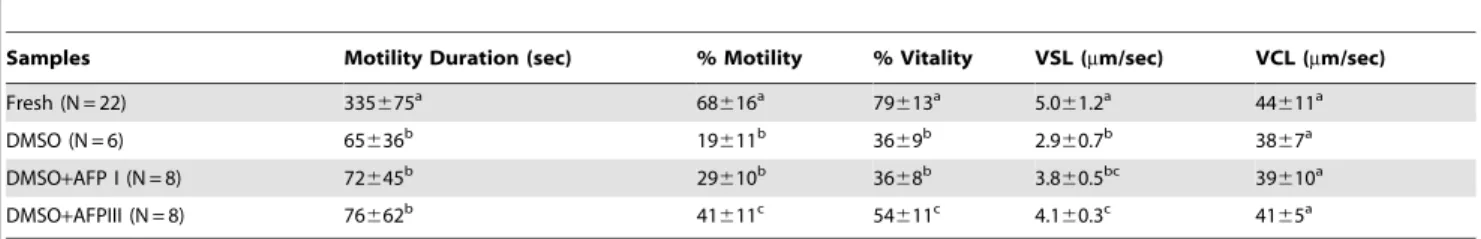
As reported in Table 2, sea bream semen cryopreserved in DMSO-containing extender showed, after thawing, a significant reduction of motility rate and duration, viability and straight-line velocity (VSL) with respect to fresh semen. The addition of AFPIII to DMSO extender improved the protection against freezing with respect to DMSO or DMSO plus AFPI. A percentage increase of motility rate and viability, and VSL was observed as compared to DMSO alone. No significant changes were found by the addition
of AFPI to DMSO extender. In agreement with Beiraoet al[5]
curvilinear velocity (VCL) seems to be almost unaffected under the tested experimental conditions. Motility duration shows a signif-icant reduction after cryopreservation procedure in all conditions. Fertilization trials were performed using fresh and DMSO-AFPIII cryopreserved gametes (since DMSO-AFPIII as cryoprotectant gave the best results in terms of sperm quality and proteins protection after cryopreservation). Our results demonstrated that there were no differences between fertilization ability obtained
with fresh (7769%) and cryopreserved spermatozoa (7367%).
Effect of antifreeze proteins on 2DE protein profiles of Isolated flagella and head plasma membranes from sea bream spermatozoa
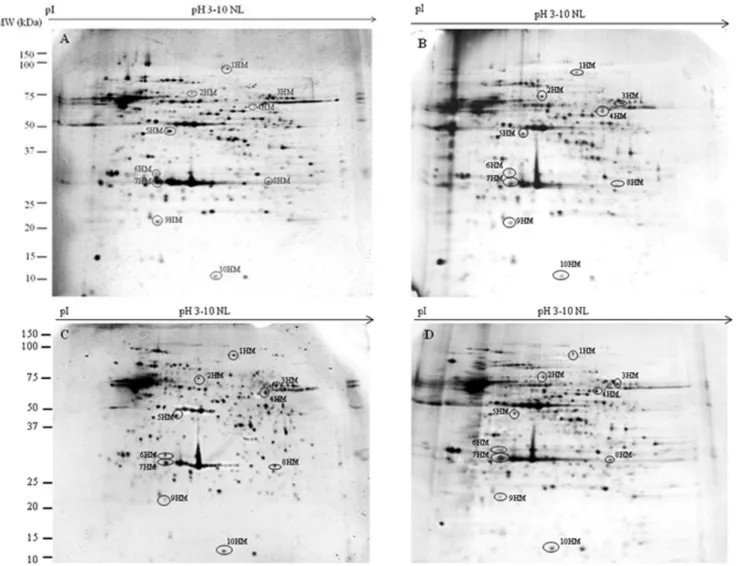
To evaluate the ability of DMSO, AFPI and AFPIII to protect plasma membrane proteins during the freezing-thawing proce-dure, we compared the 2DE-profiles of proteins extracted from isolated flagella and heads of spermatozoa before and after the freezing-thawing (in DMSO or DMSO+AFPI or DMSO+AFPIII) procedure. Representative 2DE gels of proteins extracted from isolated flagella and plasma membrane heads of fresh and cryopreserved semen are shown in Figures 1 (flagella) and 2 (head plasma membranes).
between 10 and 150 kDa and the isoelectric points ranged between 3.0 and 10 pH units.
Quantification and statistical analysis using Mann–Whitney test
(significance defined as p,0.05) showed that the normalized
volume of 19 out of 270 protein spots obtained from flagella and 10 out of 150 protein spots extracted from sperm heads plasma
membranes were$20% significantly different between at least one
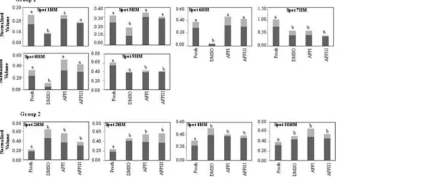
of the 3 cryopreserved groups and the fresh sperm. The gel location of these spots in sperm flagella and head plasma membranes is shown in Figures 1 and 2, respectively, while the corresponding normalized volumes, under the different experi-mental conditions, are reported in Figures 3 and 4. Following the freezing-thawing procedure, among the 19 highlighted spots from the flagella showed in Figure 1, 12 showed a significant decrease Table 2.Effects of different cryoprotectants on different motility and viability parameters in sea bream spermatozoa before freezing (fresh sperm) and after different thawing conditions.
Samples Motility Duration (sec) % Motility % Vitality VSL (mm/sec) VCL (mm/sec)
Fresh (N = 22) 335675a 68
616a 79
613a 5.0
61.2a 44
611a
DMSO (N = 6) 65636b 19
611b 36
69b 2.9
60.7b 38
67a
DMSO+AFP I (N = 8) 72645b 29
610b 36
68b 3.8
60.5bc 39
610a
DMSO+AFPIII (N = 8) 76662b 41
611c 54
611c 4.1
60.3c 41
65a
Values are expressed as means6SD for % motility, motility duration and % vitality and as means6SEM for VCL and VSL. Within each column, values with different superscript letters indicate significant statistical differences (P,0.05).
VSL: Velocity Straight Line. VCL: curvilinear velocity.
doi:10.1371/journal.pone.0099992.t002
Figure 1. Representative 2-D gels of proteins extracted from flagella isolated from fresh (A), cryopreserved with DMSO (B), with
DMSO plus AFPI (C) and with DMSO plus AFPIII (D) gilthead sea bream spermatozoa.Extracted flagellar proteins (60mg) were loaded
onto non-linear IPG strips pH 3–10, and following isoelectric focusing were separated by 12.5% SDS–PAGE. Gels were silver stained as described in Materials and Methods. Spots that changed their expression after cryopreservation procedure are highlighted with a circle.
(group 1 of Figure 3) while 7 showed a significant increase (group 2 of Figure 3). The results demonstrate that DMSO, DMSO+AFPI and DMSO+AFPIII act differently on the expression of each of these spots. The use of DMSO as cryoprotectant in the absence of AFPs determined the decrease of all spots of group 1 and the
increase of all spots of group 2. In the presence of AFPI (1mg/ml)
the freezing-thawing procedure determined the significant de-crease of 3 out of 12 spots within the group 1 and the inde-crease of 5 out of 7 within the group 2. Noteworthy, the addition of AFPIII
(1mg/ml) to DMSO extender determined the significant decrease
of only 1 spot belonging to group 1 and the increase of only 3 spots within group 2.
The results shown in Figure 4 demonstrate that the freezing-thawing procedure differently affects the expression of protein spots in the head membrane when different cryoprotectants were added to the extender medium. The cryopreservation protocol carried out in DMSO extender significantly decreased 6 spots of group 1 and increased 4 out of 4 spots of group 2. In the presence of AFPI or AFPIII only 2 out of 6 spots were significantly decreased and 4 out of 4 were increased.
Identification of proteins
Within the 29 protein spots (19 from flagella and 10 from head plasma membranes) which changed their expression following the freezing-thawing procedure, 18 (13 from flagella and 5 from head plasma membranes) were successfully detected by silver staining on 2DE preparative gels. Next, the detected spots were subjected to nanoHPLC ESI-Q-Tof MS/MS analysis for identification. Table 3 reports the results of such analyses with the identified 6 proteins clustered according to their putative function or their involvement in common metabolic pathways. 4 spots (7F1: alcohol dehydrogenase III; 10F1: glyceraldehyde-3-phosphate dehydroge-nase; 11F1: cytosolic malate dehydrogenase and 14F1: axonemal dyenein light intermediate polyopeptide 1) were identified in flagella. The spots 15Fl (from flagella) and 7HM (from head plasma membranes) were both identified as carbonic anhydrase while spots 19F1 (from flagella) and 10HM (from head plasma membranes) were identified as superoxide dismutase [Cu-Zn].
Figure 2. Representative 2-D gels of proteins extracted from the plasma membrane head isolated from fresh (A), cryopreserved
with DMSO (B), with DMSO plus AFPI (C) and with DMSO plus AFPIII (D) gilthead sea bream spermatozoa.Extracted flagellar proteins
Discussion
In the present study we demonstrate that the addition of AFPIII
(1mg/ml) to the extender medium significantly increased, with
respect to control (DMSO extender), viability, motility rate and VSL of thawed spermatozoa. Moreover, we analysed, for the first time in sperm, the effect of AFPs on the profile of proteins
extracted from isolated flagella and heads plasma membranes from sea bream spermatozoa. The results of the 2DE experiments suggest a higher ability of AFPIII, as compared to AFPI, to protect proteins during the freezing-thawing procedure. Studies of three-dimensional structures of both AFPs revealed important
differ-ences between them. AFPI is a small hydrophobica-helix, whose
direct interaction with phospholipids was suggested by Ingliset al.
Figure 3. Differences in relative abundance of 19 flagella spots in fresh and cryopreserved sea bream spermatozoa. Spots are
highlighted in Figure 1 and thexaxis identified the experimental conditions (fresh semen or cryopreserved with DMSO, with DMSO plus AFPI and with DMSO plus AFPIII). They-axis corresponds to the intensity of spots expressed as normalized spot volume. Spot intensity is expressed as mean (dark bars)6SD (light grey bars) of normalized spot volume (N = 18, that correspond to 6 different fish and 3 replicates for each). Different letters in the same graph indicate that mean values are significantly different by Mann–Whitney test (p,0.05). The spots showing a decrease of their expression after cryopreservation procedure are reported in the group 1, and those showing an increase are reported in the group 2.
doi:10.1371/journal.pone.0099992.g003
Figure 4. Differences in relative abundance of 10 sperm head membrane spots in fresh and cryopreserved sea bream spermatozoa. Spots are highlighted in Figure 2 and thexaxis identified the experimental conditions (fresh semen or cryopreserved with DMSO, with DMSO plus AFPI and with DMSO plus AFPIII). They-axis corresponds to the intensity of spots expressed as normalized spot volume. Spot intensity is expressed as mean (dark bars)6SD (light grey bars) of normalized spot volume (N = 18, that correspond to 6 different fish and 3 replicates for each). Different letters in the same graph indicate that mean values are significantly different by Mann–Whitney test (p,0.05). The spots showing a decrease of their expression after cryopreservation procedure are reported in the group 1, and those showing an increase are reported in the group 2.
Spot Number
Protein name
Acc.
No.a Species MascotScoreb Uniquepetidesc Peptidesequenced % SCe pIf MWg pIh MWi
7Fl Alcohol dehydrogenase class-3 gi|5902742 Sparus aurata 78 2 ITQGQGLLPDK 5 6.71 40930 6.41 43000
LVEDYMSK
10Fl Glyceraldehyde 3-phosphate dehydrogenase, testis-specific
gi|15146358 Pagrus major 463 8 VVAINDPFIDLK 32 6.36 36381 6.22 37000
YHGEVSEEDGK
VVVSAPSPDAPMFVMGVNEEK
GAHQNIIPASTGAAK
LTGMAFR
VPVADVSVVDLTCR
LISWYDNEFGYSHR
11Fl cytosolic malate dehydrogenase thermolabile form
gi|14583131 Sphyraena idiastes 114 2 VLVVGNPANTNCLIAAK 8 6.60 36463 6.37 36500
MDATAAELIEER
14Fl Axonemal dynein light intermediate polypeptide 1
gi|30172570 Mus musculus 42 1 ELYSQCFDELIR 4 8.21 29947 6.46 31000
15Fl = 7HM Carbonic anhydrase gi|213514954 Salmo salar Oncorhynchus mykiss.
145 3 QFHFHWGASDDR FPCELHLVHWNTK IGAANPR
12 7.6 28823 5.48 28000
gi|185135824
19Fl = 10HM Superoxide dismutase [Cu-Zn] gi|27462182 Pagrus major 131 1 HVGDLGNVTAGADNVAK 11 5.7 16167 6.00 13000
aNCBInr. accession number.
bMascot score obtained from MS/MS ion search against NCBInr [the Mascot score for an MS/MS match is based on the absolute probability (P) that the observed match between the experimental data and the database sequence is a random event, the reported score is210 Log(P) and the significance threshold is p,0.05; all the ion scores are higher than the threshold values (see also www.matrixscience.com).
cNumber of unique identifed peptides. dAmino acid sequences of identified peptides. eSequence coverage of the identified protein. fTheoretical p
I. gTheoretical MW. hExperimental pI. iExperimental MW.
doi:10.1371/journal.pone.0099992.t003
Fish
Sperm
Cryopre
servation
with
AFPs
ONE
|
www.ploson
e.org
8
June
2014
|
Volume
9
|
Issue
6
|
[27] working with liposomes. AFPIII is a globular protein with several hydrophilic and hydrophobic domains [37]. A direct interaction between AFPIII and the plasma membrane lipids has been indicated in our recent study [5]. In sea bream spermatozoa we showed that the addition of AFPIII to DMSO may avoid changes, which usually occur by freezing with DMSO alone, in membrane phospholipids composition as well as in the saturation/ unsaturation degree of their component fatty acids. Activities and expression of several proteins are well known to be affected by the lipid micro domain surrounding membrane proteins [38]. This aspect should be taken into account when examining the protein expression of sperm cryopreserved in the different experimental conditions tested in the present study, i.e. in DMSO without or with AFPs. In addition, the higher ability of AFPIII (with respect to AFPI) to protect spermatozoa during the freezing-thawing procedure could be the result of the above reported mechanism or/and to a better sinergy between AFPIII and DMSO (with respect to the sinergy between AFPI and DMSO).
The observed decrease in protein abundance may be due either to degradation following freezing-thawing stress [12], or leakage of proteins from spermatozoa to the extracellular medium, as reported in human [34], boar [32] and bull sperm [33], while the observed increase of some protein spots could be due to one or more post-translation modifications (phosphorylation, acetylation, glycation, etc.) following the cryopreservation procedure, as we demonstrated in gilthead sea bream spermatozoa [39] or be a consequence of the freezing/thawing procedure and/or exposure to cryoprotectants on the regulation of mRNA translation, since, as it has been demonstrated in mammals, spermatozoa are not transcriptionally and translationally dormant cells [40–42].
In the present study the protein profiles were obtained by using both isolated flagella (Fig. 1) and head plasma membranes (Fig. 2) of sea bream spermatozoa. In our previous paper [12] the protein profile was obtained by using proteins extracted from the whole spermatozoa of sea bass. Due to the different starting samples (spermatozoa obtained from different fish species; whole sperma-tozoa and isolated head plasma membranes and flagella) and also to differences in sample preparation (amount of DMSO used, incubation time, isolation procedure, etc.) it is very difficult to compare the effect of the cryopreservation procedure on the detected protein markers obtained in the present and in our previous work.
Among the six identified proteins spot 7Fl was an Alcohol dehydrogenase class-III (ADH III). Spot 10Fl was identified as Glyceraldehyde 3-phosphate dehydrogenase (GAPDH). This protein is expressed in sperm at specific stages of spermiogenesis and can still be detected in mature spermatozoa of vertebrates [43]. Variations in the expression of GAPDH block the progressive motility of spermatozoa [44]. Thus, it can be hypothesized that the observed decrease of sperm motility after the freezing-thawing procedure (Table 2) could be attributed, at least partially, to the reduction of GAPDH expression. Protein spot 11Fl matched with the cytosolic malate dehydrogenase thermolabile form (MDH), which was previously found in the mid-piece of ram, boar and buffalo spermatozoa [45]. Interestingly,
ADHIII, GAPDH and MDH show as common feature to be
linked to the bioenergetic system of the cell. NADH+H+
is a product of both the ADHIII and GAPDH activities. In mammalian spermatozoa the transfer of reduced equivalents from the cytosol to the mitochondria occurs by the Malate-Aspartate shuttle, in which two isoforms of MDH, cytosolic and mitochon-drial, are operative. By this shuttle, the hydrogen ions of the cofactor NADH produced in the cytosol can reach the electron transport chain in the mitochondria, and generate ATP by the oxydative phosphorylation (OXPHOS) system. Note that motility of fresh spermatozoa mainly depends on sperm ATP synthesized by mitochondrial OXPHOS [46]. Therefore, the observed reduction in GAPDH and MDH expression in cryopreserved sperm (in the presence of DMSO) may contribute to the reduced sperm motility observed after freezing-thawing procedure.
Spot 14Fl was identified as axonemal dynein light intermediate polypeptide 1 that belongs to the inner dynein arm light chain family and may play a dynamic role in flagellar motility [47].
The protein spots 15Fl-7HM matched with carbonic anhydrase (CA). In flatfish species CA plays an important role in the
regulation of sperm motility [48] but inS. aurata, to the best of our
knowledge, its role is unknown.
The spots protein 19Fl-10HM matched with superoxide dismutase [Cu-Zn] (CuZn-SOD). Oxygen free radicals have been implicated in a variety of circumstances relevant to the function of mammal spermatozoa; they may exert both toxic actions and participate in physiological processes [49]. It has been demon-strated that human spermatozoa are exceptionally well equipped with CuZn-SOD [49], and a relationship between loss of motility and peroxidation of spermatozoa lipids has been evidenced [50,51]. The increase in CuZn-SOD we observed (at the flagella and heads level) could be a consequence of oxidative stress during the cryopreservation procedure.
The present study represents the first evidence in fish on the ability of AFPs to affect the protein expression of two different spermatozoa domains, flagella and the plasma membranes. The addition of AFPIII to the cryopreservation medium was particu-larly active in increasing the quality of the thawed spermatozoa. Our results are complementary to those of a previous study on the effects AFPs on membrane lipid composition [5], and demon-strated that during the freezing–thawing procedure, AFPs –
mainly AFPIII - act by exerting a stabilizing effect onS. aurata
sperm conservation by reducing the cryopreservation-induced changes in the protein expression pattern.
These results may prove useful in elucidating the molecular mechanism underlying alterations associated with sperm cryo-preservation, and may have implications in the development of sperm conservation strategies.
Author Contributions
Conceived and designed the experiments: LZ JB MPH SV. Performed the experiments: LZ JB RS AG. Analyzed the data: LZ SV. Contributed reagents/materials/analysis tools: LZ JB AG. Wrote the paper: LZ SV.
References
1. Rana KJ (1995) Preservation of gametes. In: Bromage NR, Roberts RJ, editors. Broodstock Management and Eggs and Larval Quality. Cambridge, UK: Cambridge University Press. 53–76.
2. Suquet MD, Dreanno C, Fauvel C, Cosson J, Billard R (2000) Cryopreservation of sperm in marine fish. Aquacult Res 31: 231–243.
3. Fabbrocini A, Lubrano Lavadera S, Rispoli S, Sansone G (2000) Cryopreser-vation of sea breamSparus aurataspermatozoa. Cryobiology 40: 46–53.
4. Cabrita E, Robles V, Cun˜ado S, Wallace JC, Sarasquete C, et al. (2005) Evaluation of gilthead sea bream,Sparus aurata, sperm quality after cryopres-ervation in 5 ml macrotubes. Cryobiology 50: 273–84.
5. Beira˜o J, Zilli L, Vilella S, Cabrita E, Schiavone R, et al. (2012) Improving sperm cryopreservation with antifreeze proteins: effect on gilthead seabream
6. Labbe C, Martoriati A, Devaux A, Maisse G (2001) Effect of sperm cryopreservation on sperm DNA stability and progeny development in rainbow trout. Mol Reprod Dev 60: 397–404.
7. Zilli L, Schiavone R, Zonno V, Storelli C, Vilella S (2003) Evaluation of DNA damage inDicentrarchus labraxsperm following cryopreservation. Cryobiology 47: 227–235.
8. Pe´rez-Cerezales S, Martı´nez-Pa´ramo S, Cabrita E, Martı´nez-Pastor F, Herra´ez MP (2009) Evaluation of oxidative DNA damage promoted by storage in sperm from sex-reversed rainbow trout. Theriogenology 71: 605–613.
9. Maldjian A, Pizzi F, Gliozzi T, Cerolini S, Penny P, et al. (2005) Changes in sperm quality and lipid composition during cryopreservation of boar semen. Theriogenology 63: 411–421.
10. Mu¨ller K, Mu¨ller P, Pincemy G, Kurz A, Labbe C (2008) Characterization of sperm plasma membrane properties after cholesterol modification: consequences for cryopreservation of rainbow trout spermatozoa. Biol Reprod 78: 390–399. 11. Linhart O, Rodina M, Cosson J (2000) Cryopreservation of sperm in common
carp Cyprinus carpio: sperm motility and hatching success of embryos.
Cryobiology 41: 241–250.
12. Zilli L, Schiavone R, Zonno V, Rossano R, Storelli C, et al. (2005) Effect of cryopreservation on sea bass sperm proteins. Biol Reprod 72: 1262–1267. 13. Rodina M, Gela D, Kocour M, Alavi SM, Hulak M, et al. (2007)
Cryopreservation of tench, Tinca tinca, sperm: sperm motility and hatching success of embryos. Theriogenology 67: 931–940.
14. Beira˜o J, Cabrita E, Pe´rez-Cerezales S, Martı´nez-Pa´ramo S, Herra´ez MP (2011) Effect of cryopreservation on fish sperm subpopulations. Cryobiology 62: 22–31. 15. Gwo JC, Arnold CR (1992) Cryopreservation of Atlantic croaker spermatozoa:
evaluation of morphological changes. J Exp Zool 264: 444–453.
16. Pe´rez-Cerezales S, Martı´nez-Pa´ramo S, Beira˜o J, Herra´ez MP (2010) Evaluation of DNA damage as a quality marker for rainbow trout sperm cryopreservation an use of LDL as cryoprotectant. Theriogenology 74: 282–289.
17. Suquet MD, Dreanno C, Petton B, Normant Y, Omnes MH, et al. (1998) Long-term effects of the cryopreservation of turbot (Psetta maxima) spermatozoa. Aquat Living Resour 11: 45–48.
18. De Vries AL, Komatsu SK, Feeney RE (1970) Chemical and physical properties of freezing pointdepressing glycoproteins from Antarctic fishes. J Biol Chem 245: 2901–2908.
19. Duman JG, De Vries AL (1974) Freezing resistance in winter flounder,
Pseudopleuronectes americanus.Nature 247: 237–238.
20. Hew CL, Slaughter D, Joshi SB, Fletcher GL, Ananthanarayanan VS (1984) Antifreeze polypeptides from the Newfoundland ocean pout, Macrozoarece
americanus; presence of multiple compositionally diverse components. J Comp
Physiol B 155: 81–88.
21. De Vries AL (1988) The role of antifreeze glycoproteins and peptides in the freeze avoidance of antarctic fishes. Comp Biochem Physiol 90: 611–621. 22. Rubinsky B, Arav A, Fletcher GL (1991) Hypothermic protection–a
fundamen-tal property of ‘antifreeze’ proteins. Biochem Biophys Res Commun 180: 566– 567.
23. Hansen T, Smith F, Brockbank KGM (1993) AFP type III protein attenuates cell recoveries. Transplantation Proc 25: 3182–3184.
24. Wang JH (2000) A coomprehensive evaluation of the effect and mechanisms of antifreeze proteins during low temperature preservation. Cryobiology 41: 1–9. 25. Tomczak MM, Hincha DK, Estrada SD, Wolkers WF, Crowe LM, et al. (2002)
A mechanism for stabilization of membranes at low temperatures by an antifreeze protein. Biophys J 82: 874–881.
26. Venketesh S, Dayananda C (2008) Properties, potentials, and prospects of antifreeze proteins. Crit Rev Biotechnol 28: 57–82.
27. Inglis SR, Turner JJ, Harding MM (2006) Applications of type I antifreeze proteins: studies with model membranes & cryoprotectant properties. Curr Protein Pept Sci 7: 509–522.
28. Younis AI, Rooks B, Khan S, Gould KG (1998) The effects of antifreeze peptide III (AFP) and insulin transferrin selenium (ITS) on cryopreservation of chimpanzee (Pan troglodytes) spermatozoa. J Androl 19: 207–214.
29. Prathalingam NS, Holt WV, Revell SG, Mirczuk S, Fleck RA, et al. (2006) Impact of antifreeze proteins and antifreeze glycoproteins on bovine sperm during freeze-thaw. Theriogenology 66: 1894–1900.
30. Karanova MV, Tsvetkova LI (1994) Cryoprotective properties of antifreeze glycoproteins upon freezing of fish sperm. Izv Akad Nauk Biol 5: 818–827. 31. Karanova MV, Zsvetkova LI, Petropavlov NN (1997) Effect of antifreeze
glycoproteins on quality of cryoconserved carp sperm (Russian). Biofizika 42: 725–728.
32. Huang SY, Kuo YH, Lee WC, Tsou HL, Lee YP, et al. (1999) Substantial decrease of heat-shock protein 90 precedes the decline of sperm motility during cooling of boar spermatozoa. Theriogenology 51: 1007–1016.
33. Lessard C, Parent S, Leclerc P, Bailey JL, Sullivan R (2000) Cryopreservation alters the levels of the bull sperm surface protein P25b. J Androl 21: 700–707. 34. Cao WL, Wang YX, Xiang ZQ, Li Z (2003) Cryopreservation-induced decrease
in heat- shock protein 90 in human spermatozoa and its mechanism. Asian J Androl 5: 43–46.
35. Barbato F, Canese S, Moretti F, Misiti S, Laconi F, et al. (1998) Preliminary experien ces for cryopreservation ofSparus aurataandDiplodus puntazzosemen. In: Bartley DM, Basurco B, editors. Genetics and Breeding of Mediterranean Aquaculture Species. Zaragoza: CIHEAM (Cahiers Options Me´diterrane´ennes; n. 34) 281–287.
36. Taurino F, Stanca E, Siculella L, Trentadue R, Papa S, et al. (2012) Mitochondrial proteome analysis reveals depression of the Ndufs3 subunit and activity of complex I in diabetic rat brain. J Proteomics 75: 2331–2341. 37. Howard EI, Blakeley MF, Haertlein M, Petit-Haertlein I, Mitschler A, et al.
(2011) Neutron structure of type-III antifreeze protein allows the reconstruction of AFP–ice interface. J Mol Recognit 24: 724–732.
38. Pike LJ (2004) Lipid rafts: heterogeneity on the high seas. Biochem J, 378: 281– 292.
39. Zilli L, Schiavone R, Storelli C, Vilella S (2008) Effect of cryopreservation on phosphorylation state of proteins involved in sperm motility initiation in sea bream. Cryobiology 57: 150–155.
40. Ostermeier GC, Goodrich RJ, Moldenhauer JS, Diamond MP, Krawetz SA (2005) A suite of novel human spermatozoal RNAs. J Androl 26: 70–74. 41. Hamatani T (2012) Human spermatozoal RNAs. Fertil Steril 97: 275–281. 42. Das PJ, McCarthy F, Vishnoi M, Paria N, Gresham C, et al. (2013) Stallion
sperm transcriptome comprises functionally coherent coding and regulatory RNAs as revealed by microarray analysis and RNA-seq. PloS one 8: e56535. 43. Feiden S, Wolfrum U, Wegenr G, Kamp G (2008) Expression and
compartmentalisation of the glycolytic enzymes GAPDH and pyruvate kinase in boar spermatogenesis. Reprod Fertil Dev 20: 713–723.
44. Miki K, Ou W, Goulding E, Willis WD, Bunch DO, et al. (2004) Glyceraldehyde 3-phosphate dehydrogenase-S, a sperm-specific glycolytic enzyme, is required for sperm motility and male fertility. Proceed Nat Acad Sci 101: 16501–16506.
45. Kohsaka T, Takahara H, Tagami S, Sasada H, Masaki J (1992) A new technique for the precise location of lactate and malate dehydrogenases in goat, boar and water buffalo spermatozoa using gel incubation film. J Reprod Fertil 95: 201–209.
46. Perchec G, Jeulin C, Cosson J, Andre´ F, Billard R (1995) Relationship between sperm ATP content and motility of carp spermatozoza. J Cell Sci 108: 747–753. 47. Kastury K, Taylor WE, Shen R, Arver S, Gutierrez M, et al. (1997) Complementary deoxyribonucleic acid cloning and characterization of a putative human axonemal dynein light chain gene. J Clin Endocrinol Metab 82: 93047–93053.
48. Inaba K, Dre´anno C, Cosson J (2003) Control of flatfish sperm motility by CO2
and carbonic anhydrase. Cell Mot Cytoskel 55: 174–187.
49. Peeker R, Abramsson L, Marklund SL (1997) Superoxide dismutase isoenzymes in human seminal plasma and spermatozoa. Mol Hum Reprod 3: 1061–1066. 50. Alvarez JG, Storey BT (1982) Spontaneous lipid peroxidation in rabbit
epididymal spermatozoa. Its effect on sperm motility. Biol Reprod 27: 1102– 1108.