online | memorias.ioc.fiocruz.br Kinetoplastid flagellates (Euglenozoa and
Kineto-plastea) are a mixed group of free-living organisms and mono and dixenous parasites that colonise a variety of eu-karyotes, preferentially insects (Wallace 1966, Fernandes et al. 1993, Vickerman 1994, Podlipaev et al. 2004). The most studied kinetoplastids belong to the family Trypano-somatidae, which is characterised by a single flagellum and a relatively small kinetoplast, resembling a part of a single-branched mitochondrion of the cell containing a large mass of mitochondrial DNA (McGhee & Cosgrove 1980, Maslov et al. 1996, 2001, Merzlyak et al. 2001).
Trypanosomatids of the genera Trypanosoma, En-dotrypanum and Leishmania are dixenous parasites of vertebrates (Fernandes et al. 1993, Merzlyak et al. 2001, Podlipaev 2001). The remaining members of the family include monoxenous parasites of invertebrates (genera Blastocrithidia, Crithidia, Leptomonas, Herpetomonas and Wallaceina) and dixenous parasites of plants and in-sects (genus Phytomonas) (Dollet et al. 2000, Maslov et al. 2001, Podlipaev et al. 2004). Since the early 1960s, life-cycle information combined with morphological features such as cell shape and dimension and the
rela-tive positions of the nucleus and kinetoplast have been used to develop the current taxonomic system (Hoare 1964, Hoare & Wallace 1966, Wallace 1966).
Although morphological characteristics are often re-liable for identifying a genus, they are inadequate for identifying the species, mainly due to the high variabil-ity of morphology and their sensitivvariabil-ity to environmen-tal factors or culture conditions (Merzlyak et al. 2001). As stated by Yurchenko et al. (2006), the validity of most of the described trypanosomatid species is ques-tionable, with the notable exception of those of medical and veterinary importance (Trypanosoma and Leishma-nia), which have been studied extensively. Moreover, as shown by recent molecular phylogenetic studies (Maslov et al. 1996, Merzlyak et al. 2001, Podlipaev et al. 2004, Simpson et al. 2006, Yurchenko et al. 2006), most mor-phological genera are polyphyletic.
One of the main concerns in the field of trypanoso-matid research is the scarcity of information about the diversity of this group. It has been estimated that of the approximately one million insect species described, only ~2,500 have been examined for the potential presence of trypanosomatids (Momen 2001, Podlipaev 2001). For instance, of approximately 23,000 identified species of Hemiptera, one of the most studied orders, only 500-600 have been examined for flagellate parasites (Teixeira et al. 2000). Moreover, the current system does not take into ac-count genetic and evolutionary parameters; consequently, a great number of synonyms or descriptions of organisms of dubious origin have been generated (Podlipaev 2001). Financial support: COLCIENCIAS (021-2005)
+ Corresponding author: [email protected] Received 24 October 2010
Accepted 26 January 2011
Morphological and molecular description of
Blastocrithidia cyrtomeni
sp. nov. (Kinetoplastea: Trypanosomatidae) associated with
Cyrtomenus bergi
Froeschner (Hemiptera: Cydnidae) from Colombia
Ana Milena Caicedo1/+, Gerardo Gallego2, Jaime Eduardo Muñoz3, Harold Suárez2,
Gerardo Andrés Torres4, Humberto Carvajal5, Fanny Caro De Carvajal5,
Andrés Mauricio Posso3, Dmitriv Maslov6, James Montoya-Lerma1
1Biology Department 5Parasitology Laboratory, Universidad del Valle, Cali, Valle, Colombia 2Biodiversity and Biotechnology Program, International Center of Tropical Agriculture, Cali, Valle, Colombia 3Molecular Biology Laboratory, Universidad Nacional de Colombia,
Palmira, Valle, Colombia 4Microscopy Laboratory, Universidad del Cauca, Popayán, Cauca, Colombia 6Department of Biology, University of California, Riverside, California, USA
A new trypanosomatid species, Blastocrithidia cyrtomeni, is herein described using morphological and molecu-lar data. It was found parasitising the alimentary tract of the insect host Cyrtomenus bergi, a polyphagous pest. The morphology of B. cyrtomeni was investigated using light and transmission microscopy and molecular phylogeny was inferred from the sequences of spliced leader RNA (SL rRNA) - 5S rRNA gene repeats and the 18S small subunit (SSU) rRNA gene. Epimastigotes of variable size with straphanger cysts adhering to the middle of the flagellum were observed in the intestinal tract, hemolymph and Malpighian tubules. Kinetoplasts were always observed anterior to the nucleus. The ultrastructure of longitudinal sections of epimastigotes showed the flagellum arising laterally from a relatively shallow flagellar pocket near the kinetoplast. SL RNA and 5S rRNA gene repeats were positive in all cases, producing a 0.8-kb band. The amplicons were 797-803 bp long with > 98.5% identity, indicating that they originated from the same organism. According to the sequence analysis of the SL-5S rRNA gene repeats and the 18S SSU rRNA gene, B. cyrtomeni is different from all other known species or isolates of Trypanosomatidae. Both analyses indicate that among known species, it is most closely related to Blastocrithidia triatomae.
To overcome the aforementioned taxonomic limi-tations, biochemical, ultrastructural, serological and nutritional approaches to discriminate and define taxa were proposed 20 years ago (Wallace et al. 1983). The most recent characterisations and analyses of biodiver-sity of new trypanosomatid species are based on the use of molecular methods and phylogenetic analysis of gene sequences such as spliced leader RNA (SL rRNA), 5S rRNA and glycosomal glyceraldehyde phosphate dehydrogenase, which have proven to be powerful for discriminating among genera and species (Podlipaev et al. 2004, Yurchenko et al. 2006, Maslov et al. 2007, Svobodová et al. 2007). Recently, small subunit (SSU) rRNA gene-based phylogenies have been used to estab-lish major natural groups within a family (Merzlyak et al. 2001). Although useful in individual cases, these new criteria have not been applied uniformly (Podlipaev et al. 2004, Yurchenko et al. 2006). Therefore, it is highly advisable to use and compare at least two of these meth-ods when a new species is described.
In recent studies, we observed that Cyrtomenus bergi Froeschner, a polyphagous Cydnidae pest, is naturally infected with trypanosomatid flagellates in their salivary glands, intestinal tract and hemolymph (AM Caicedo, unpublished observations). In this study, we report the isolation of the trypanosomatid parasites from C. bergi as well as their morphological description and molecular characterisation. Based on these analyses, this organism has been identified as a new species of Blastocrithidia.
MATEriALS AnD METHoDS
Maintenance of insect hosts - Approximately 5,000 specimens of the burrowing bug C. bergi were collected in onion fields in Pereira (Risaralda, Colombia) from 2004-2008. The insects were maintained in an insectarium (23 ± 2ºC, relative humidity 65 ± 5%, L12:D12) in sterilised loamy clay soil and sand, mixed in a 3:1 proportion, at a moisture level approximating field conditions (33.5% gra-vimetric soil water) and fed on sprouting maize kernels.
Light microscopy - Hemolymph taken from fifth instar nymphs and adults was re-suspended in phos-phate-buffered saline (PBS). Drops of the suspension were smeared on slides, methanol dried and stained with Wright-Giemsa at room temperature (RT) for 24 h. Slides were examined at 100x with a Nikon Microphot. Measures and photographs of different morph types were taken using an NIS elements Nikon camera (v. 2.3). Wright-Giemsa-stained microscope slides, representing gut smears of the original host infected with the Try-panosomatidae isolate and described herein as Blas-tocrithidia cyrtomeni, were deposited with the Depart-ment of Microbiology of the Universidad del Valle, Cali (Valle de Cauca Province, Colombia).
Electron microscopy - Flagellates collected from the mid and hindgut of C. bergi adults were centrifuged at 10,000 g, washed in 0.1 M PBS and fixed in 2.5% glutar-aldehyde in 5 mM HCl and 0.1 M cacodylate buffer, first for 1 h at RT and then for at least 24 h at 4ºC. Samples were post-fixed in 2% osmium tetroxide in the same buf-fer for 2 h at RT. After dehydration in an ethanol
gradi-ent series, the cells were embedded in resin. Thin ultra-microtomed sections (0.7-0.8 µm) were stained with lead citrate and uranyl acetate and examined under a JEOL JEM 1010 microscope.
DNA extraction, polymerase chain reaction (PCR) amplification and sequencing - Pooled gut and hemo-lymph samples from five infected insects were homoge-nised and the debris was removed by centrifugation. Total DNA was extracted according to Rotureau et al. (2005) and Westenberger et al. (2004).
The SL rRNA gene repeats were amplified with the primers ME-1 5’-TTCTGTACTTTATTGGTA and
ME-2 5’-CAATAAAGTACAGAAACTG (Podlipaev
et al. 2004). The 5S rRNA gene repeats were amplified with the primers 5S-L 5’-CCGTCCGATTTGTGAAGT-TAAGC and 5S-R 5’-TAACTTCACAAATCGGACG-GGAT. Amplifications were done with Taq polymerase. The thermal cycling profile for both 5S rRNA and SL RNA genes consisted of the following steps: initial de-naturation at 94ºC for 2 min, 40 cycles of denaturing at 94ºC for 30 s, annealing at 50ºC for 30 s and extension at 72ºC for 30 s, followed by a final extension at 72ºC for 12 min. SSU rRNA genes were amplified with the primers S-762 5’-GACTTTTGCTTCCTCTA(A/T)TG and S-763 5’-CATATGCTTGTTTCAAGGAC (Maslov et al. 1996). Taq DNA polymerase was used for amplification and ther-mal cycling conditions were as follows: initial denaturation at 95ºC for 5 min, 5 cycles at 95ºC for 1 min, annealing at 45ºC for 30 s and extension at 65ºC for 1 min, 35 cycles at 95ºC for 1 min, 50ºC for 30 s and 72ºC for 1 min, and a final extension at 65ºC for 30 min (Maslov et al. 1996).
The amplified SL and 5S products were extracted from agarose gel using a QIAquick gel extraction kit (QUIGEN, Valencia, CA) and cloned into the pGEM-T Easy Vector (Promega, Madison, WI). The clones were sequenced at a commercial facility (Macrogen, Korea). The SSU rRNA gene amplification product was gel-purified by the same procedure and directly sequenced using a set of conserved internal primers (Maslov et al. 1996) at the University of California-Riverside IIGB Core Instrumentation facility.
The sequences were deposited in GenBankTM under
the following accessions: SL RNA gene and 5S rRNA gene repeat unit: FJ916991 and FJ916990 and SSU rRNA gene: FJ916992.
us-ing Modeltest, v. 3.06 (Posada & Crandall 1998), which corresponded to the general time-reversible model with a proportion of invariable sites I = 0.5029 and the gamma distribution shape parameter Г = 0.5416. The analyses involved maximum likelihood, distance and parsimony implemented with PAUP* 4.0 (Swofford 1998).
rESuLTS
Trypanosomatid infection (100% prevalence) was detected in C. bergi adult populations collected in onion fields in La Florida, Pereira. However, no obvious mor-phological or behavioural symptoms of infection were observed in individual hosts. Light microscopy revealed trypanosomatids within the intestinal tract, hemolymph and Malpighian tubules. Attempts were made to estab-lish a stable culture starting from hemolymph samples in several media, including NNN, LIT and Schneider, but these were not successful.
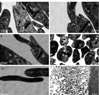
Light microscopy - Hemolymph and midgut smears revealed epimastigotes of variable size with straphanger cysts adhering to the middle of the flagellum. The cells varied in size and shape, from slender to rounded forms with short and long flagella. Some promastigotes could also be seen (Figs 1A, B, 8). The kinetoplast was always observed anterior to the nucleus.
Three forms of epimastigotes were seen: Form I, with a short flagellum and rounded body (Fig. 2A, B), Form II, with a slender body, pointed ends, a short flagellum and two to three spiral twists in the body (Fig. 3), and Form III, representing apparently dividing epimastigotes, with a sec-ond long flagellum separated from the cell body (Fig. 4).
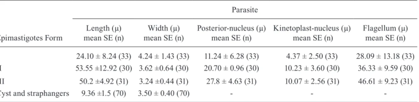
The morphometry of the three main morph types observed is summarised in Table I. The most noticeable difference between Forms I and III was flagellum length, with the latter being longer than the former. These forms also showed at least two-three twists, and the posterior end of Form I was more pointed than Form III (Figs 1A, 3, 4).
Furthermore, the presence of flagellar cysts (strap-hangers) was observed in Forms I and III, with groups of four-five cysts adhering to the middle of the flagellum (Figs 1A, 5). In addition, relatively small cells, appar-ently generated by unequal division, were also observed as flagellar attachments (Figs 1A, 5), suggesting that they represent immature cysts. Cyst forms varied from small and round, to bacillus-like cells, and to those with anterior pointed ends. In all forms, the kinetoplast was anterior to the nucleus. The width of the mature strap-hangers ranged from 2.42-4.43 µm, the length ranged from 5.86-11.93 µm.
Transmission (TEM) and scanning (SEM) electron microscopy - The ultrastructure of longitudinal sec-tions of epimastigotes isolated from midgut showed that the flagellum arises laterally from a relatively shallow flagellar pocket near the kinetoplast. The typical disk-shaped kinetoplast was located next to the bottom of the flagellar pocket (Fig. 6). We observed the longitudinal division of epimastigotes with kinetoplasts and bi-fla-gellum (Fig. 7). Few promastigotes were seen with the flagellum arising in front of the kinetoplast in the midg-ut (Fig. 8). Different forms of amastigotes with some acidocalcisomes and basophilic granules were also ob-served throughout the cell body (Fig. 9). The flagellum was supported by a nonprominent paraflagellar rod (Fig. 10). The parasites were attached to the intestine by the flagellum and different forms of parasites were observed (Fig. 11). In general, the nucleus was found mainly in the middle or close to the posterior end of the cell body, covering most of the cell width. The apparent absence of an undulated membrane, one of the features of epimas-tigotes, is noteworthy.
Figs1-5: morphology of Blastocrithidia cyrtomeni sp. nov. from in-fected hosts as revealed by light microscopy. 1A: different forms (small body with straphangers with typical division forms); 1B: slender with needle-like posterior end with short flagella; 2A: epi and promastigote forms (up); 2B: slender body, short flagellum, body with three spiral twists; 3: rounded forms with short flagellum; 4: groups of epimastig-otes with straphangers at the middle of the flagellum; 5: a cell with a second flagellum, apparently during an early stage of cell division.
Using SEM, it was possible to observe numerous parasites adhered to the intestinal wall by their flagella (Fig. 12). Typical elongated epimastigotes with strap-hanger cysts were also observed on the midgut of C. bergi adults (Fig. 13).
PCR amplification and phylogenetic analysis - DNA was extracted from the midgut of C. bergi adults, fourth and fifth instar nymphs and eggs. PCR amplification of SL RNA gene repeats was positive in all cases, produc-ing a 0.8 kb band correspondproduc-ing to a monomeric repeat unit, often accompanied by higher molecular weight bands, possibly representing repeat dimers and trimers. Similarly, PCR amplification with 5S rRNA primers re-sulted in a monomeric amplicon of the same size and higher molecular weight products that were apparently oligomeric (Fig. 14).
Cloning and sequencing of the SL RNA and 5S rRNA monomeric amplicons showed that they have the same re-peat unit, wherein individual SL RNA genes were inter-spersed with 5S rRNA genes, forming a linkage arrange-ment often observed in trypanosomatids (Westenberger et al. 2004). Individual repeats, derived from more than 30 sequenced amplicons, were 797-803 bp long and > 98.5% identical to each other, indicating that they all originated from the same organism (Thomas et al. 2005).
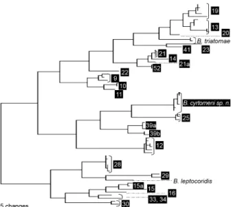
Seven individual SL repeat units were compared with the database of the repeats available from other Trypano-somatidae (Maslov et al. 2007) using neighbour-joining cluster analysis. The trypanosomatid from C. bergi clus-tered with members of the B. triatomae-Blastocrithidia leptocoridis clade (not shown). A part of this analysis, covering the Blastocrithidia group, is shown in Fig. 15. The repeats from the C. bergi parasite are unique and clearly distinct from B. triatomae (59.2% identity level) and B. leptocoridis (55.8% identity). The C. bergi parasite is most closely related to the organisms from Typing Unit 25,an unnamed trypanosomatid species found in two spe-cies of Leptoscelis (Hemiptera: Coreidae) from Costa Rica and Ecuador. However, the relatively low repeat identity level (~63%) can clearly separate these two organisms.
The identification of the new parasite as Blastocrithidia was verified by the analyses of SSU rRNA gene sequenc-es and comparison with selected members of the major known phylogenetic clades of the Trypanosomatidae. The general topology of the resulting trees was analogous to those derived in previous studies (Merzlyak et al. 2001,
TABLE I
Morphometric analysis of Blastocrithidia cyrtomeni - midgut
Epimastigotes Form
Parasite
Length (µ) mean SE (n)
Width (µ) mean SE (n)
Posterior-nucleus (µ) mean SE (n)
Kinetoplast-nucleus (µ) mean SE (n)
Flagellum (µ) mean SE (n)
I 24.10 ± 8.24 (33) 4.24 ± 1.43 (33) 11.24 ± 6.28 (33) 4.37 ± 2.50 (33) 28.09 ± 13.18 (33) II 53.55 ±12.92 (30) 3.62 ±0.64 (30) 20.70 ± 0.96 (30) 10.23 ± 3.60 (30) 36.33 ± 9.59 (30)
III 50.2 ±4.92 (31) 3.24 ±0.44 (31) 27.8 ± 4.63 (31) 10.07 ± 2.56 (31) 46.61 ± 9.23 (31) Cyst and straphangers 9.36 ±1.5 (70) 3.50 ± 0.40 (70) - -
-Figs 12-13:scanning electronic microscopy of Blastocrithidia cyrtome-ni sp. nov. in midgut of Cyrtomenus bergi adults. Most elongated forms adhered by flagellum to the intestine wall (12). Epimastigotes form with straphanger cysts and round body cell with long flagellum (13). Yurchenko et al. 2006, 2008) (data not shown). In all analyses, the C. bergi parasites were found to be closely associated with B. triatomae (the only available represen-tative of the aforementioned clade), with 100% bootstrap support in all the methods used (Fig. 16).
Based on the presence of epimastigotes and flagel-lar cysts and on the molecuflagel-lar phylogenetic analyses, we concluded that the parasite from C. bergi is a new trypanosomatid species that should be assigned to the genus Blastocrithidia.
Taxonomic summary - Class: Kinetoplastea Honig-berg 1963 emends. Vickerman 1976. Subclass: Metaki-netoplastina Vickerman 2004. Order: Trypanosomatida Kent 1880 stat. nov. Hollande 1952. Family: Trypanoso-matidae Doflein 1951. B. cyrtomeni sp. nov. Caicedo, Gal-lego, Muñoz, Montoya, Carvajal & Maslov et al. 2009.
Type host - Salivary glands, intestinal tract and hemo-lymph of C. bergi Froeschner, 1960 (Hemiptera: Cydnidae).
Type locality - La Florida, Pereira, Risaralda Provin-ce, Colombia (4º45’38.65’’N 75º36’57.49’’W).
Type data - Microscope slides (MUV 001-006), des-ignated as hapantotype, were deposited in the type col-lection of the School of Microbiology, Universidad del Valle, Cali, Colombia.
Species characters - The species is mainly defined by molecular phylogenetic criteria. The new species is clearly distinguished from the other known trypanoso-matids, including the closely related B. triatomae, by the SL RNA - 5S rRNA gene repeat sequences (GenBankTM
accessions FJ916991, FJ916990).
Fig.14:amplification of the 5S rRNA (A) and spliced leader RNA (B) genes of Blastocrithidia cyrtomeni sp. nov. M: molecular marker [1 kb plus DNA Ladder (Invitrogen)]; 1: midgut of female; 2: midgut of female; 3: egg; 4: midgut of female; 5: midgut of the fifth instar nymph; 6: midgut of fourth instar nymph; 7: in vitro.
Fig. 15: neighbor-joining analyses of the conserved regions of the spliced leader RNA gene repeats from Blastocrithidia cyrtomeni along with its closest relatives, which include Blastocrithidia triatomae,
Blastocrithidia leptocoriodis and several unnamed Blastocorithidia
spp represented by the respective Typing Units. The unit designations (in black boxes) correspond to the trypanosomatid samples described by Maslov et al. (2007) and Westenberger et al. (2004). The tree is unrooted. The bar represents 0.01 substitutions per site.
Fig.16:bootstrap majority consensus maximum likelihood small sub-unit rRNA gene tree of the selected Trypanosomatidae with emphasis on the position of Blastocrithidia cyrtomeni sp. nov. The tree (-Ln = 9403.73836) was inferred by assuming the general time-reversible model of sequence evolution with I = 0.5029 and Г= 0.5416. The boot -strap percent values shown were derived from 100 replicates with maximum likelihood (1st value), 1,000 replicates with minimum evo-lution (2nd value) and 1,000 replicates with parsimony (3rd value). Asterisks mean bootstrap values below 50%.
Remarks - Epimastigotes often display straphanger cysts adhered to the middle of the flagellum in clusters of two-four. The species has proven to be refractory to cultivation in the media tested.
Etymology - The species name is derived from the insect host.
DiSCuSSion
The new species of Blastocrithidia Laird, 1959, named B. cyrtomeni sp. nov., was isolated from hemo-lymph and the intestinal tract of C. bergi. Identification of this trypanosomatid was based on morphological descriptions using light and electron microscopic tech-niques and supported by analyses of molecular markers and phylogeny (Maslov et al. 1996, 2007, Merzlyak et al. 2001, Yurchenko et al. 2006).
Blastocrithidia familiaris Gibbs (1950), B. leptocoridis McCulloch (1915), Blastocrithidia ortheae Uribe (1926), Blastocrithidia sandoni Gibbs (1951) and Blastocrithidia euchisti Hanson and McGhee, 1961 (Cerisola et al. 1971). In B. triatomae, Cerisola et al. (1971) mentioned the cysts as a remarkable feature, describing them as small bodies of variable size hanging from the flagellum in most specimens.
As mentioned by Merzlyak et al. (2001), the origi-nal identification of Blastocrithidia gerricola from the gut of Gerris lacustris was based on the epimastigotes observed with some rare promastigotes. This is also the case with B. cyrtomeni, with epimastigotes being the predominant forms and few promastigotes observed.
We did not observe the “cytoplasmatic bridge” men-tioned by Schaub and Böker (1986) in their microscopic studies of B. triatomae, nor were we able to find cysts connected to the posterior end of epimastigotes as re-ported by the authors in three cases.
The posterior end of members of the Blastocrithidia species is a variable feature. In B. cyrtomeni, it was ob-served as needle-like in light SEM and TEM images (Figs 1B, 13). In contrast, Schaub and Böker (1986) observed that the broad posterior end was rolled up in a spiral in the first SEM images of B. triatomae and B. gerridis.
B. cyrtomeni has nearly the same body length as B. gerridis (Wallace 1966) and is almost double the average size of B. triatomae (Cerisola et al. 1971). The flagel-lum length is the most noticeable difference among these three species; it is shortest in B. gerridis and longest in B. cyrtomeni (Table II).
Common features among these three Blastocrithidia species are the position of the kinetoplast, which is ante-rior to the nucleus, and the width of the nucleus, which is almost the same as that of the body (Wallace 1966, Cerisola et al. 1971).
An undulated membrane in the epimastigotes was not observed. Thus far, only small forms of B. leptotry-panoides (Hollande 1922) have been reported without an undulating membrane (Wallace 1966).
In Leptomonas wallacei, a trypanosomatid from the lygaeid bug Oncopeltus fasciatus, straphanger cysts ad-here to the beginning of the base of the flagellar pocket (Romeiro et al. 2001). The dimensions of the straphang-ers of B. cyrtomeni are almost three times larger in length and width (9.36 and 3.50 µm, respectively), than those of B. triatomae (3.4 and 1.9 µm, respectively) (Ce-risola et al. 1971).
Although we observed oval cells of B. cyrtomeni ad-hered to the epithelium of the insect’s midgut (Fig. 11) but free in the hemolymph, we were unable to elucidate whether these forms represent intermediate develop-mental stages toward the formation of draught-resistant cysts, such as in B. triatomae, a parasite of Triatominae bugs (Schaub et al. 1990).
We also do not know whether the host mounts a re-sponse aimed at reducing the growth rate of the parasite population. There is no evidence of a successful response to eliminate the parasite from the body of the host (com-plete recovery), but it is likely that the insect regulates the parasite population at some permanent steady-state
level. We found a special case of direct transmission (transovarial), in which the infection is transferred by a parent to its unborn offspring. Therefore, despite the fact that B. cyrtomeni has an extremely high rate of di-rect reproduction within the host, it is unlikely that this microparasite is a pathogen.
Using PCR amplification of SL rRNA genes from 16 samples of parasites isolated from the intestinal tract and eggs of C. bergi, it was possible to identify B. cyrtomeni sp. nov. using the approach proposed by Maslov et al. (2007), who established that a single repeat of SL is suf-ficient to serve as a marker for a natural clone or species of trypanosomatid and to distinguish among closely re-lated trypanosomatids.
It is critical to establish the presence and prevalence of these monoxenous trypanosomatids in other insect species and to evaluate in greater detail whether the association between insect pests and trypanosomatids could be regulating the action of microorganisms used for biological control.
ACKnoWLEDGEMEnTS
To Manuel Castrillón, for measuring the parasites, to Meleny Ramírez, for helping to obtain permanent slides, to Efren, for transmission microscopy pictures, to José Arroyabe, for scan-ning microscopy pictures, to Nydia Betancourth, for helping with the figures, and to Dra Tina Trenczek and Dr Gustavo Vallejo, for the first instructions at the beginning of the research.
rEFErEnCES
Cerisola JA, del Prado CE, Rohwedder R, Bozzini JP 1971. Blas-tocrithidia triatomae n. sp. found in Triatoma infestans from Ar-gentina. J Protozool18: 503-506.
Dollet M, Sturm NR, Sanchez Moreno M, Campbell DA 2000. 5S ribo-somal RNA gene repeat sequences define at least eight groups of plant trypanosomatids (Phytomonas spp.): Phloem-restricted patho-gens form a distinct section. J Eukaryot Microbiol47: 569-574.
Fernandes AP, Nelson K, Beverely SM 1993. Evolution of nuclear ribo-somal RNA in kinetoplastid protozoa: perspectives on the age and origins of parasitism. Proc Natl Acad Sci USA90: 11608-11612.
Hoare CA 1964. Morphological and taxonomical studies on mam-malian trypanosomes. x Revision of the systematics. J Proto-zool 11: 200-210.
TABLE II
Dimensions of Blastocrithidia species
Species
Body length (µ)
Flagellum length (µ)
Blastocrithidia gerridis 17.5 70 (48.6) 1.75-2.7 (2.3) Blastocrithidia gallardoi 30-38 39-90
Blastocrithidia triatomae 14.8-32 (25) 19.5-28.5 (22.5)
Blastocrithidia lituri, Blastocrithidia nalipi, Blastocrithidia spinigeri, Blastocrithidia vacuolata
20
-Blastocrithidia apiomerusi 34 20
Blastocrithidia cyrtomeni 53.5 ± 12.9 36.3 ± 9.5
Hoare CA, Wallace FG 1966. Developmental stages of trypanosoma-tid flagellates: a new terminology. Nature212: 1385-1386.
Maslov DA, Lukes J, Jirku M, Simpson L 1996. Phylogeny of try-Phylogeny of try-panosomes as inferred from the small and large subunit rRNAs: implications for the evolution of parasitism in the trypanosomatid protozoa. Mol Biochem Parasitol75: 197-205.
Maslov DA, Podlipaev SA, Lukes J 2001. Phylogeny of the kineto-plastida: taxonomic problems and insights into the evolution of parasitism. Mem Inst Oswaldo Cruz96: 397-402.
Maslov DA, Westenberger SJ, xing x, Campbell DA, Sturm NR 2007. Discovery and barcoding by analysis of spliced leader RNA gene sequences of new isolates of Trypanosomatidae from Heteroptera in Costa Rica and Ecuador. J Eukaryot Microbiol54: 57-65.
McGhee RB, Cosgrove WB 1980. Biology and physiology of the low-er Trypanosomatidae. Microbiol Rev 44: 140-173.
Merzlyak E, Yurchenko A, Kolesnikov A, Alexandrov K, Podlipaev S, Maslov DA 2001. Diversity and phylogeny of insect trypano-somatid based on small subunit rRNA genes: Polyphyly of Lepto-monas and Blastocrithidia. J Eukaryot Microbiol48: 163-171.
Momen H 2001. Some current problems in the systematics of trypano-somatids. Int J Parasitol31: 640-642.
Podlipaev S 2001. The more insect trypanosomatids under study, the more diverse Trypanosomatidae appears. Int J Parasitol31: 648-652.
Podlipaev S, Sturm N, Fiala I, Fernandes O, Westenberger SM, Campbell D, Lukes J 2004. Diversity of insect trypanosomatids assessed from the spliced leader RNA and 5S rRNA genes and intergenic regions. J Eukaryot Microbiol 51: 283-290.
Posada D, Crandall KA 1998. Modeltest: testing the model of DNA substitution. Bioinformatics14: 817-818.
Romeiro A, Solé-Cava A, Sousa MA, de Souza W, Attias M 2001. Ultrastructural and biochemical characterization of promastigote and cystic forms of Leptomonas wallacei n. sp. isolated from the intestine of its natural host Oncopeltus fasciatus (Hemiptera: Ly-gaeidae). J Eukaryot Microbiol47: 208-220.
Rotureau B, Gego A, Carme B 2005. Trypanosomatid protozoa: a simplified DNA isolation procedure. Exp Parasitol111: 205-209.
Schaub GA, Böker CA 1986. Scanning electron microscopic studies of Blastocrithidia triatomae (Trypanosomatidae) in the rectum of
Triatoma infestans (Reduviidae). J Protozool33: 266-270.
Schaub GA, Reduth D, Pudney M 1990. The peculiarities of Blas-tocrithidia triatomae. Parasitol Today6: 361-363.
Simpson AGB, Stevens JR and Lukes J 2006. The evolution and di-versity of kinetoplastid flagellates. Trends Parasitol 22: 168-174.
Svobodová M, Zidková L, Cepieka I, Obornik M, Lukes J, Votypka J 2007. Sergeia podlipaevi gen. nov., sp. nov. (Trypanosomatida, Kinetoplastida): a parasite of the biting midges (Ceratopogoni-dae, Diptera). Int J Syst Evol Microbiol57: 423-432.
Swofford DL 1998. PAUP* 4.0: phylogenetic analysis using parsi-mony (and other methods), beta version, Sinauer Associates, Sunderland, MA.
Teixeira MMG, Serrano MG, Camargo EP 2000. New data from old trypanosomatid preparations. Parasitol Today16: 261-263.
Thomas S, Westenberger SJ, Campbell DA, Sturm NR 2005. In-tragenomic spliced leader RNA array analysis of kinetoplastids reveals unexpected transcribed region diversity in Trypanosoma cruzi. Gene352: 100-108.
Thompson JD, Gibson TJ, Plewniak F, Jeanmougin F, Higgins DG 1997. The CLUSTALx windows interface: flexible strategies for multiple sequence alignment aided by quality analysis tools.
Nucleic Acids Res25: 4876-4882.
Vickerman K 1994. The evolutionary expansion of the trypanosoma-tid flagellates. Int J Parasitol24: 1317-1331.
Wallace FG 1966. The trypanosomatid parasites of insects and arach-nids. Exp Parasitol18: 124-193.
Wallace FG, Camargo EP, McGhee RB, Roitman I 1983. Guidelines for the description of new species of lower trypanosomatids.
J Protozool 30: 308-313.
Westenberger SJ, Strum NR, Yanega D, Podlipaev SA, Zeledon R, Campbell DA, Maslov DA 2004. Trypanosomatid biodiversity in Costa Rica: genotyping of parasites from Heteroptera using the spliced leader RNA gene. Parasitology129: 537-547.
Yurchenko VA, Lukeš J, Tesařová M, Jirků M, Maslov DA 2008. Mor -phological discordance of the new trypanosomatid species phyloge-netically associated with the genus Crithidia. Protist159: 99-114.